Skip to product information
1 of 8

PAX8 Recombinant Rabbit mAb (SDT-R027)

PAX8 Recombinant Rabbit mAb (SDT-R027)

Catalog Number: S0B2071 Application: WB, IHC-P, FCM, ChIP Reactivity: Hu, Ms, Rt Conjugation: Brand: Starter
Price:
Regular price $100 USD
Regular price Sale price $100 USD
Size:

For shipping services or bulk orders, you may request a quotation.
Secure checkout with
View full details

Product Details

Product Specification


Host Rabbit
Antigen PAX8
Immunogen Recombinant Protein
Location Nucleus
Accession Q06710
Clone Number SDT-R027
Antibody Type Rabbit mAb
Application WB, IHC-P, FCM, ChIP
Reactivity Hu, Ms, Rt
Purification Protein A
Concentration 2 mg/ml
Physical Appearance Liquid
Storage Buffer

PBS, 40% Glycerol, 0.05%BSA, 0.03% Proclin 300

Stability & Storage

12 months from date of receipt / reconstitution, -20 °C as supplied

Dilution


application dilution species
IHC-P 1:1000 Hu, Ms, Rt
FCM 1:250
WB 1:500 Hu
ChIP 1:20-1:50 Hu

Background

Paired box gene 8, also known as PAX8, is a protein which in humans is encoded by the PAX8 gene. This gene is a member of the paired box (PAX) family of transcription factors. Members of this gene family typically encode proteins which contain a paired box domain, an octapeptide, and a paired-type homeodomain. The PAX gene family has an important role in the formation of tissues and organs during embryonic development and maintaining the normal function of some cells after birth. PAX8 (and PAX2) is one of the important regulators of urogenital system morphogenesis.

Picture

Validation Data

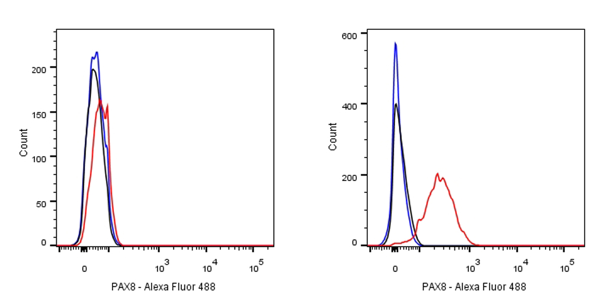
Flow cytometric analysis of HeLa (left) / SK-OV-3 (right) cells labelling PAX8 antibody at 1/250 dilution (0.1ug)/ (red) compared with a Rabbit monoclonal IgG (Black) isotype control and an unlabelled control (cells without incubation with primary antibody and secondary antibody) (Blue).

Goat Anti-Rabbit IgG Alexa Fluor® 488 at 1/1000 dilution was used as the secondary antibody.

Negative control: HeLa

Western Blot

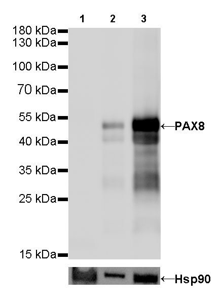
WB result of PAX8 Rabbit mAb                

Primary antibody: PAX8 Rabbit mAb at 1/500 dilution
Lane 1: Hela whole cell lysate 20 µg
Lane 2: SK-OV-3 whole cell lysate 20 µg
Lane 3: OVCAR-3 whole cell lysate 20 µg
Negative control: Hela whole cell lysate    

Secondary antibody: Goat Anti-Rabbit IgG, (H+L), HRP conjugated at 1/10000 dilution
Predicted MW: 48 kDa
Observed MW: 48 kDa
Exposure time: 30s

ChIP

Chromatin immunoprecipitation (ChIP) was performed on OVCAR-3 cells cross - linked with 1% formaldehyde for 10 min, then chromatin was fragmented by sonication. 
Parallel reactions used PAX8 Recombinant Rabbit mAb (SDT-R027) and Rabbit mAb IgG Isotype Control (SDT-R173) at 1:50 for immunoprecipitation. 
Post - immunoprecipitation, both samples were washed, eluted, and cross - links reversed. Purified DNA was analyzed by qPCR.
qPCR showed the enrichment of CTGF, E2F1 and SAT-α in PAX8 Recombinant Rabbit mAb (SDT-R027)-immunoprecipitated sample.

Immunohistochemistry

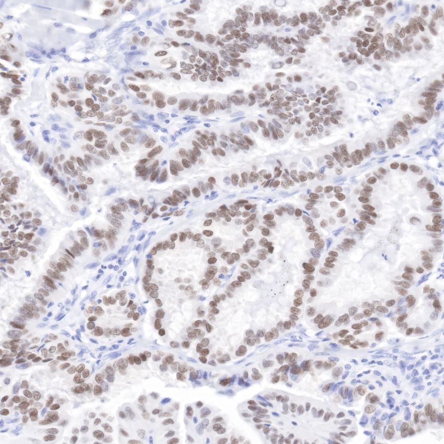
IHC shows positive staining in paraffin-embedded human thyroid cancer.

Anti-PAX8 antibody was used at 1/1000 dilution, followed by a Goat Anti-Rabbit IgG H&L (HRP) ready to use. Counterstained with hematoxylin.

Heat mediated antigen retrieval with Tris/EDTA buffer pH9.0 was performed before commencing with IHC staining protocol.

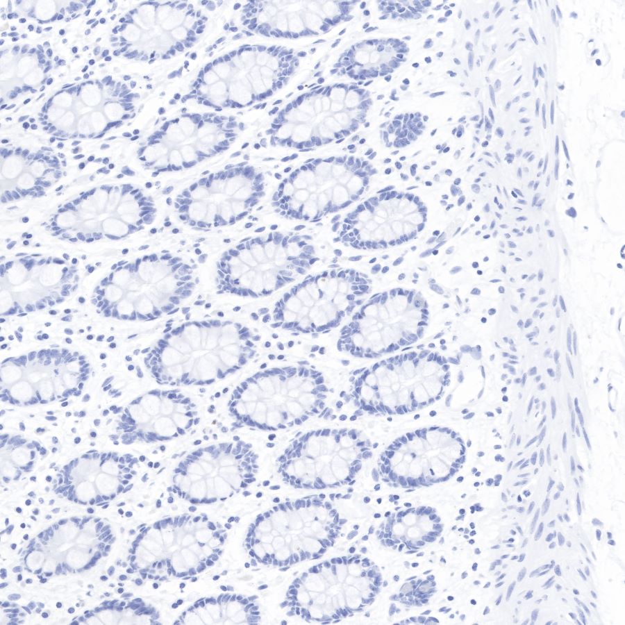
Negative control: IHC shows negative staining in paraffin-embedded human colon.

Anti-PAX8 antibody was used at 1/1000 dilution, followed by a Goat Anti-Rabbit IgG H&L (HRP) ready to use. Counterstained with hematoxylin.

Heat mediated antigen retrieval with Tris/EDTA buffer pH9.0 was performed before commencing with IHC staining protocol.

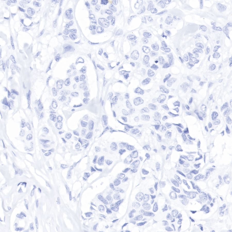
Negative control: IHC shows negative staining in paraffin-embedded human breast cancer.

Anti-PAX8 antibody was used at 1/1000 dilution, followed by a Goat Anti-Rabbit IgG H&L (HRP) ready to use.

Counterstained with hematoxylin.

Heat mediated antigen retrieval with Tris/EDTA buffer pH9.0 was performed before commencing with IHC staining protocol.

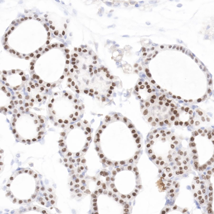
IHC shows positive staining in paraffin-embedded mouse thyroid.

Anti-PAX8 antibody was used at 1/1000 dilution, followed by a Goat Anti-Rabbit IgG H&L (HRP) ready to use.

Counterstained with hematoxylin.

Heat mediated antigen retrieval with Tris/EDTA buffer pH9.0 was performed before commencing with IHC staining protocol.

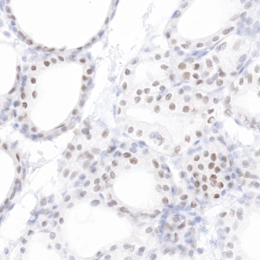
IHC shows positive staining in paraffin-embedded rat thyroid. Anti-PAX8 antibody was used at 1/1000 dilution, followed by a Goat Anti-Rabbit IgG H&L (HRP) ready to use.

Counterstained with hematoxylin.

Heat mediated antigen retrieval with Tris/EDTA buffer pH9.0 was performed before commencing with IHC staining protocol.