Mouse C5a ELISA Kit
Mouse C5a ELISA Kit
Product Details
Product Details
Product Specification
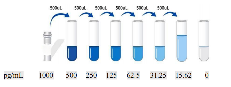
| Usage | I. Sample Handling and Requirements: 1. The detection range of the kit is not equivalent to the concentration range of the analyte in the sample. Before the experiment, it is recommended to estimate the analyte concentration in the sample based on relevant literature and conduct preliminary experiments to determine the actual concentration. If the analyte concentration in the sample is too high or too low, please dilute or concentrate the sample appropriately. 2. If the sample type to be tested is not listed in the instructions, it is recommended to conduct preliminary experiments to verify the validity of the assay. 3. Serum: Collect whole blood in a serum separator tube and incubate at room temperature for 2 hours or at 2-8°C overnight. Then, centrifuge at 1000×g for 20 minutes. The supernatant can be collected or stored at -20°C or -80°C. Avoid repeated freezing and thawing. 4. Plasma: Collect the sample using EDTA or heparin as an anticoagulant. Centrifuge the sample at 1000 × g for 15 minutes at 2-8°C within 30 minutes of collection. The supernatant can be assayed or stored at -20°C or -80°C, but avoid repeated freezing and thawing. 5. Tissue Homogenization: Rinse the tissue with pre-chilled PBS (0.01M, pH 7.4) to remove residual blood (lysed red blood cells in the homogenate may affect test results). Weigh the tissue and mince it. Add the minced tissue to the appropriate volume of PBS (generally a 1:9 weight-to-volume ratio, e.g., 1 g of tissue sample to 9 mL of PBS. The specific volume can be adjusted based on experimental needs and recorded. It is recommended to add protease inhibitors to the PBS) in a glass homogenizer and thoroughly grind on ice or in a homogenizer. To further lyse tissue cells, the homogenate can be sonicated or repeatedly frozen and thawed. Finally, centrifuge the homogenate at 5000×g for 5-10 minutes, and remove the supernatant for analysis. 6. Cell Culture Supernatant: Centrifuge at 1000×g for 20 minutes. Remove the supernatant for analysis, or store at -20°C or -80°C, but avoid repeated freeze-thaw cycles. 7. Cell Lysis Buffer: Gently wash adherent cells with ice-cold PBS, then trypsinize and harvest by centrifugation at 1000×g for 5 minutes. Suspension cells can be harvested directly by centrifugation. Wash the harvested cells three times with ice-cold PBS and resuspend in 150-200 μL of PBS per 1×10^6 cells (it is recommended to add protease inhibitors to the PBS; if the cell count is very low, reduce the PBS volume appropriately). Disrupt the cells by repeated freeze-thaw cycles or sonication. Centrifuge the extract at 1500×g for 10 minutes at 2-8°C, and remove the supernatant for analysis. 8. Other Biological Samples: Centrifuge at 1000×g for 20 minutes, remove the supernatant, and proceed to testing. 9. Sample Appearance: Samples should be clear and transparent, and suspended matter should be removed by centrifugation. 10. Sample Storage: Samples collected for testing within one week can be stored at 4°C. If testing cannot be performed promptly, aliquot the sample into single-use portions and freeze at -20°C (for testing within one month) or -80°C (for testing within six months). Avoid repeated freeze-thaw cycles. Hemolysis of the sample can affect the final test results, so hemolyzed samples are not suitable for this test. 2. Sample Dilution Protocol: Please estimate the sample concentration range in advance. If your test sample requires dilution, the following dilution protocol is recommended: 100-fold dilution: One-step dilution. Add 5uL of sample to 495uL of universal diluent for a 100-fold dilution. To dilute 1000-fold: perform a two-step dilution. Add 5uL of sample to 95uL of universal diluent for a 20-fold dilution. Then, add 5uL of the 20-fold diluted sample to 245uL of universal diluent for a 50-fold dilution, for a total of 1000-fold dilution. To dilute 100,000-fold: perform a three-step dilution. Add 5µL of sample to 195µL of universal diluent and dilute 40-fold. Then, add 5µL of the 40-fold diluted sample to 245µL of universal diluent and dilute 50-fold. Finally, add 5µL of the 2000-fold diluted sample to 245µL of universal diluent and dilute 50-fold, for a total dilution of 100,000-fold. For each dilution step, add at least 3µL of sample, and the dilution factor should not exceed 100-fold. Mix thoroughly at each dilution step to avoid foaming. III. Equipment required for the experiment: 1. Microplate reader (450nm) 2. High-precision pipettes and tips: 0.5-10uL, 5-50uL, 20-200uL, 200-1000uL 3. 37°C constant temperature box 4. Distilled or deionized water IV. Preparation before testing: 1. Please take out the test kit from the refrigerator 10 minutes in advance and equilibrate it to room temperature. 2. Preparation of Gradient Standard Working Solution: Add 1 mL of Universal Diluent to the lyophilized standard. Let stand for 15 minutes to completely dissolve, then gently mix (concentration 1000 pg/mL). Then dilute to the following concentrations: 1000 pg/mL, 500 pg/mL, 250 pg/mL, 125 pg/mL, 62.5 pg/mL, 31.25 pg/mL, 15.62 pg/mL, and 0 pg/mL. Serial Dilution Method: Add 500 μL of Universal Diluent to each of seven EP tubes. Pipette 500 μL of the 1000 pg/mL standard working solution into the first EP tube and mix thoroughly to make a 500 pg/mL standard working solution. Repeat this procedure for subsequent tubes. The last tube is directly used as a blank well, and there is no need to draw liquid from the penultimate tube, as shown in the figure below.
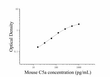
3. Preparation of biotinylated detection antibody working solution: 15 minutes before use, centrifuge the concentrated biotinylated antibody at 1000×g for 1 minute, and dilute the 100× concentrated biotinylated antibody to a 1× working concentration with universal diluent (e.g., 10uL concentrate + 990uL 4. Preparation of enzyme conjugate working solution: 15 minutes before use, centrifuge 100 μL of concentrated enzyme conjugate at 1000 × g for 1 minute. Dilute 100 μL of concentrated HRP enzyme conjugate with universal diluent to a 1 × working concentration (e.g., 10 μL concentrate + 990 μL universal diluent). Prepare and use immediately. 5. Preparation of 1× Wash Buffer: Dissolve 10 mL of 20× Wash Buffer in 190 mL of distilled water. (Concentrated Wash Buffer removed from the refrigerator may crystallize; this is normal. Allow to stand at room temperature until the crystals have completely dissolved before preparing the solution.) V. Procedure: 1. Remove the desired strips from the aluminum foil bag after equilibration at room temperature for 10 minutes. Seal the remaining strips in a ziplock bag and return them to 4°C. 2. Sample Addition: Add 100 μL of sample or standard of varying concentrations to the corresponding wells. Add 100 μL of Universal Diluent to the blank well. Cover with film and incubate at 37°C for 60 minutes. (Recommendation: Dilute the sample to be tested at least 1-fold with universal diluent before adding it to the ELISA plate for testing. This will minimize the impact of matrix effects on the test results. The final calculation of sample concentration should be multiplied by the corresponding dilution factor. It is recommended to run duplicate wells for all samples and standards.) 3. Adding Biotinylated Antibody: Remove the ELISA plate and discard the liquid. Do not wash. Add 100 μL of Biotinylated Antibody Working Solution directly to each well. Cover with a film sealer and incubate at 37°C for 60 minutes. 4. Washing: Discard the liquid. Add 300 μL of 1x Wash Solution to each well. Let stand for 1 minute. Shake off the wash solution and pat dry on absorbent paper. Repeat this process three times (a plate washer can also be used). 5. Adding Enzyme Conjugate Working Solution: Add 100 μL of Enzyme Conjugate Working Solution to each well. Cover with a film sealer and incubate at 37°C for 30 minutes. 6. Washing: Discard the liquid and wash the plate five times according to the washing method in step 4. 7. Add substrate: Add 90 μL of TMB to each well, cover with sealing film, and incubate at 37°C in the dark for 15 minutes. 8. Add stop solution: Remove the ELISA plate and add 50 μL of stop solution directly to each well. Immediately measure the OD value of each well at a wavelength of 450 nm. VI. Calculation of experimental results: 1. Calculate the average OD value of the standard and sample replicates and subtract the OD value of the blank well as the correction value. Plot the standard curve for the four-parameter logistic function on double-logarithmic graph paper, using concentration as the horizontal axis and OD value as the vertical axis.
Note : This graph is for reference only. The sample content should be calculated based on the standard curve drawn based on the experimental data. VII. Kit Performance:
4. Sensitivity: 6.9pg/mL /> 8. Problem analysis:
|