Lambda Light chain Recombinant Rabbit mAb (SDT-326-22)
Lambda Light chain Recombinant Rabbit mAb (SDT-326-22)
Price:
Regular price
$100.00 USD
Regular price
Sale price
$100.00 USD
Unit price
per
For shipping services or bulk orders, you may request a quotation.
Secure checkout with

View full details
Product Details
Product Details
Product Specification
| Host | Rabbit |
| Antigen | Lambda Light chain |
| Synonyms | Immunoglobulin lambda constant 1, Ig lambda chain C region MGC, Ig lambda-1 chain C region |
| Immunogen | Recombinant Protein |
| Location | Secreted, Cell membrane |
| Accession | P0CG04 |
| Clone Number | SDT-326-22 |
| Antibody Type | Recombinant mAb |
| Application | WB, IHC-P, ICC |
| Reactivity | Hu |
| Purification | Protein A |
| Concentration | 0.5 mg/ml |
| Physical Appearance | Liquid |
| Storage Buffer | PBS, 40% Glycerol, 0.05% BSA, 0.03% Proclin 300 |
| Stability & Storage | 12 months from date of receipt / reconstitution, -20 °C as supplied |
Dilution
| application | dilution | species |
| WB | 1:1000-1:5000 | |
| IHC-P | 1:5000 | |
| ICC | 1:2000 |
Background
There are two types of light chains: kappa and lambda chains. The ratio or proportion between the kappa and lambda light chains indicates an excess production of one chain over the other, and therefore can be used as an indication of disease progression or remission.
Picture
Picture
Western Blot
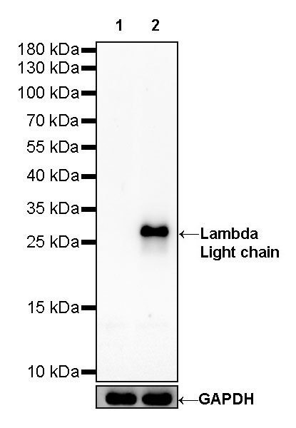
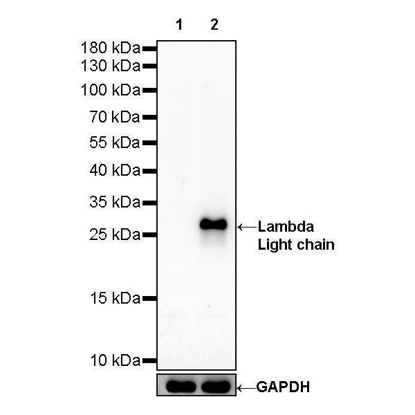
WB result of Lambda Light chain Rabbit mAb Primary antibody: Lambda Light chain Rabbit mAb at 1/1000 dilution Lane 1: Jurkat whole cell lysate 20 µg Lane 2: Ramos whole cell lysate 20 µg Negative control: Jurkat whole cell lysate Secondary antibody: Goat Anti-Rabbit IgG, (H+L), HRP conjugated at 1/10000 dilution Predicted MW: 25kDa Observed MW: 27kDa
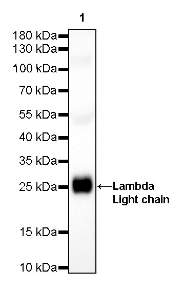
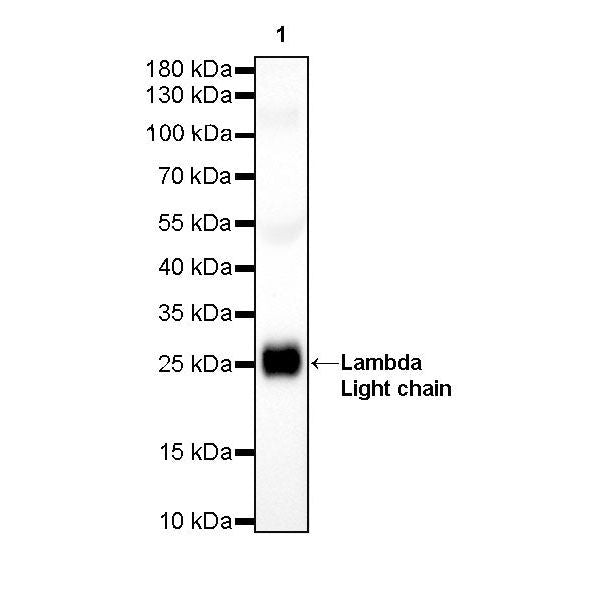
WB result of Lambda Light chain Rabbit mAb Primary antibody: Lambda Light chain Rabbit mAb at 1/5000 dilution Lane 1: human serum lysate 20 µg Secondary antibody: Goat Anti-Rabbit IgG, (H+L), HRP conjugated at 1/10000 dilution Predicted MW: 25kDa Observed MW: 25kDa
Immunohistochemistry
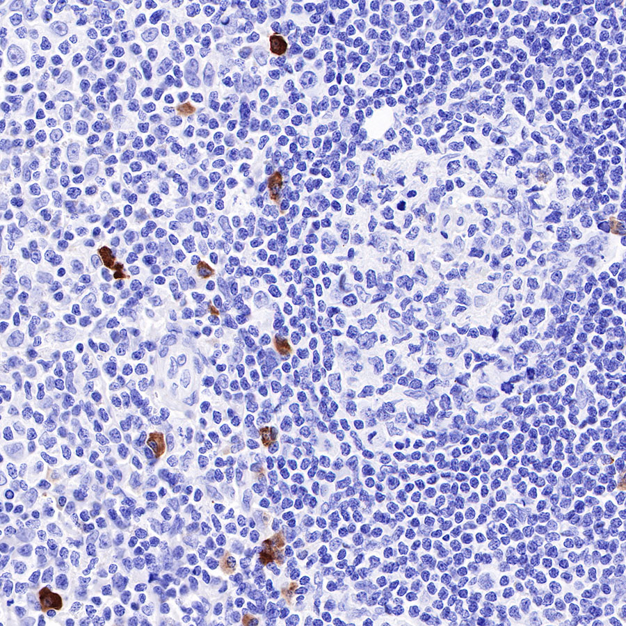
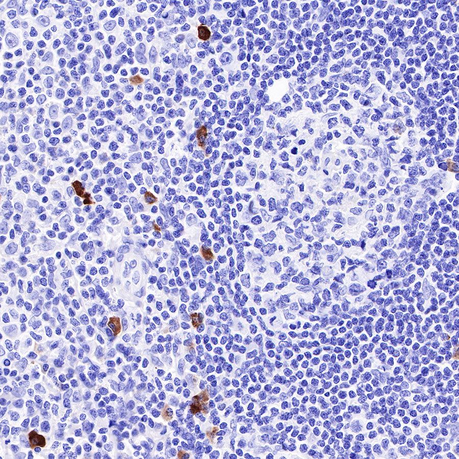
IHC shows positive staining in paraffin-embedded human tonsil. Anti-Lambda Light chain antibody was used at 1/5000 dilution, followed by a HRP Polymer for Mouse & Rabbit IgG (ready to use). Counterstained with hematoxylin. Heat mediated antigen retrieval with Tris/EDTA buffer pH9.0 was performed before commencing with IHC staining protocol.

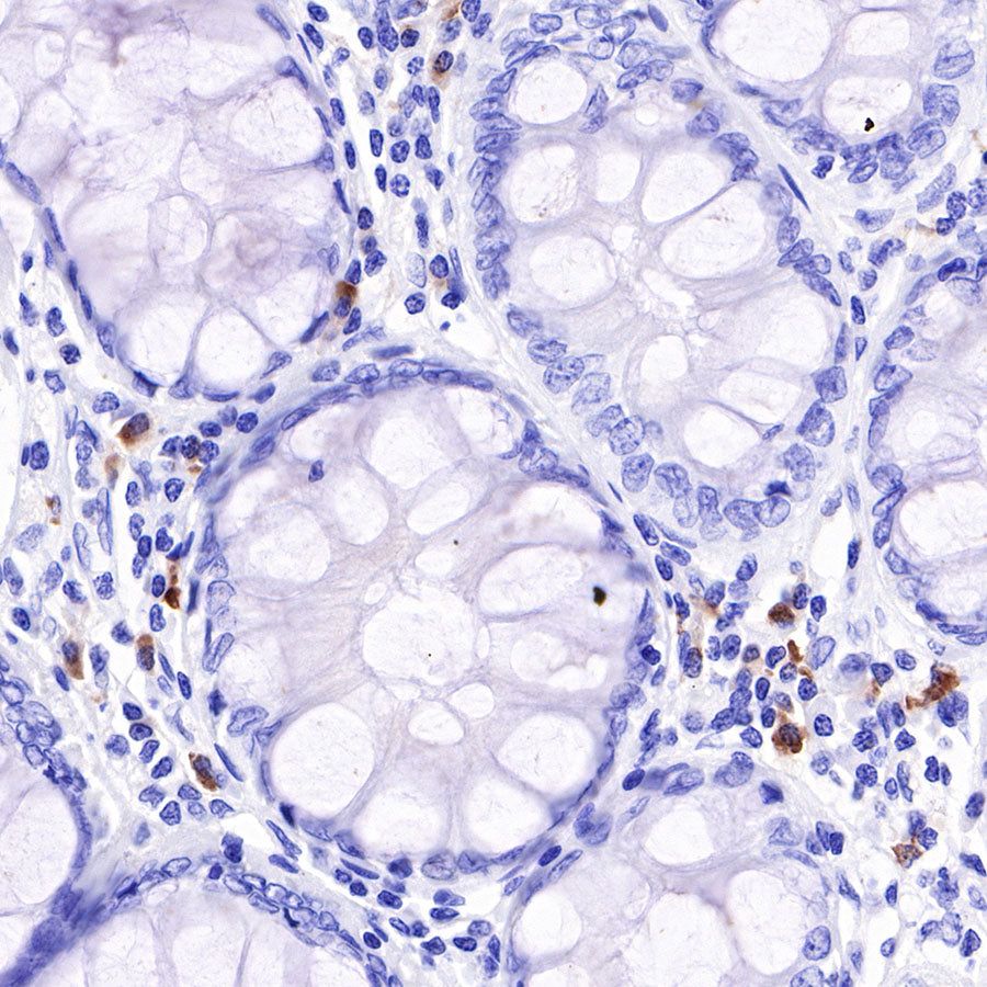
IHC shows positive staining in paraffin-embedded human colon. Anti-Lambda Light chain antibody was used at 1/5000 dilution, followed by a HRP Polymer for Mouse & Rabbit IgG (ready to use). Counterstained with hematoxylin. Heat mediated antigen retrieval with Tris/EDTA buffer pH9.0 was performed before commencing with IHC staining protocol.
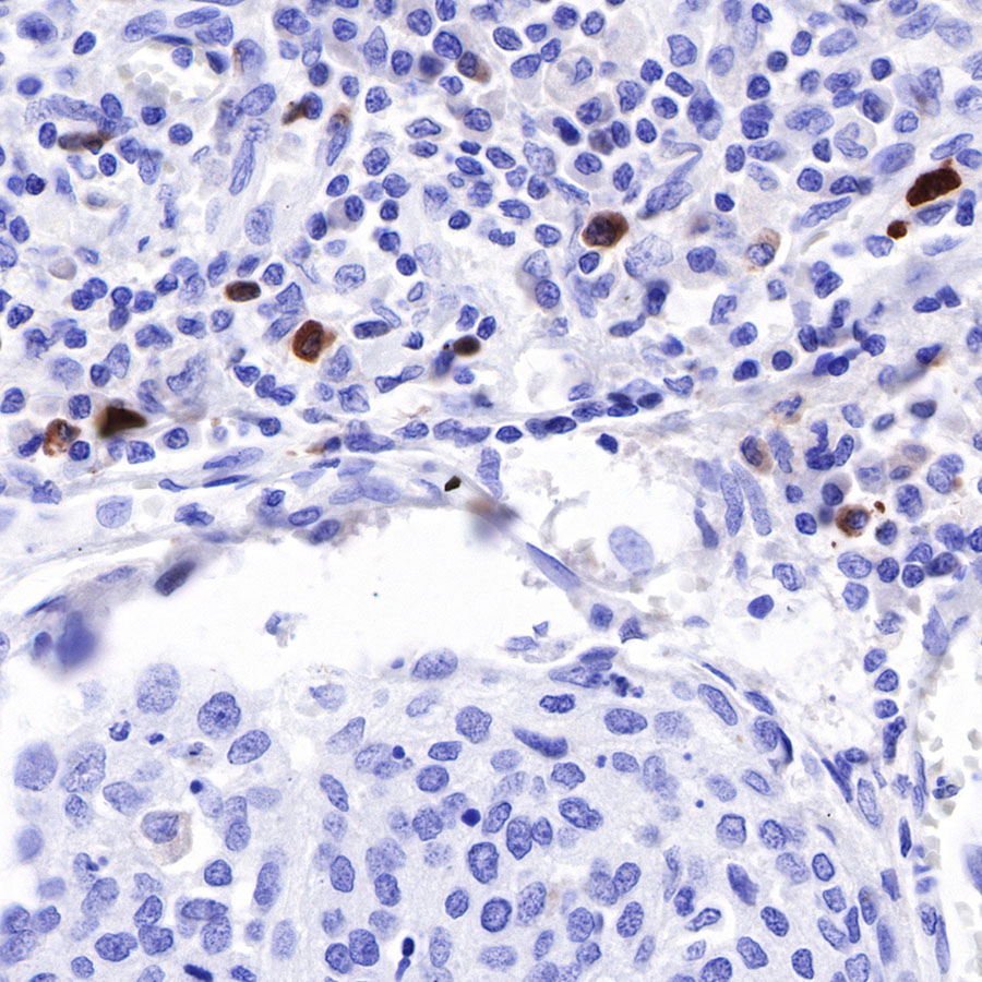
IHC shows positive staining in paraffin-embedded human cervical squamous cell carcinoma. Anti-Lambda Light chain antibody was used at 1/5000 dilution, followed by a HRP Polymer for Mouse & Rabbit IgG (ready to use). Counterstained with hematoxylin. Heat mediated antigen retrieval with Tris/EDTA buffer pH9.0 was performed before commencing with IHC staining protocol.
Immunocytochemistry
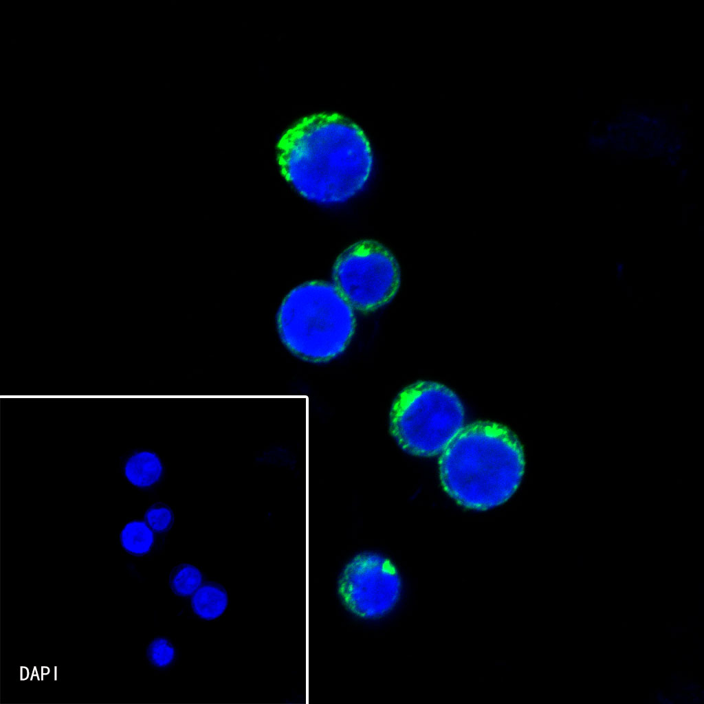
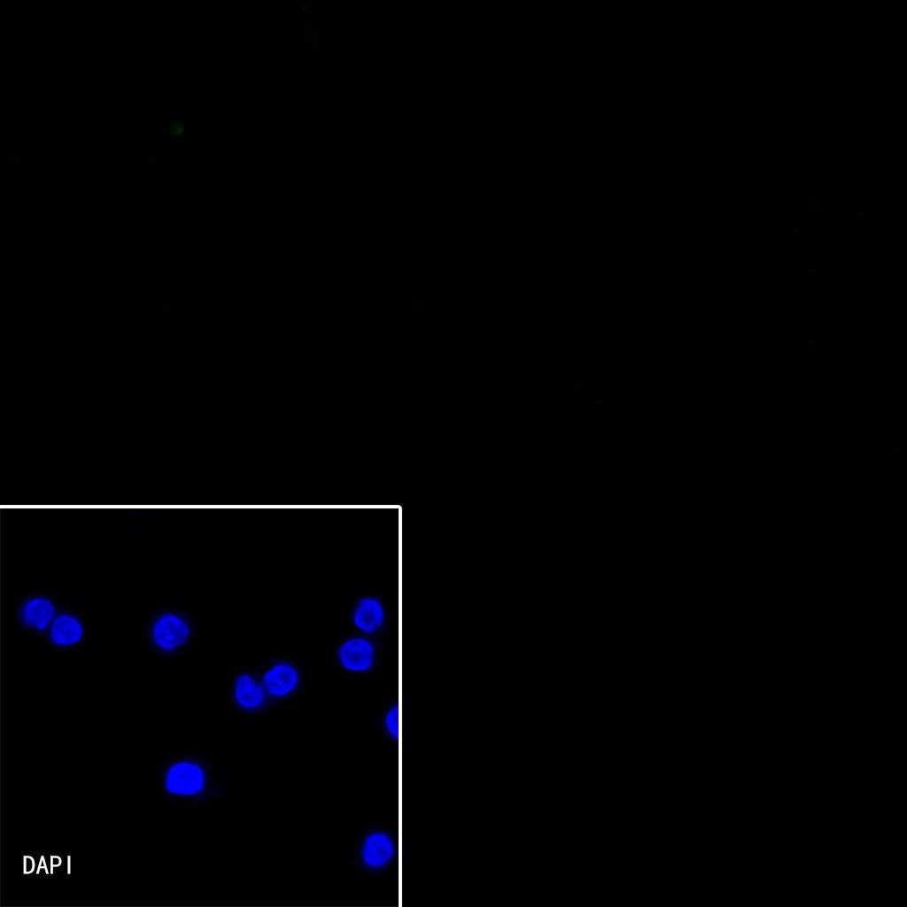
ICC shows positive staining in Ramos cells. Anti-Lambda Light chain antibody was used at 1/2000 dilution (Green) and incubated overnight at 4°C. Goat polyclonal Antibody to Rabbit IgG - H&L (Alexa Fluor® 488) was used as secondary antibody at 1/1000 dilution. The cells were fixed with 100% ice-cold methanol and permeabilized with 0.1% PBS-Triton X-100. Nuclei were counterstained with DAPI (Blue).

Negative control: ICC shows negative staining in Jurkat cells. Anti-Lambda Light chain antibody was used at 1/2000 dilution and incubated overnight at 4°C. Goat polyclonal Antibody to Rabbit IgG - H&L (Alexa Fluor® 488) was used as secondary antibody at 1/1000 dilution. The cells were fixed with 100% ice-cold methanol and permeabilized with 0.1% PBS-Triton X-100. Nuclei were counterstained with DAPI (Blue).