Primary antibody: Ku80 Rabbit mAb at 1/1000 dilution Lane 1: HeLa whole cell lysate 20 µg Lane 2: MCF7 whole cell lysate 20 µg Lane 3: A549 whole cell lysate 20 µg Lane 4: U937 whole cell lysate 20 µg Lane 5: HepG2 whole cell lysate 20 µg Secondary antibody: Goat Anti-Rabbit IgG, (H+L), HRP conjugated at 1/10000 dilution Predicted MW: 83 kDa Observed MW: 86 kDa
Ku80 Recombinant Rabbit mAb (S-R226)
Ku80 Recombinant Rabbit mAb (S-R226)
Price:
Regular price
$100.00 USD
Regular price
Sale price
$100.00 USD
Unit price
per
For shipping services or bulk orders, you may request a quotation.
Secure checkout with

View full details
Product Details
Product Details
Product Specification
| Host | Rabbit |
| Antigen | Ku80 |
| Synonyms | X-ray repair cross-complementing protein 5, 86 kDa subunit of Ku antigen, ATP-dependent DNA helicase 2 subunit 2, ATP-dependent DNA helicase II 80 kDa subunit, CTC85, CTCBF, XRCC5, Ku86, Lupus Ku autoantigen protein p86, Nuclear factor IV, Thyroid-lupus autoantigen (TLAA) |
| Location | Nucleus, Nucleolus |
| Accession | P13010 |
| Clone Number | S-R226 |
| Antibody Type | Recombinant mAb |
| Application | WB, IHC-P, ICC, ICFCM, IP |
| Reactivity | Hu |
| Purification | Protein A |
| Concentration | 0.5 mg/ml |
| Conjugation | Unconjugated |
| Physical Appearance | Liquid |
| Storage Buffer | PBS, 40% Glycerol, 0.05%BSA, 0.03% Proclin 300 |
| Stability & Storage | 12 months from date of receipt / reconstitution, -20 °C as supplied |
Dilution
| application | dilution | species |
| WB | 1:1000 | null |
| IHC | 1:500 | null |
| ICFCM | 1:500 | null |
| ICC | 1:500 | null |
| IP | 1:50 | null |
Background
Ku80 is a protein that, in humans, is encoded by the XRCC5 gene. Together, Ku70 and Ku80 make up the Ku heterodimer, which binds to DNA double-strand break ends and is required for the non-homologous end joining (NHEJ) pathway of DNA repair. It is also required for V(D)J recombination, which utilizes the NHEJ pathway to promote antigen diversity in the mammalian immune system. In addition to its role in NHEJ, Ku is required for telomere length maintenance and subtelomeric gene silencing. Ku80 protein expression was found to be deficient in melanoma. In addition, low expression of Ku80 was found in 15% of adenocarcinoma type and 32% of squamous cell type non-small cell lung cancers, and this was correlated with hypermethylation of the XRCC5 promoter.
Picture
Picture
Western Blot
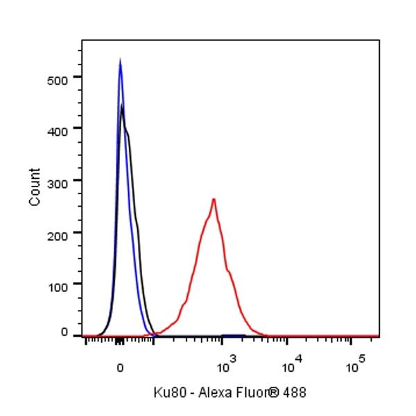
FC
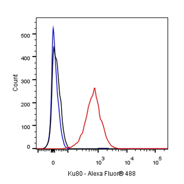
Flow cytometric analysis of 4% PFA fixed 90% methanol permeabilized HeLa (Human cervix adenocarcinoma epithelial cell) cells labelling Ku80 antibody at 1/500 dilution (0.1 μg)/ (Red) compared with a Rabbit monoclonal IgG (Black) isotype control and an unlabelled control (cells without incubation with primary antibody and secondary antibody) (Blue). Goat Anti - Rabbit IgG Alexa Fluor® 488 was used as the secondary antibody.
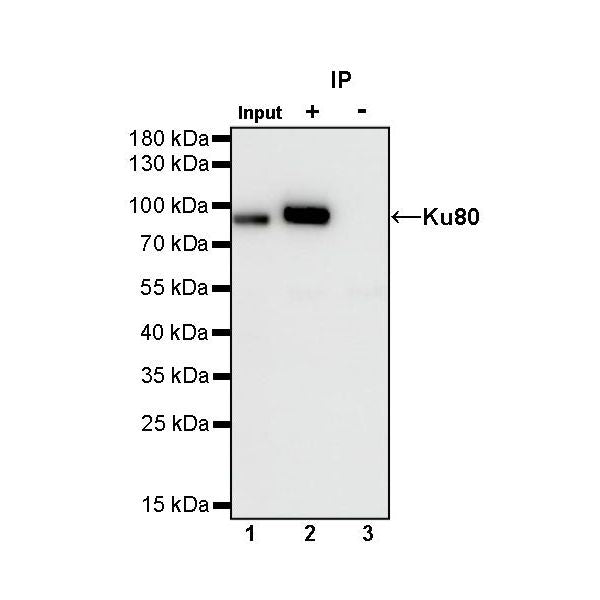
IP
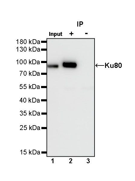
Ku80 Rabbit mAb at 1/50 dilution (1 µg) immunoprecipitating Ku80 in 0.4 mg HeLa whole cell lysate.
Western blot was performed on the immunoprecipitate using Ku80 Rabbit mAb at 1/1000 dilution.
Secondary antibody (HRP) for IP was used at 1/400 dilution.
Lane 1: HeLa whole cell lysate 20 µg (Input)
Lane 2: Ku80 Rabbit mAb IP in HeLa whole cell lysate
Lane 3: Rabbit monoclonal IgG IP in HeLa whole cell lysate
Predicted MW: 83 kDa
Observed MW: 86 kDa
Immunohistochemistry
IHC shows positive staining in paraffin-embedded human colon. Anti-Ku80 antibody was used at 1/500 dilution, followed by a HRP Polymer for Mouse & Rabbit IgG (ready to use). Counterstained with hematoxylin. Heat mediated antigen retrieval with Tris/EDTA buffer pH9.0 was performed before commencing with IHC staining protocol.
IHC shows positive staining in paraffin-embedded human tonsil. Anti-Ku80 antibody was used at 1/500 dilution, followed by a HRP Polymer for Mouse & Rabbit IgG (ready to use). Counterstained with hematoxylin. Heat mediated antigen retrieval with Tris/EDTA buffer pH9.0 was performed before commencing with IHC staining protocol.
IHC shows positive staining in paraffin-embedded human cervical squamous cell carcinoma. Anti-Ku80 antibody was used at 1/500 dilution, followed by a HRP Polymer for Mouse & Rabbit IgG (ready to use). Counterstained with hematoxylin. Heat mediated antigen retrieval with Tris/EDTA buffer pH9.0 was performed before commencing with IHC staining protocol.
IHC shows positive staining in paraffin-embedded human colon cancer. Anti-Ku80 antibody was used at 1/500 dilution, followed by a HRP Polymer for Mouse & Rabbit IgG (ready to use). Counterstained with hematoxylin. Heat mediated antigen retrieval with Tris/EDTA buffer pH9.0 was performed before commencing with IHC staining protocol.
IHC shows positive staining in paraffin-embedded human lung cancer. Anti-Ku80 antibody was used at 1/500 dilution, followed by a HRP Polymer for Mouse & Rabbit IgG (ready to use). Counterstained with hematoxylin. Heat mediated antigen retrieval with Tris/EDTA buffer pH9.0 was performed before commencing with IHC staining protocol.
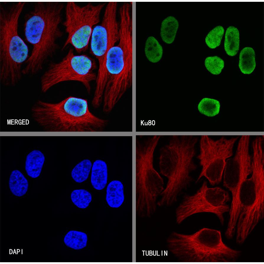
Immunocytochemistry
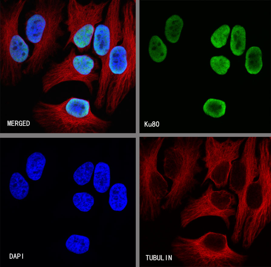
ICC shows positive staining in HeLa cells. Anti-Ku80 antibody was used at 1/500 dilution (Green) and incubated overnight at 4°C. Goat polyclonal Antibody to Rabbit IgG - H&L (Alexa Fluor® 488) was used as secondary antibody at 1/1000 dilution. The cells were fixed with 4%PFA and permeabilized with 0.1% PBS-Triton X-100. Nuclei were counterstained with DAPI (Blue). Counterstain with tubulin (red).