WB result of KRAS Rabbit mAb
Primary antibody: KRAS Rabbit mAb at 1/2000 dilution
Lane 1: SW480 whole cell lysate 20 µg
Lane 2: Caco-2 whole cell lysate 20 µg
Lane 3: HCT 116 whole cell lysate 20 µg
Secondary antibody: Goat Anti-Rabbit IgG, (H+L), HRP conjugated at 1/10000 dilution
Predicted MW: 22 kDa
Observed MW: 21, 23 kDa
KRAS Recombinant Rabbit mAb (S-R291)
KRAS Recombinant Rabbit mAb (S-R291)
Price:
Regular price
$100.00 USD
Regular price
Sale price
$100.00 USD
Unit price
per
For shipping services or bulk orders, you may request a quotation.
Secure checkout with

View full details
Product Details
Product Details
Product Specification
| Host | Rabbit |
| Antigen | KRAS |
| Synonyms | GTPase KRas, K-Ras 2, Ki-Ras, c-K-ras, c-Ki-ras, KRAS2, RASK2 |
| Location | Cytoplasm, Cell membrane |
| Accession | P01116 |
| Clone Number | S-R291 |
| Antibody Type | Recombinant mAb |
| Isotype | IgG |
| Application | WB, ICC, ICFCM, IP |
| Reactivity | Hu, Ms, Rt |
| Purification | Protein A |
| Concentration | 0.5 mg/ml |
| Conjugation | Unconjugated |
| Physical Appearance | Liquid |
| Storage Buffer | PBS, 40% Glycerol, 0.05%BSA, 0.03% Proclin 300 |
| Stability & Storage | 12 months from date of receipt / reconstitution, -20 °C as supplied |
Dilution
| application | dilution | species |
| WB | 1:2000 | null |
| ICC | 1:500 | null |
| ICFCM | 1:50 | null |
| IP | 1:50 | null |
Background
The K-Ras protein is a GTPase, a class of enzymes which convert the nucleotide guanosine triphosphate (GTP) into guanosine diphosphate (GDP). In this way the K-Ras protein acts like a switch that is turned on and off by the GTP and GDP molecules. To transmit signals, it must be turned on by attaching (binding) to a molecule of GTP. The K-Ras protein is turned off (inactivated) when it converts the GTP to GDP. When the protein is bound to GDP, it does not relay signals to the nucleus. Like other members of the ras subfamily of GTPases, the K-Ras protein is an early player in many signal transduction pathways. Once it is allosterically activated, it recruits and activates proteins necessary for the propagation of growth factors, as well as other cell signaling receptors like c-Raf and PI 3-kinase.
Picture
Picture
Western Blot
WB result of KRAS Rabbit mAb
Primary antibody: KRAS Rabbit mAb at 1/2000 dilution
Lane 1: NIH/3T3 whole cell lysate 20 µg
Secondary antibody: Goat Anti-Rabbit IgG, (H+L), HRP conjugated at 1/10000 dilution
Predicted MW: 22 kDa
Observed MW: 21, 23 kDa
WB result of KRAS Rabbit mAb
Primary antibody: KRAS Rabbit mAb at 1/2000 dilution
Lane 1: PC-12 whole cell lysate 20 µg
Secondary antibody: Goat Anti-Rabbit IgG, (H+L), HRP conjugated at 1/10000 dilution
Predicted MW: 22 kDa
Observed MW: 21 kDa
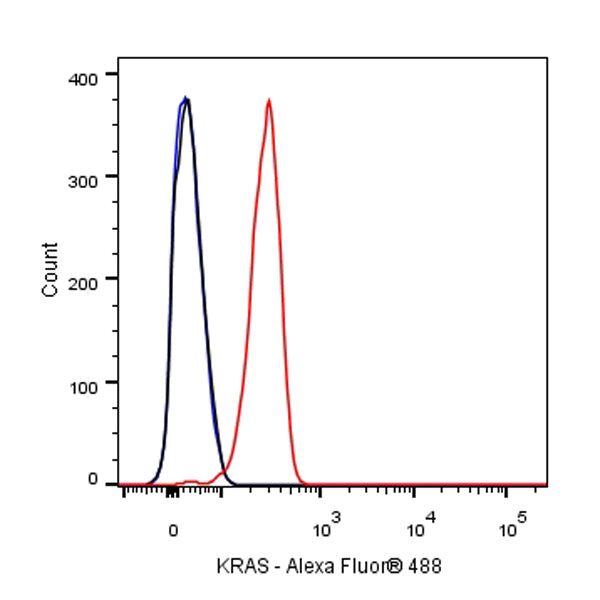
FC
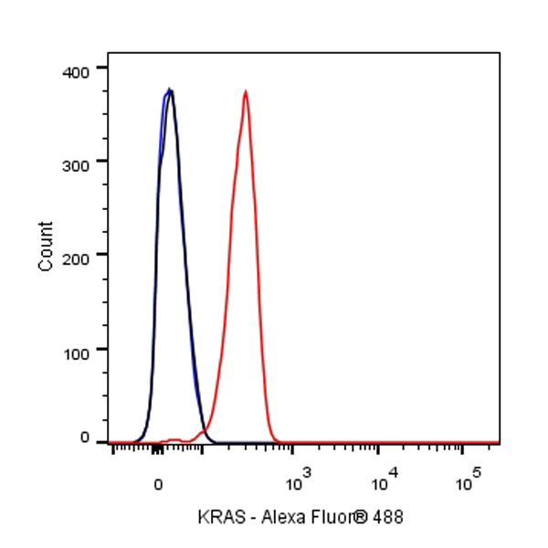
Flow cytometric analysis of 4% PFA fixed 90% methanol permeabilized NIH/3T3 (Mouse embryonic fibroblast) cells labelling KRAS antibody at 1/50 dilution (1 μg)/ (Red) compared with a Rabbit monoclonal IgG (Black) isotype control and an unlabelled control (cells without incubation with primary antibody and secondary antibody) (Blue). Goat Anti - Rabbit IgG Alexa Fluor® 488 was used as the secondary antibody.
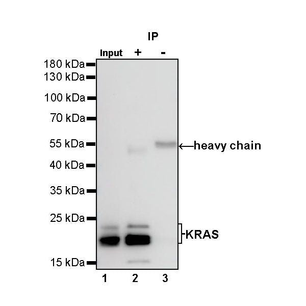
IP
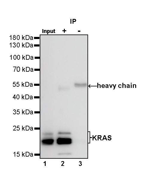
KRAS Rabbit mAb at 1/50 dilution (1 µg) immunoprecipitating KRAS in 0.4 mg HCT-116 whole cell lysate.
Western blot was performed on the immunoprecipitate using KRAS Rabbit mAb at 1/1000 dilution.
Secondary antibody (HRP) for IP was used at 1/400 dilution.
Lane 1: HCT-116 whole cell lysate 20 µg (Input)
Lane 2: KRAS Rabbit mAb IP in HCT-116 whole cell lysate
Lane 3: Rabbit monoclonal IgG IP in HCT-116 whole cell lysate
Predicted MW: 22 kDa
Observed MW: 21, 23 kDa
Immunocytochemistry
ICC shows positive staining in HeLa cells. Anti-KRAS antibody was used at 1/500 dilution (Green) and incubated overnight at 4°C. Goat polyclonal Antibody to Rabbit IgG - H&L (Alexa Fluor® 488) was used as secondary antibody at 1/1000 dilution. The cells were fixed with 4% PFA and permeabilized with 0.1% PBS-Triton X-100. Nuclei were counterstained with DAPI (Blue). Counterstain with tubulin (red).
ICC shows positive staining in NIH/3T3 cells. Anti-KRAS antibody was used at 1/500 dilution (Green) and incubated overnight at 4°C. Goat polyclonal Antibody to Rabbit IgG - H&L (Alexa Fluor® 488) was used as secondary antibody at 1/1000 dilution. The cells were fixed with 4% PFA and permeabilized with 0.1% PBS-Triton X-100. Nuclei were counterstained with DAPI (Blue). Counterstain with tubulin (red).