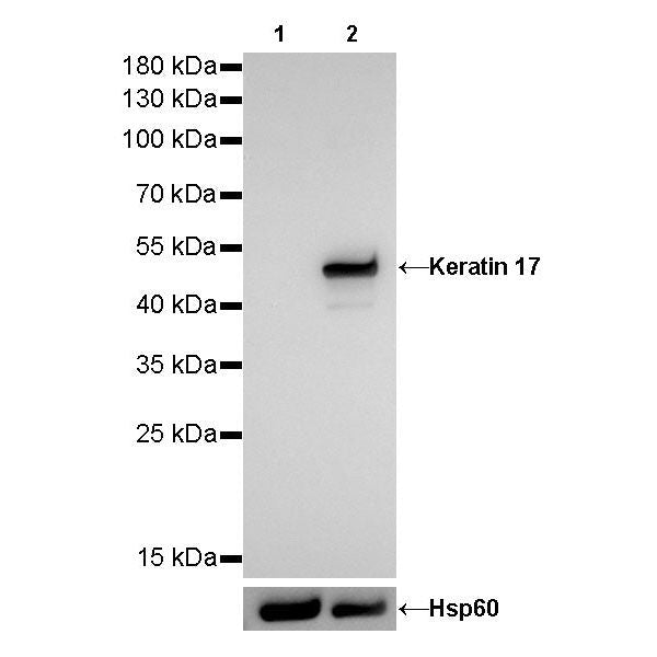
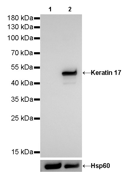
WB result of Keratin 17 Rabbit mAb
Primary antibody: Keratin 17 Rabbit mAb at 1/1250 dilution
Lane 1: MOLT-4 whole cell lysate 5 µg
Lane 2: HeLa whole cell lysate 5 µg
Secondary antibody: Goat Anti-Rabbit IgG, (H+L), HRP conjugated at 1/10000 dilution
Predicted MW: 48 kDa
Observed MW: 48 kDa
Exposure time: 7.5s
Keratin 17 Recombinant Rabbit mAb (SDT-011-43)
Keratin 17 Recombinant Rabbit mAb (SDT-011-43)
Price:
Regular price
$100.00 USD
Regular price
Sale price
$100.00 USD
Unit price
per
For shipping services or bulk orders, you may request a quotation.
Secure checkout with

View full details
Product Details
Product Details
Product Specification
| Host | Rabbit |
| Antigen | Keratin 17 |
| Synonyms | Cytokeratin-17, CK-17 |
| Immunogen | Synthetic Peptide |
| Location | Cytoplasm |
| Accession | Q04695 |
| Clone Number | SDT-011-43 |
| Antibody Type | Rabbit mAb |
| Application | WB, IHC-P, ICC |
| Reactivity | Hu, Ms |
| Predicted Reactivity | Gor, Pr, Cz, Or, Bv, Sh, Rt, Ct, Rb, Dg, Hdg, Pr, Bb, Hr |
| Purification | Protein A |
| Concentration | 0.125 mg/ml |
| Physical Appearance | Liquid |
| Storage Buffer | PBS, 40% Glycerol, 0.05%BSA, 0.03% Proclin 300 |
| Stability & Storage | 12 months from date of receipt / reconstitution, -20 °C as supplied |
Dilution
| application | dilution | species |
| IHC-P | 1:1000 | |
| WB | 1:250-1:1500 | |
| ICC | 1:125 |
Background
Keratins (cytokeratins) are intermediate filament proteins that are mainly expressed in epithelial cells. Keratin 17 is involved in wound healing and cell growth, two processes that require rapid cytoskeletal remodeling. CK17 maybe an excellent marker for the identification of squamous cell carcinomas in various tissues including the cervix, lung and oral cavity.
Picture
Picture
Western Blot
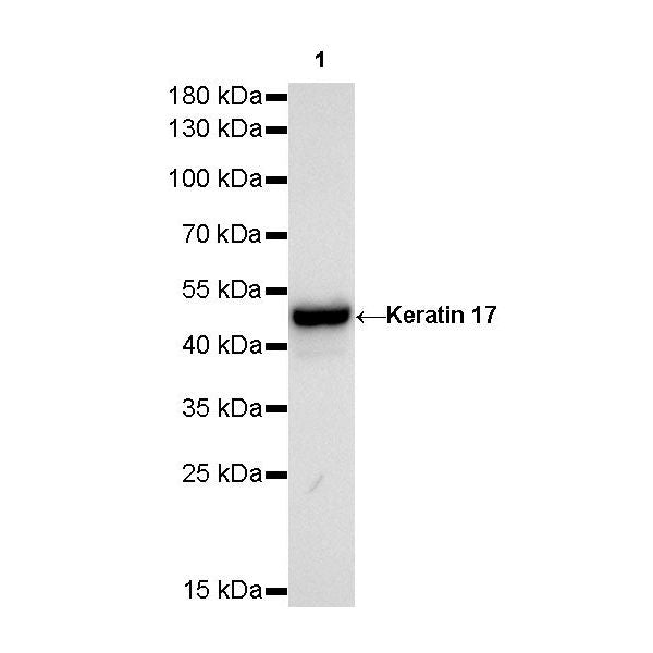
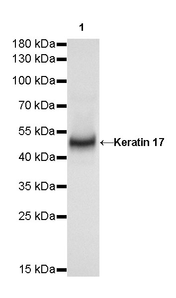
WB result of Keratin 17 Rabbit mAb
Primary antibody: Keratin 17 Rabbit mAb at 1/250 dilution
Lane 1: A431 whole cell lysate 20 µg
Secondary antibody: Goat Anti-Rabbit IgG, (H+L), HRP conjugated at 1/10000 dilution
Predicted MW: 48 kDa
Observed MW: 48 kDa
Exposure time: 30s
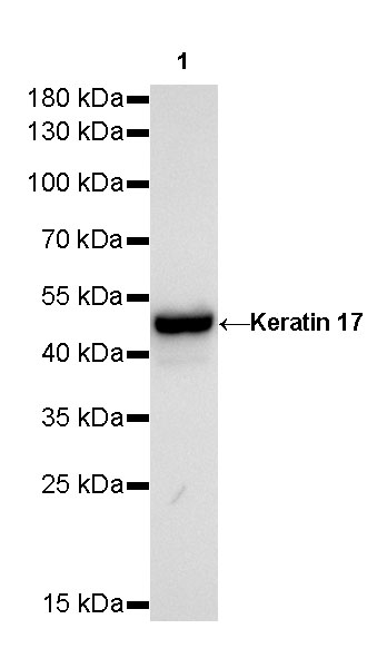
WB result of Keratin 17 Rabbit mAb
Primary antibody: Keratin 17 Rabbit mAb at 1/250 dilution
Lane 1: mouse skin lysate 20 µg
Secondary antibody: Goat Anti-Rabbit IgG, (H+L), HRP conjugated at 1/10000 dilution
Predicted MW: 48 kDa
Observed MW: 48 kDa
Exposure time: 30s
Immunohistochemistry
IHC shows positive staining in paraffin-embedded human breast cancer. Anti-Keratin 17 antibody was used at 1/1000 dilution, followed by a HRP Polymer for Mouse & Rabbit IgG (ready to use). Counterstained with hematoxylin. Heat mediated antigen retrieval with Tris/EDTA buffer pH9.0 was performed before commencing with IHC staining protocol.
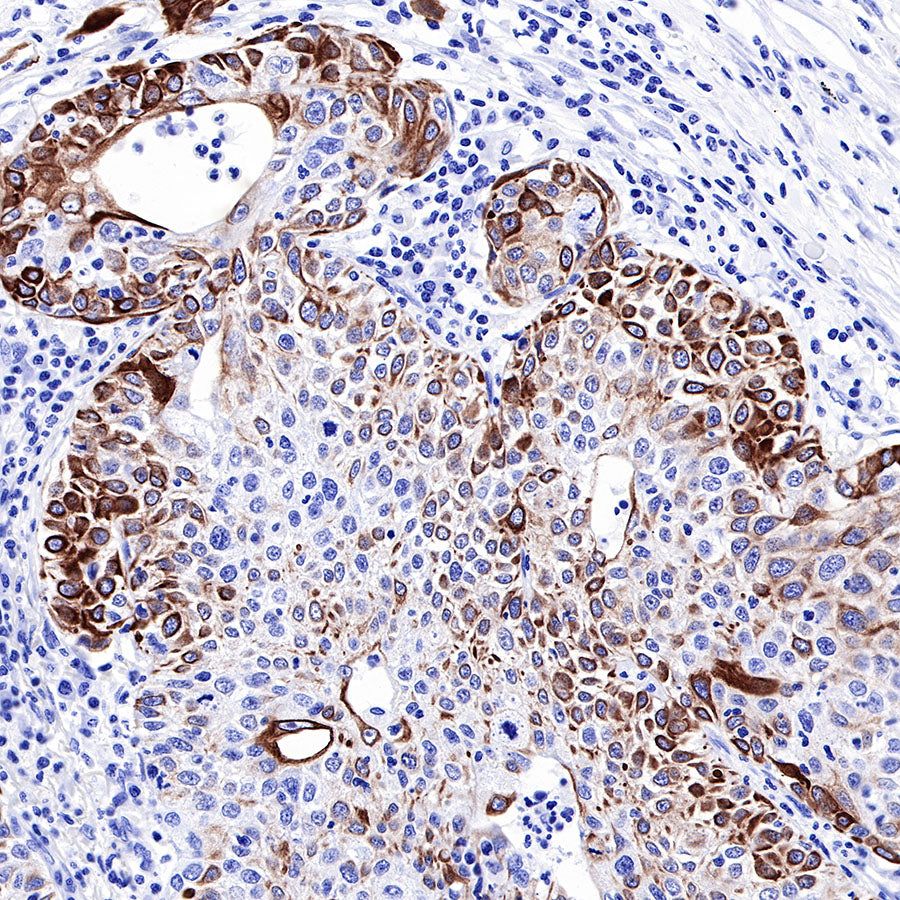
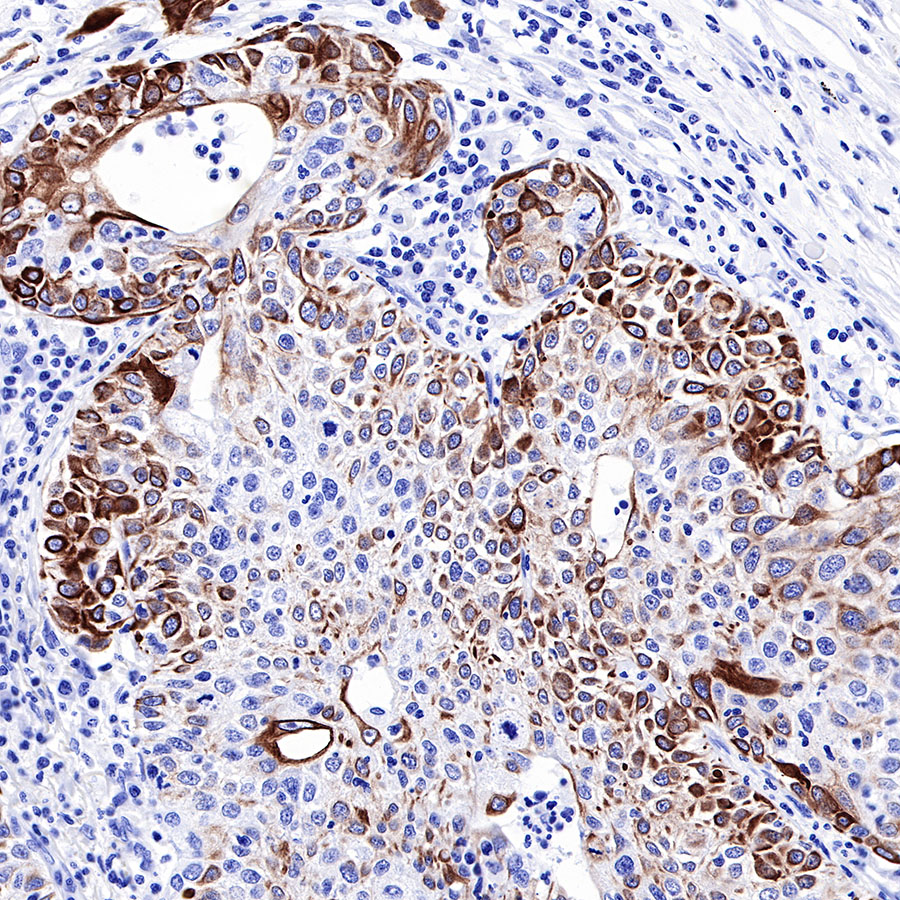
IHC shows positive staining in paraffin-embedded human cervical carcinoma. Anti-Keratin 17 antibody was used at 1/1000 dilution, followed by a HRP Polymer for Mouse & Rabbit IgG (ready to use). Counterstained with hematoxylin. Heat mediated antigen retrieval with Tris/EDTA buffer pH9.0 was performed before commencing with IHC staining protocol.
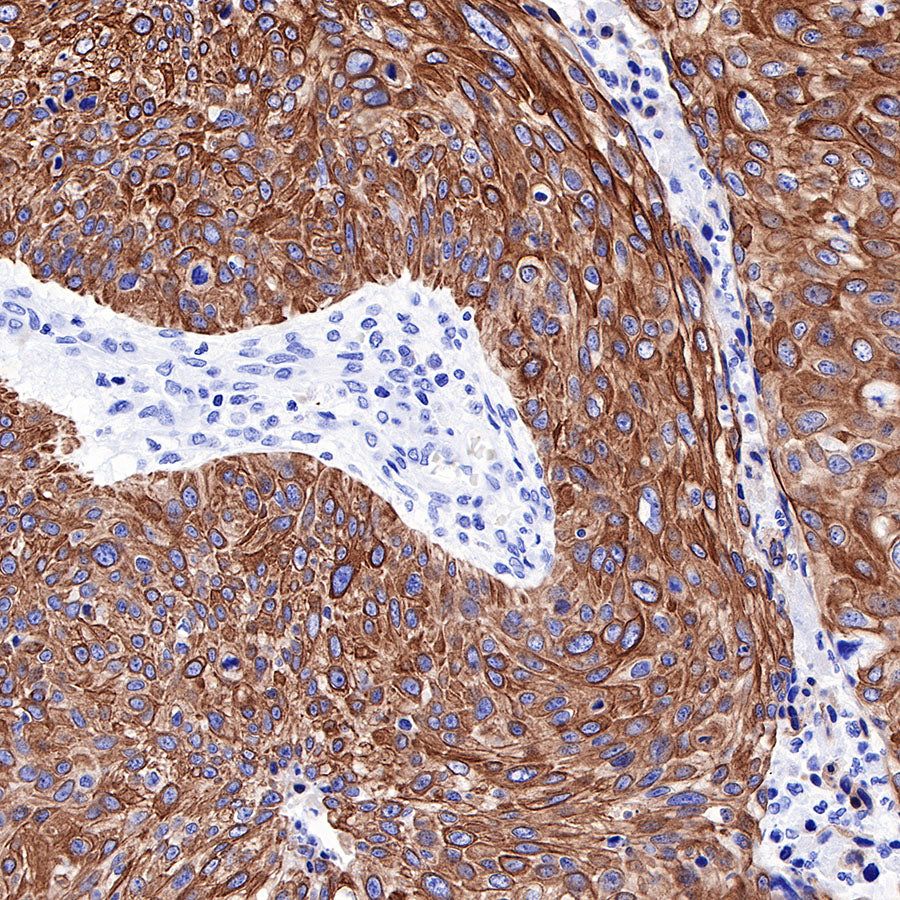
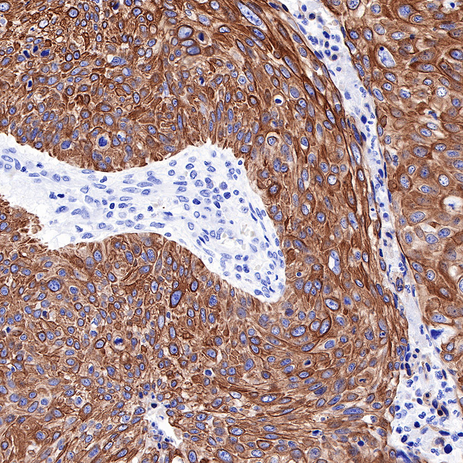
IHC shows positive staining in paraffin-embedded human lung squamous cell carcinoma. Anti-Keratin 17 antibody was used at 1/1000 dilution, followed by a HRP Polymer for Mouse & Rabbit IgG (ready to use). Counterstained with hematoxylin. Heat mediated antigen retrieval with Tris/EDTA buffer pH9.0 was performed before commencing with IHC staining protocol.
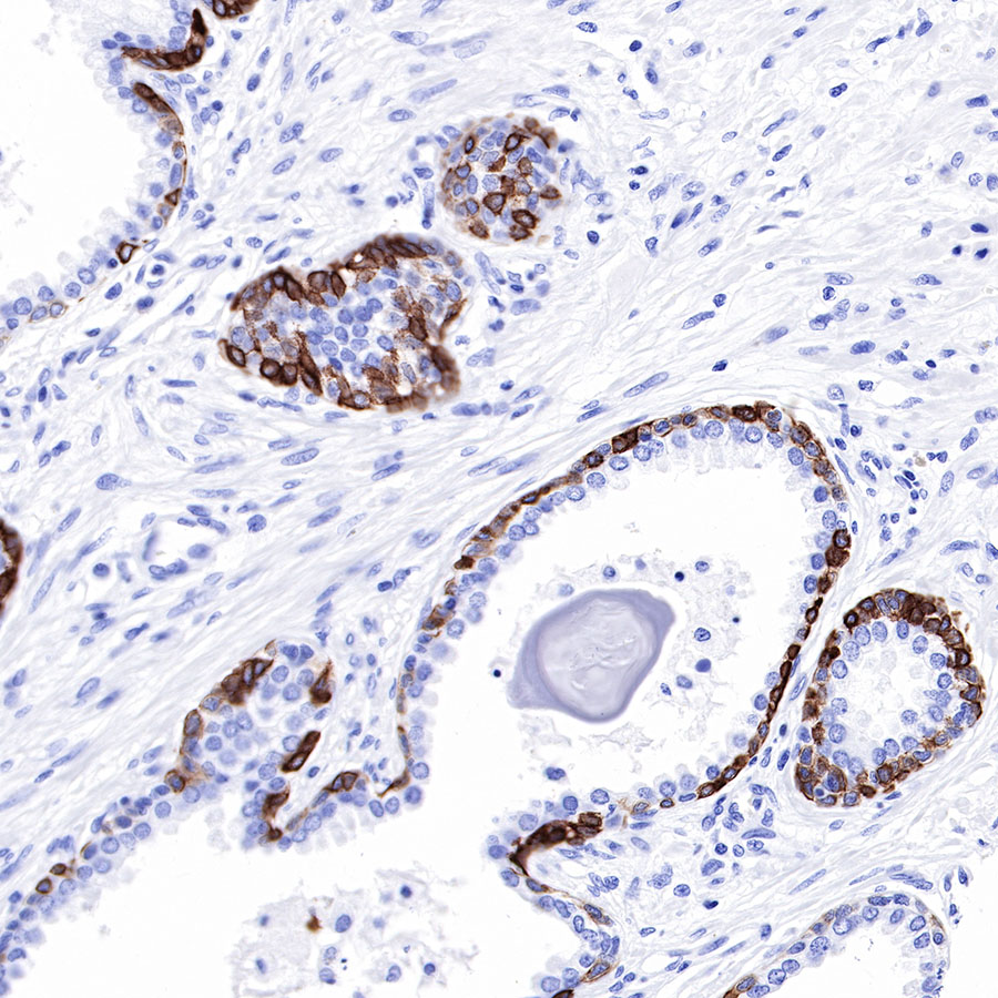
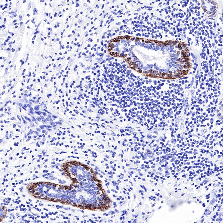
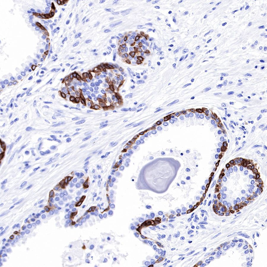
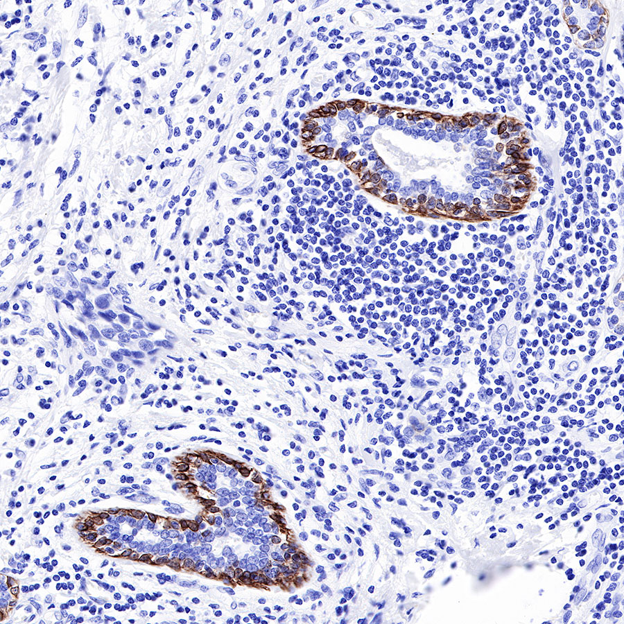
IHC shows positive staining in paraffin-embedded human prostatic hyperplasia. Anti-Keratin 17 antibody was used at 1/1000 dilution, followed by a HRP Polymer for Mouse & Rabbit IgG (ready to use). Counterstained with hematoxylin. Heat mediated antigen retrieval with Tris/EDTA buffer pH9.0 was performed before commencing with IHC staining protocol.

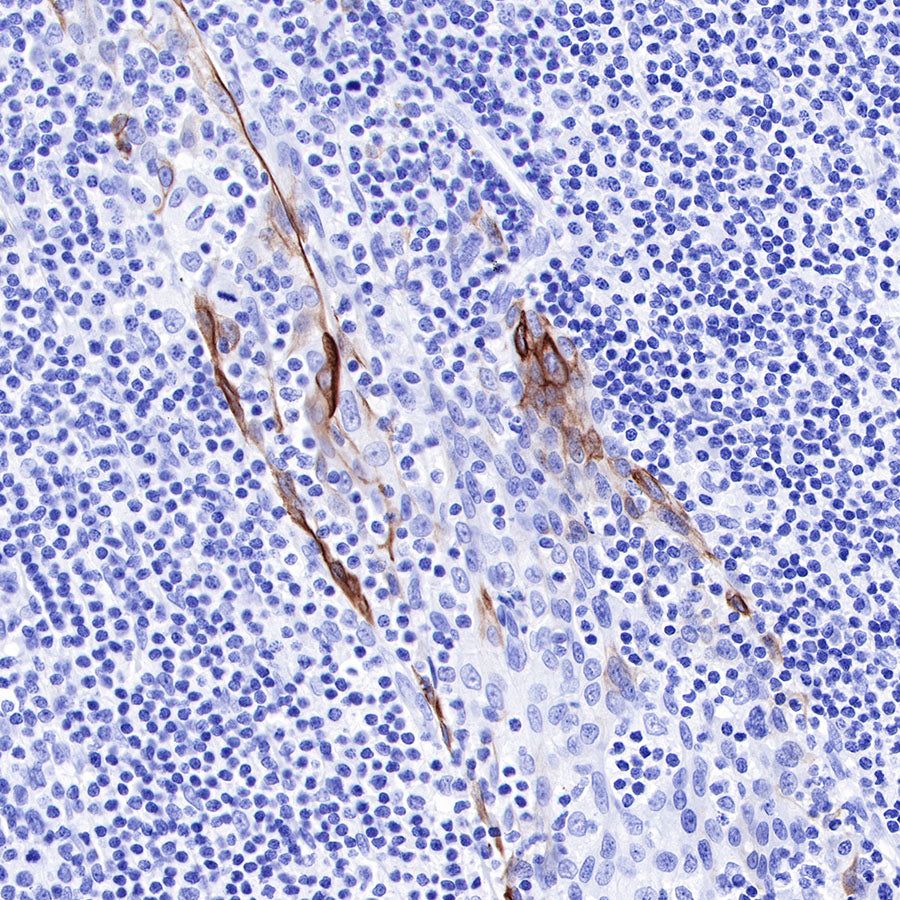
IHC shows positive staining in paraffin-embedded human tonsil. Anti-Keratin 17 antibody was used at 1/1000 dilution, followed by a HRP Polymer for Mouse & Rabbit IgG (ready to use). Counterstained with hematoxylin. Heat mediated antigen retrieval with Tris/EDTA buffer pH9.0 was performed before commencing with IHC staining protocol.
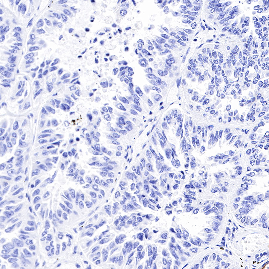
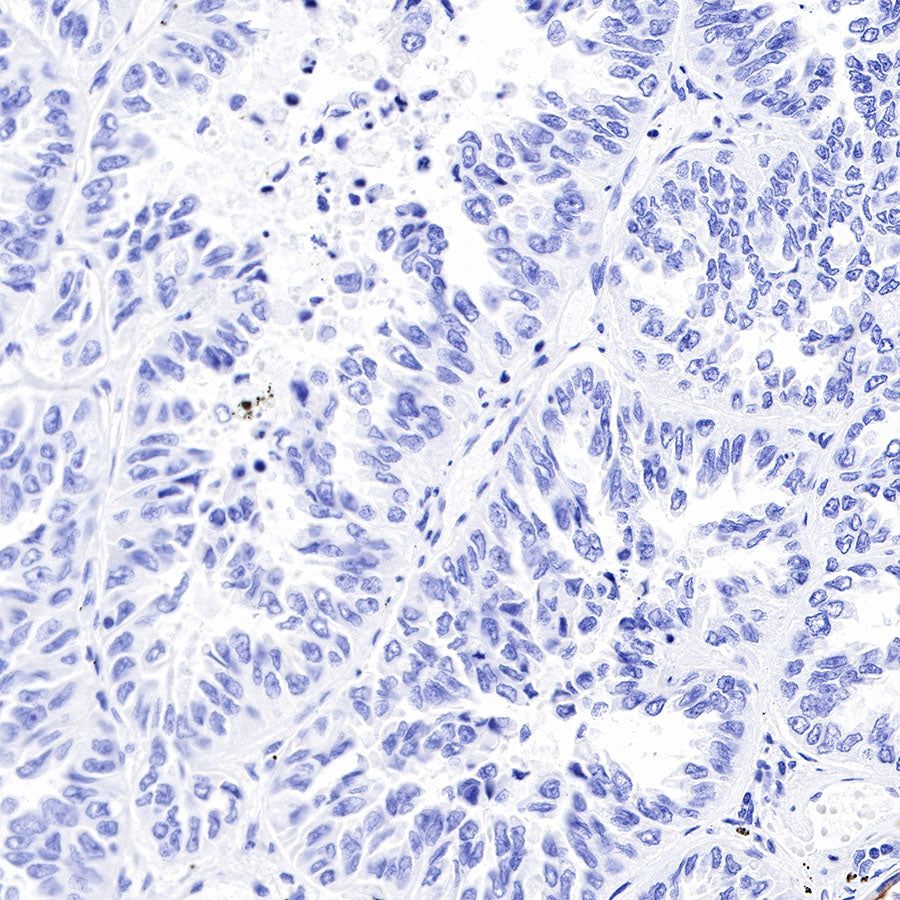
Negative control: IHC shows negative staining in paraffin-embedded human lung adenocarcinoma. Anti-Keratin 17 antibody was used at 1/1000 dilution, followed by a HRP Polymer for Mouse & Rabbit IgG (ready to use). Counterstained with hematoxylin. Heat mediated antigen retrieval with Tris/EDTA buffer pH9.0 was performed before commencing with IHC staining protocol.
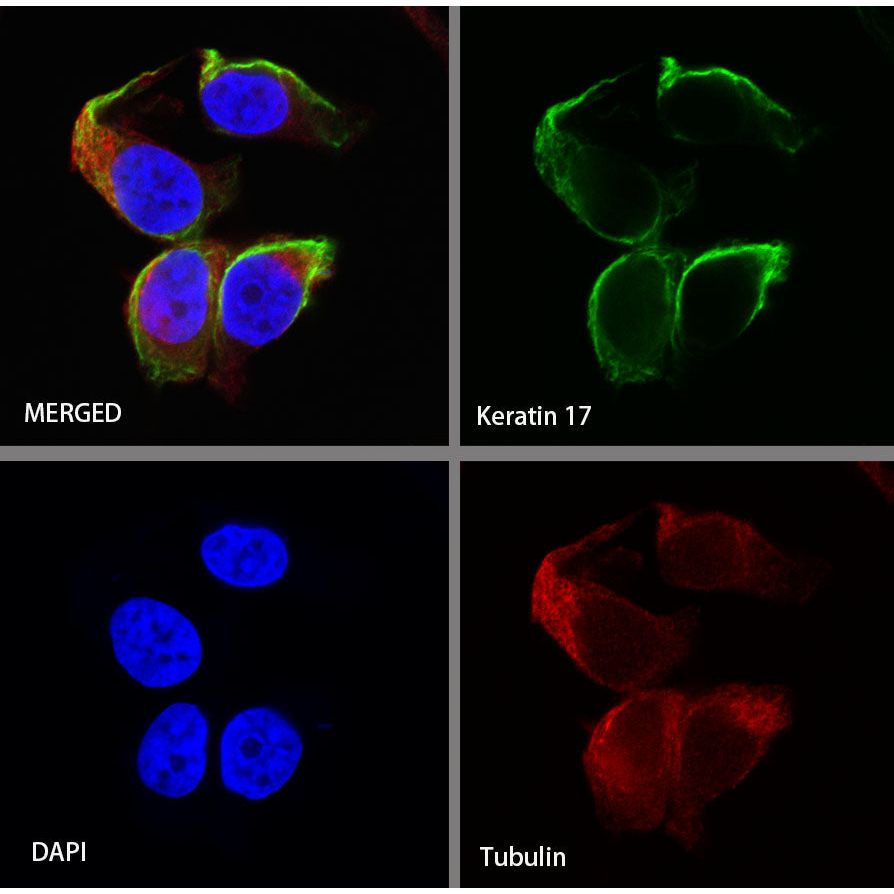
Immunocytochemistry
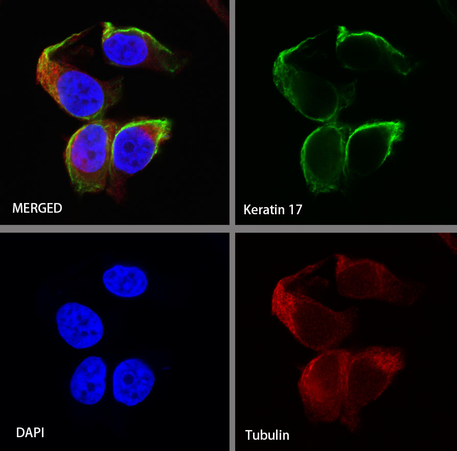
ICC shows positive staining in HeLa cells. Anti-Keratin 17 antibody was used at 1/125 dilution (Green) and incubated overnight at 4°C. Goat polyclonal Antibody to Rabbit IgG - H&L (Alexa Fluor® 488) was used as secondary antibody at 1/1000 dilution. The cells were fixed with 4% PFA and permeabilized with 0.1% PBS-Triton X-100. Nuclei were counterstained with DAPI (Blue). Counterstain with tubulin (red).