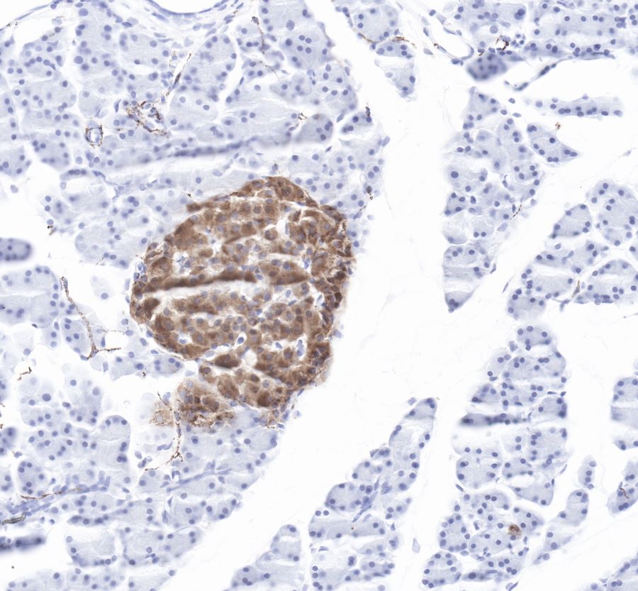
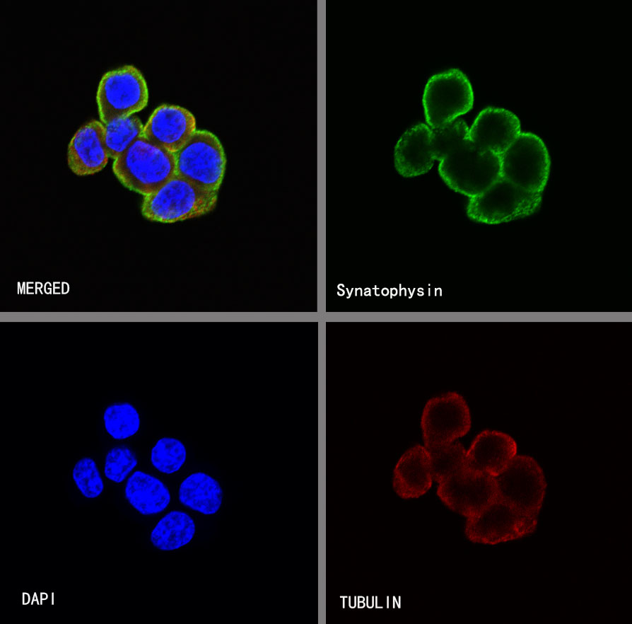
Product Specification
| Host |
Rabbit |
| Antigen |
Synaptophysin |
| Synonyms |
Major synaptic vesicle protein p38,SYP |
| Immunogen |
Synthetic Peptide |
| Location |
Synaptosome, Intracellular |
| Accession |
P08247 |
| Clone Number |
SDT-137-21 |
| Antibody Type |
Rabbit mAb |
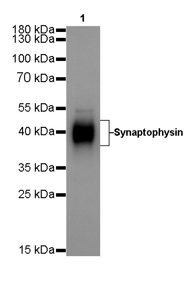
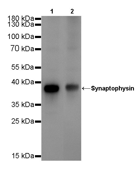
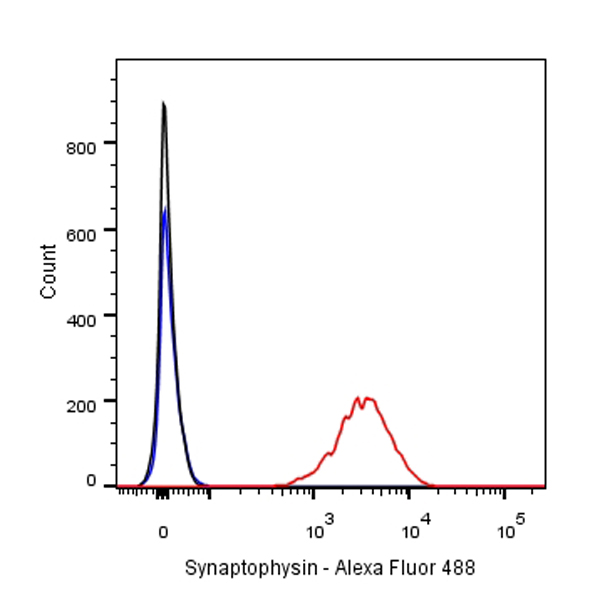
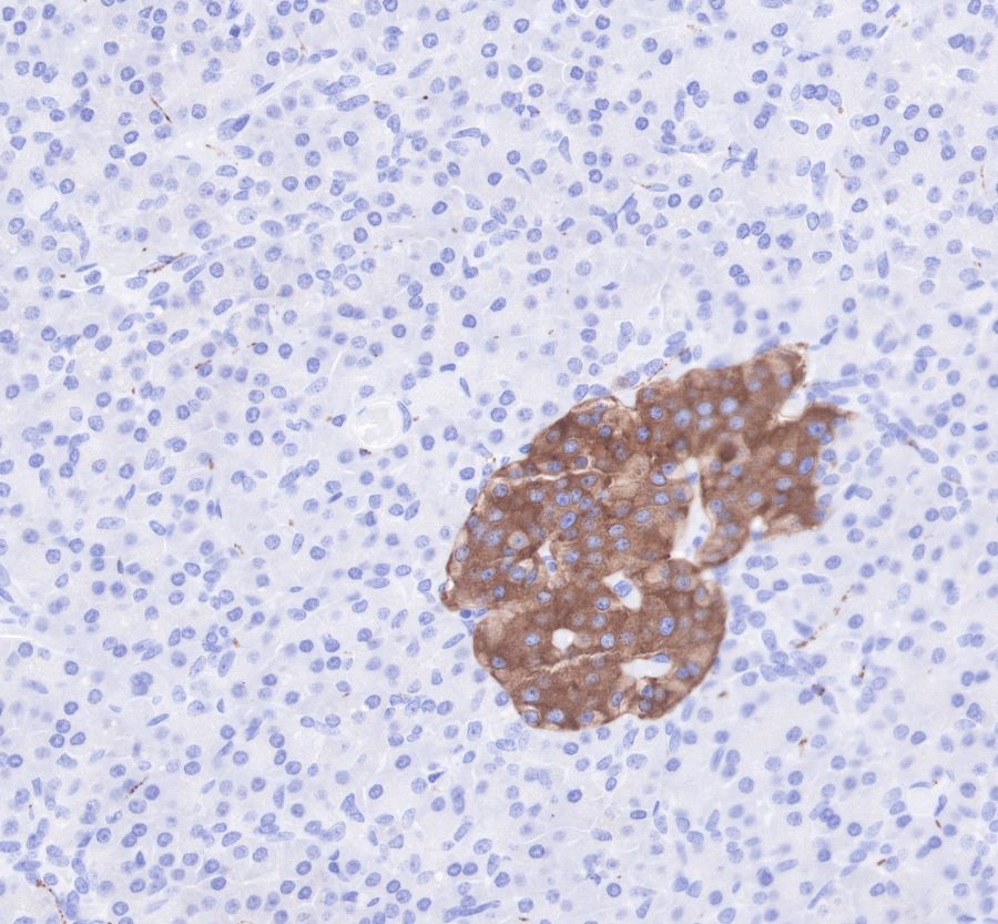
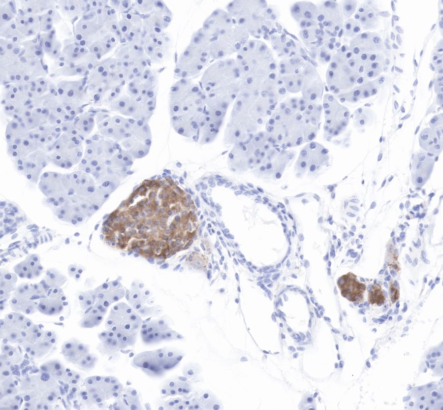
| Application |
WB, IHC-P, ICC, ICFCM |
| Reactivity |
Hu, Ms, Rt |
| Predicted Reactivity |
Bv, Sq |
| Purification |
Protein A |
| Concentration |
0.25mg/ml |
| Physical Appearance |
Liquid |
| Storage Buffer |
PBS, 40% Glycerol, 0.05%BSA, 0.03% Proclin 300 |
| Stability & Storage |
12 months from date of receipt / reconstitution, -20 °C as supplied |
Dilution
| application |
dilution |
species |
| WB |
1:500 |
|
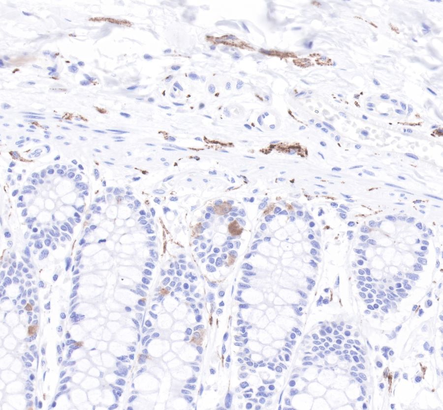
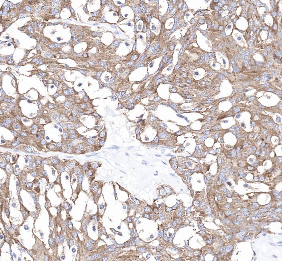
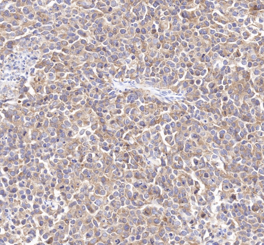
| IHC-P |
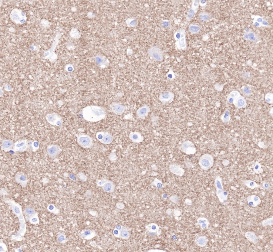
1:4000(Ms) |
|
| IHC-P |
1:1000(Hu) |
|
| ICFCM |
1:500 |
|
| ICC |
1:250 |
|
Background
Synaptophysin, also known as the major synaptic vesicle protein p38, is a protein that in humans is encoded by the SYP gene. The protein is a synaptic vesicle glycoprotein with four transmembrane domains weighing 38kDa. It is present in neuroendocrine cells and in virtually all neurons in the brain and spinal cord that participate in synaptic transmission. It acts as a marker for neuroendocrine tumors, and its ubiquity at the synapse has led to the use of synaptophysin immunostaining for quantification of synapses.