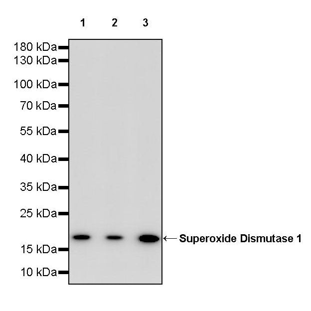
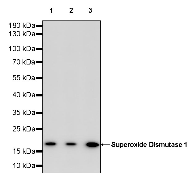
WB result of Superoxide Dismutase 1 Recombinant Rabbit mAb
Primary antibody: Superoxide Dismutase 1 Recombinant Rabbit mAb at 1/10000 dilution
Lane 1: HeLa whole cell lysate 20 µg
Lane 2: MCF7 whole cell lysate 20 µg
Lane 3: HEK-293 whole cell lysate 20 µg
Secondary antibody: Goat Anti-rabbit IgG, (H+L), HRP conjugated at 1/10000 dilution
Predicted MW: 16 kDa
Observed MW: 18 kDa
Superoxide Dismutase 1 Recombinant Rabbit mAb (S-1537-39)
Superoxide Dismutase 1 Recombinant Rabbit mAb (S-1537-39)
Price:
Regular price
$100 USD
Regular price
Sale price
$100 USD
Unit price
per
For shipping services or bulk orders, you may request a quotation.
Secure checkout with

View full details
Product Details
Product Details
Product Specification
| Host | Rabbit |
| Antigen | Superoxide Dismutase 1 |
| Immunogen | Synthetic Peptide |
| Location | Cytoplasm, Nucleus |
| Accession | P00441 |
| Clone Number | S-1537-39 |
| Antibody Type | Recombinant mAb |
| Isotype | IgG |
| Application | WB, IHC-P, ICC |
| Reactivity | Hu, Ms, Rt |
| Positive Sample | HeLa, MCF7, HEK-293, RAW264.7, rat liver |
| Predicted Reactivity | Or, CaMk, Pr, Hr, Cm, GP |
| Purification | Protein A |
| Concentration | 0.5 mg/ml |
| Conjugation | Unconjugated |
| Physical Appearance | Liquid |
| Storage Buffer | PBS, 40% Glycerol, 0.05% BSA, 0.03% Proclin 300 |
| Stability & Storage | 12 months from date of receipt / reconstitution, -20 °C as supplied |
Dilution
| application | dilution | species |
| WB | 1:10000 | Hu, Ms, Rt |
| IHC-P | 1:2000 | Hu, Ms, Rt |
| ICC | 1:500 | Hu, Ms |
Background
Superoxide Dismutase 1 (SOD1) is an essential enzyme found in the cells of nearly all living organisms. It plays a critical role in protecting cells against oxidative stress by catalyzing the dismutation of superoxide radicals into molecular oxygen and hydrogen peroxide. This process helps to prevent the damaging effects of reactive oxygen species (ROS), which can lead to cellular damage and are implicated in various diseases, including cancer, Alzheimer's, and amyotrophic lateral sclerosis (ALS). SOD1 is particularly important in the mitochondria, where it helps to maintain the balance between the production of energy and the generation of potentially harmful byproducts. The enzyme is encoded by the SOD1 gene and is a homodimeric metalloprotein, with each monomer containing a copper ion and a zinc ion at its active site. Mutations in the SOD1 gene have been linked to the development of familial ALS, highlighting the importance of this enzyme in maintaining cellular health.
Picture
Picture
Western Blot
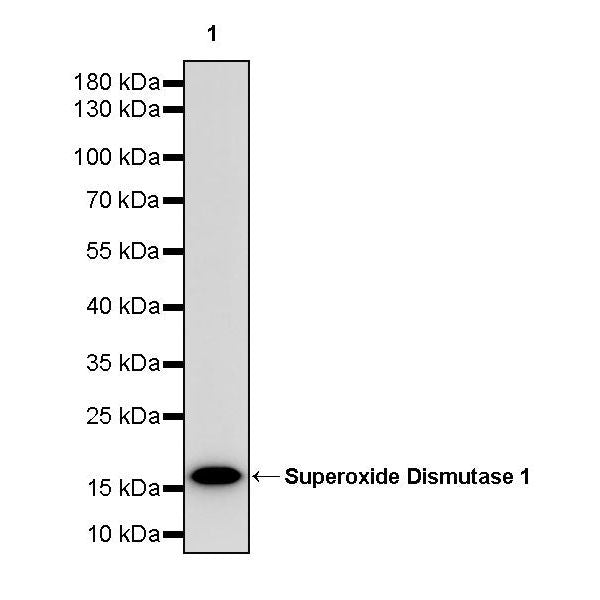
WB result of Superoxide Dismutase 1 Recombinant Rabbit mAb
Primary antibody: Superoxide Dismutase 1 Recombinant Rabbit mAb at 1/10000 dilution
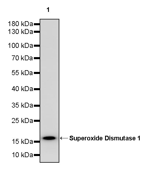
Lane 1: RAW264.7 whole cell lysate 20 µg
Secondary antibody: Goat Anti-rabbit IgG, (H+L), HRP conjugated at 1/10000 dilution
Predicted MW: 16 kDa
Observed MW: 18 kDa
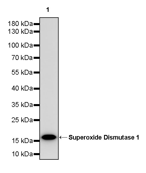
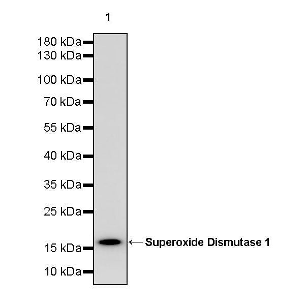
WB result of Superoxide Dismutase 1 Recombinant Rabbit mAb
Primary antibody: Superoxide Dismutase 1 Recombinant Rabbit mAb at 1/10000 dilution
Lane 1: rat liver lysate 20 µg
Secondary antibody: Goat Anti-rabbit IgG, (H+L), HRP conjugated at 1/10000 dilution
Predicted MW: 16 kDa
Observed MW: 18 kDa
Immunohistochemistry
IHC shows positive staining in paraffin-embedded human kidney. Anti-Superoxide Dismutase 1 antibody was used at 1/2000 dilution, followed by a HRP Polymer for Mouse & Rabbit IgG (ready to use). Counterstained with hematoxylin. Heat mediated antigen retrieval with Tris/EDTA buffer pH9.0 was performed before commencing with IHC staining protocol.
IHC shows positive staining in paraffin-embedded human liver. Anti-Superoxide Dismutase 1 antibody was used at 1/2000 dilution, followed by a HRP Polymer for Mouse & Rabbit IgG (ready to use). Counterstained with hematoxylin. Heat mediated antigen retrieval with Tris/EDTA buffer pH9.0 was performed before commencing with IHC staining protocol.
IHC shows positive staining in paraffin-embedded human hepatocellular carcinoma. Anti-Superoxide Dismutase 1 antibody was used at 1/2000 dilution, followed by a HRP Polymer for Mouse & Rabbit IgG (ready to use). Counterstained with hematoxylin. Heat mediated antigen retrieval with Tris/EDTA buffer pH9.0 was performed before commencing with IHC staining protocol.
IHC shows positive staining in paraffin-embedded human lung squamous cell carcinoma. Anti-Superoxide Dismutase 1 antibody was used at 1/2000 dilution, followed by a HRP Polymer for Mouse & Rabbit IgG (ready to use). Counterstained with hematoxylin. Heat mediated antigen retrieval with Tris/EDTA buffer pH9.0 was performed before commencing with IHC staining protocol.
IHC shows positive staining in paraffin-embedded mouse lung. Anti-Superoxide Dismutase 1 antibody was used at 1/2000 dilution, followed by a HRP Polymer for Mouse & Rabbit IgG (ready to use). Counterstained with hematoxylin. Heat mediated antigen retrieval with Tris/EDTA buffer pH9.0 was performed before commencing with IHC staining protocol.
IHC shows positive staining in paraffin-embedded rat cerebral cortex. Anti-Superoxide Dismutase 1 antibody was used at 1/2000 dilution, followed by a HRP Polymer for Mouse & Rabbit IgG (ready to use). Counterstained with hematoxylin. Heat mediated antigen retrieval with Tris/EDTA buffer pH9.0 was performed before commencing with IHC staining protocol.
Immunocytochemistry
ICC shows positive staining in HeLa cells. Anti- Superoxide Dismutase 1 antibody was used at 1/500 dilution (Green) and incubated overnight at 4°C. Goat polyclonal Antibody to Rabbit IgG - H&L (Alexa Fluor® 488) was used as secondary antibody at 1/1000 dilution. The cells were fixed with 100% ice-cold methanol and permeabilized with 0.1% PBS-Triton X-100. Nuclei were counterstained with DAPI (Blue). Counterstain with tubulin (Red).
ICC shows positive staining in Raw 264.7 cells. Anti- Superoxide Dismutase 1 antibody was used at 1/500 dilution (Green) and incubated overnight at 4°C. Goat polyclonal Antibody to Rabbit IgG - H&L (Alexa Fluor® 488) was used as secondary antibody at 1/1000 dilution. The cells were fixed with 4% PFA and permeabilized with 0.1% PBS-Triton X-100. Nuclei were counterstained with DAPI (Blue). Counterstain with tubulin (Red).