WB result of PAX5 Rabbit mAb Primary antibody: PAX5 Rabbit mAb at 1/1000 dilution Lane 1: Jurkat whole cell lysate 20 µg Lane 2: Raji whole cell lysate 20 µg Lane 3: Ramos whole cell lysate 20 µg Lane 4: Daudi whole cell lysate 20 µg Negative control: Jurkat whole cell lysate Secondary antibody: Goat Anti-Rabbit IgG, (H+L), HRP conjugated at 1/10000 dilution Predicted MW: 42 kDa Observed MW: 45 kDa
S-RMab® PAX5 Recombinant Rabbit mAb (SDT-R202)
S-RMab® PAX5 Recombinant Rabbit mAb (SDT-R202)
Price:
Regular price
$100 USD
Regular price
Sale price
$100 USD
Unit price
per
For shipping services or bulk orders, you may request a quotation.
Secure checkout with

View full details
Product Details
Product Details
Product Specification
| Host | Rabbit |
| Antigen | PAX5 |
| Synonyms | Paired box protein Pax-5, B-cell-specific transcription factor (BSAP), PAX-5, PAX 5 |
| Location | Nucleus |
| Accession | Q02548 |
| Clone Number | SDT-R202 |
| Antibody Type | Rabbit mAb |
| Application | WB, IHC-P, ICC, ICFCM, IP |
| Reactivity | Hu, Rt |
| Purification | Protein A |
| Concentration | 0.5 mg/ml |
| Conjugation | Unconjugated |
| Physical Appearance | Liquid |
| Storage Buffer | PBS, 40% Glycerol, 0.05% BSA, 0.03% Proclin 300 |
| Stability & Storage | 12 months from date of receipt / reconstitution, -20 °C as supplied |
Dilution
| application | dilution | species |
| WB | 1:1000 | null |
| IP | 1:50 | null |
| IHC | 1:500 | null |
| ICC | 1:500 | null |
| ICFCM | 1:50 | null |
Background
The transcription factor Pax5 is essential for commitment of lymphoid progenitors to the B lymphocyte lineage. Pax5 fulfils a dual role by repressing B lineage 'inappropriate' genes and simultaneously activating B lineage–specific genes. This transcriptional reprogramming restricts the broad signaling capacity of uncommitted progenitors to the B cell pathway, regulates cell adhesion and migration, induces VH-DJH recombination, facilitates (pre-)B cell receptor signaling and promotes development to the mature B cell stage. Conditional Pax5 inactivation in early and late B lymphocytes revealed an essential role for Pax5 in controlling the identity and function of B cells throughout B lymphopoiesis. PAX5 has also been implicated in human B cell malignancies, as it is deregulated by chromosomal translocations in a subset of acute lymphoblastic leukemias and non-Hodgkin lymphomas.
Picture
Picture
Western Blot
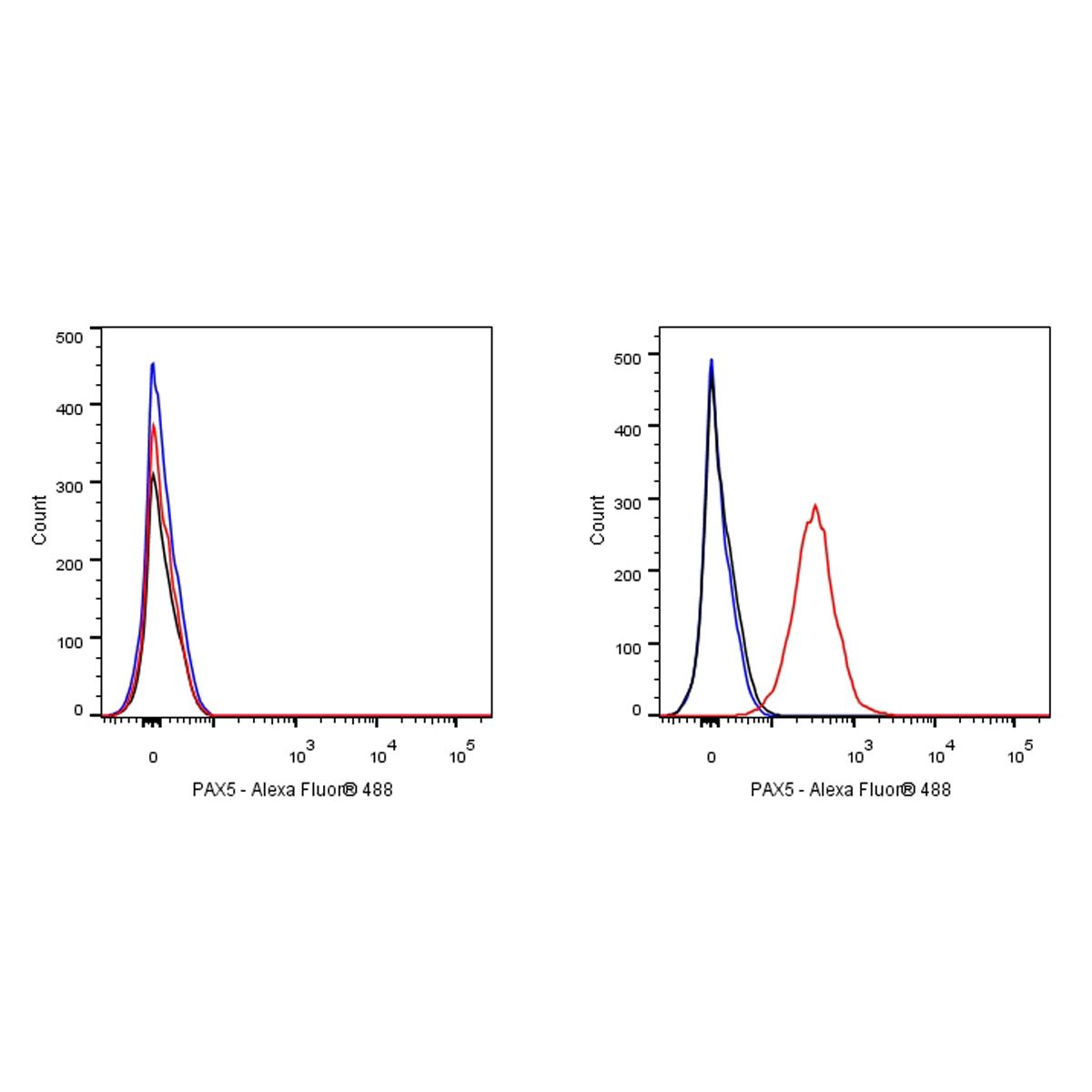
FC
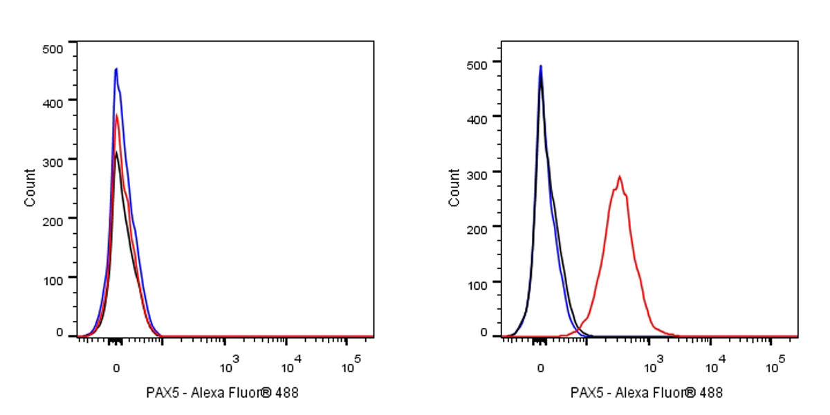
Flow cytometric analysis of 4% PFA fixed 90% methanol permeabilized Jurkat (Human T cell leukemia T lymphocyte, left) / Raji (Human Burkitt's lymphoma B lymphocyte, right) cells labelling PAX5 antibody at 1/50 dilution (1 μg) / (red) compared with a Rabbit monoclonal IgG (Black) isotype control and an unlabelled control (cells without incubation with primary antibody and secondary antibody) (Blue). Goat Anti - Rabbit IgG Alexa Fluor® 488 was used as the secondary antibody.
Negative control: Jurkat
IP
PAX5 Rabbit mAb at 1/50 dilution (1 µg) immunoprecipitating PAX5 in 0.4mg Ramos whole cell lysate. Western blot was performed on the immunoprecipitate using PAX5 Rabbit mAb at 1/1000 dilution. Secondary antibody (HRP) for IP was used at 1/400 dilution. Lane 1: Ramos whole cell lysate 20µg(input) Lane 2: PAX5 Rabbit mAb IP in Ramos whole cell lysate Lane 3: Rabbit monoclonal IgG IP in Ramos whole cell lysate Predicted MW: 42 kDa Observed MW: 45 kDa
Immunohistochemistry
IHC shows positive staining in paraffin-embedded human colon. Anti-PAX5 antibody was used at 1/500 dilution, followed by a HRP Polymer for Mouse & Rabbit IgG (ready to use). Counterstained with hematoxylin. Heat mediated antigen retrieval with Tris/EDTA buffer pH9.0 was performed before commencing with IHC staining protocol.
IHC shows positive staining in paraffin-embedded human spleen. Anti-PAX5 antibody was used at 1/500 dilution, followed by a HRP Polymer for Mouse & Rabbit IgG (ready to use). Counterstained with hematoxylin. Heat mediated antigen retrieval with Tris/EDTA buffer pH9.0 was performed before commencing with IHC staining protocol.
IHC shows positive staining in paraffin-embedded human tonsil. Anti-PAX5 antibody was used at 1/500 dilution, followed by a HRP Polymer for Mouse & Rabbit IgG (ready to use). Counterstained with hematoxylin. Heat mediated antigen retrieval with Tris/EDTA buffer pH9.0 was performed before commencing with IHC staining protocol.
IHC shows positive staining in paraffin-embedded human diffuse large B-cell lymphoma. Anti-PAX5 antibody was used at 1/500 dilution, followed by a HRP Polymer for Mouse & Rabbit IgG (ready to use). Counterstained with hematoxylin. Heat mediated antigen retrieval with Tris/EDTA buffer pH9.0 was performed before commencing with IHC staining protocol.
Negative control: IHC shows negative staining in paraffin-embedded human NK/T-cell lymphoma. Anti-PAX5 antibody was used at 1/500 dilution, followed by a HRP Polymer for Mouse & Rabbit IgG (ready to use). Counterstained with hematoxylin. Heat mediated antigen retrieval with Tris/EDTA buffer pH9.0 was performed before commencing with IHC staining protocol.
IHC shows positive staining in paraffin-embedded rat spleen. Anti-PAX5 antibody was used at 1/500 dilution, followed by a HRP Polymer for Mouse & Rabbit IgG (ready to use). Counterstained with hematoxylin. Heat mediated antigen retrieval with Tris/EDTA buffer pH9.0 was performed before commencing with IHC staining protocol.
Immunocytochemistry

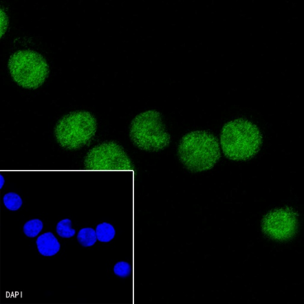
ICC shows positive staining in Ramos cells. Anti-PAX5 antibody was used
at 1/500 dilution (Green) and incubated overnight at 4°C.
Goat polyclonal Antibody to Rabbit IgG - H&L (Alexa Fluor® 488)
was used as secondary antibody at 1/1000 dilution.
The cells were fixed with 4% PFA and permeabilized with 0.1% PBS-Triton X-100.
Nuclei were counterstained with DAPI.

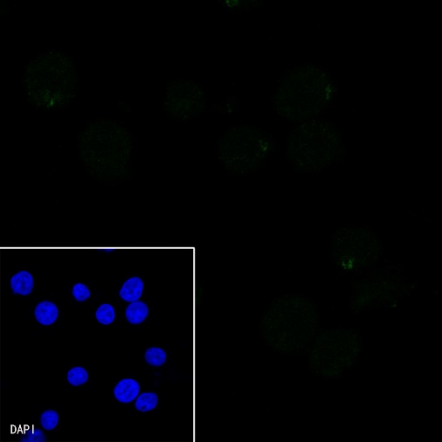
Negative control: ICC shows negative staining in Jurkat cells.
Anti-PAX5 antibody was used at 1/500 dilution and incubated overnight at 4°C.
Goat polyclonal Antibody to Rabbit IgG - H&L (Alexa Fluor® 488)
was used as secondary antibody at 1/1000 dilution.
The cells were fixed with 4% PFA and permeabilized with 0.1% PBS-Triton X-100.
Nuclei were counterstained with DAPI.