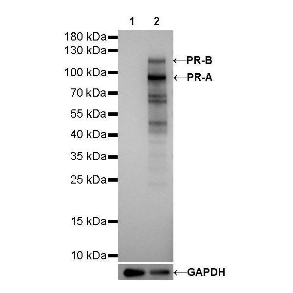
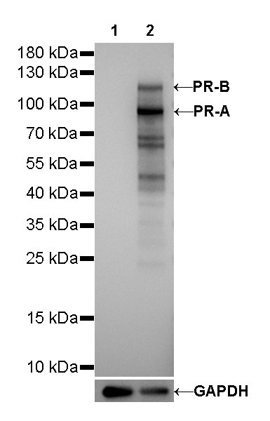
WB result of Progesterone Receptor Rabbit mAb
Primary antibody: Progesterone Receptor Rabbit mAb at 1/1000 dilution
Lane 1: MDA-MB-231 whole cell lysate 20 µg
Lane 2: T-47D whole cell lysate 20 µg
Negative control: MDA-MB-231 whole cell lysate
Secondary antibody: Goat Anti-Rabbit IgG, (H+L), HRP conjugated at 1/10000 dilution
Predicted MW: 82, 99 kDa
Observed MW: 90, 118 kDa
Progesterone Receptor Recombinant Rabbit mAb (SDT-R123)
Progesterone Receptor Recombinant Rabbit mAb (SDT-R123)
Price:
Regular price
$100 USD
Regular price
Sale price
$100 USD
Unit price
per
For shipping services or bulk orders, you may request a quotation.
Secure checkout with

View full details
Product Details
Product Details
Product Specification
| Host | Rabbit |
| Antigen | Progesterone Receptor |
| Synonyms | PR, NR3C3 |
| Immunogen | N/A |
| Location | Cytoplasm, Nucleus |
| Accession | P06401 |
| Clone Number | SDT-R123 |
| Antibody Type | Rabbit mAb |
| Application | WB, IHC-P, ICC, ICFCM |
| Reactivity | Hu |
| Purification | Protein A |
| Concentration | 0.5 mg/ml |
| Physical Appearance | Liquid |
| Storage Buffer | PBS, 40% Glycerol, 0.05% BSA, 0.03% Proclin 300 |
| Stability & Storage | 12 months from date of receipt / reconstitution, -20 °C as supplied |
Dilution
| application | dilution | species |
| WB | 1:1000 | null |
| IHC-P | 1:500 | null |
| ICFCM | 1:500 | null |
| ICC | 1:500 | null |
Background
Progesterone receptor (PR) is a master regulator in female reproductive tissues that controls developmental processes and proliferation and differentiation during the reproductive cycle and pregnancy. PR also plays a role in progression of endocrine-dependent breast cancer. As a member of the nuclear receptor family of ligand-dependent transcription factors, the main action of PR is to regulate networks of target gene expression in response to binding its cognate steroid hormone, progesterone [PMID: 27380738].
Picture
Picture
Western Blot
FC
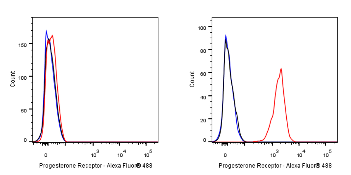
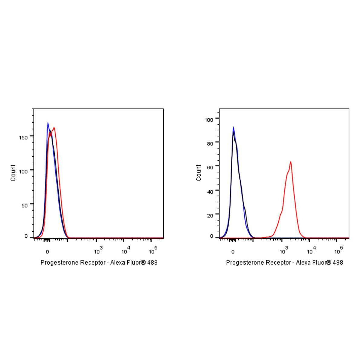
Flow cytometric analysis of 4% PFA fixed 90% methanol permeabilized MDA-MB-231 (Human breast adenocarcinoma epithelial cell) / T47D (Human ductal breast epithelial tumor epithelial cell, right) cells labelling Androgen Receptor antibody at 1/500 dilution (0.1 μg) / (red) compared with a Rabbit monoclonal IgG (Black) isotype control and an unlabelled control (cells without incubation with primary antibody and secondary antibody) (Blue). Goat Anti - Rabbit IgG Alexa Fluor® 488 was used as the secondary antibody.
Negative control: MDA-MB-231
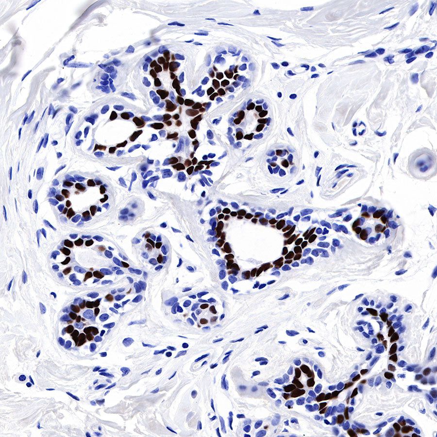
Immunohistochemistry
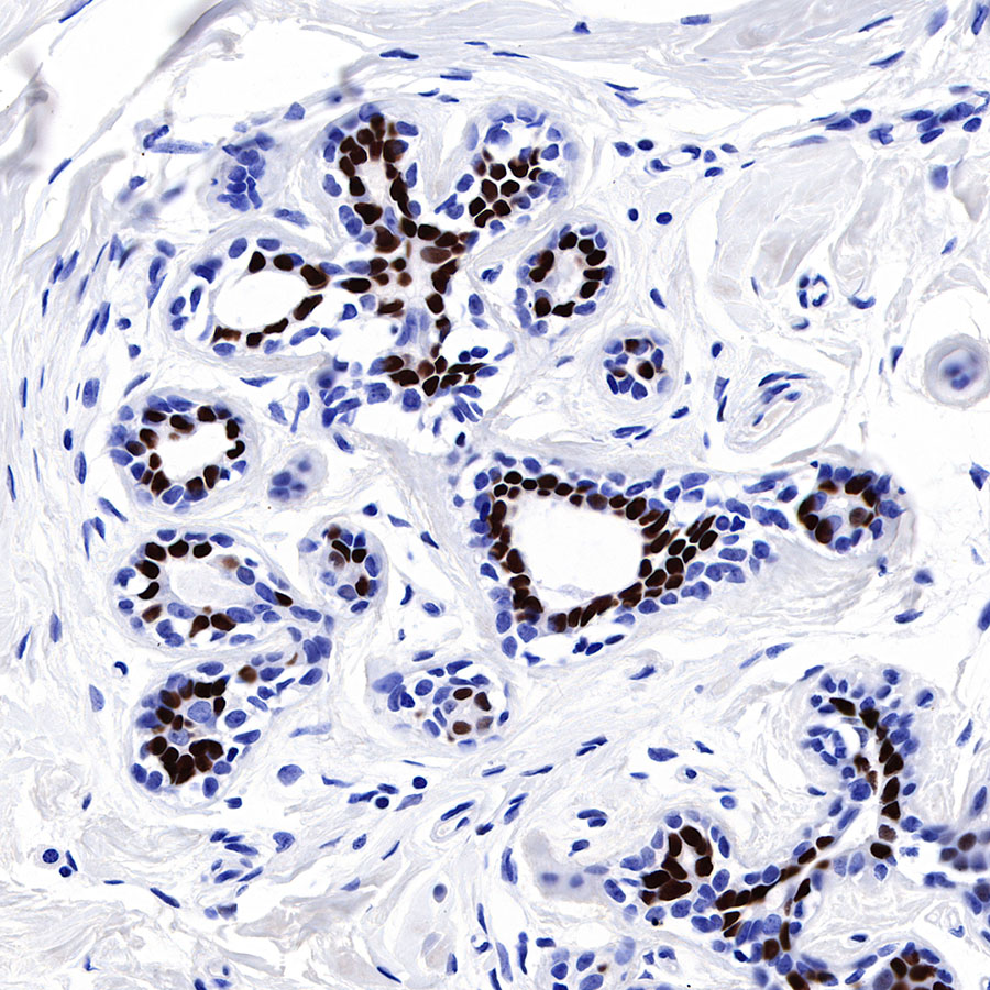
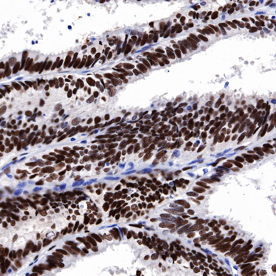
IHC shows positive staining in paraffin-embedded human breast. Anti-Progesterone Receptor antibody was used at 1/500 dilution, followed by a HRP Polymer for Mouse & Rabbit IgG (ready to use). Counterstained with hematoxylin. Heat mediated antigen retrieval with Tris/EDTA buffer pH9.0 was performed before commencing with IHC staining protocol.
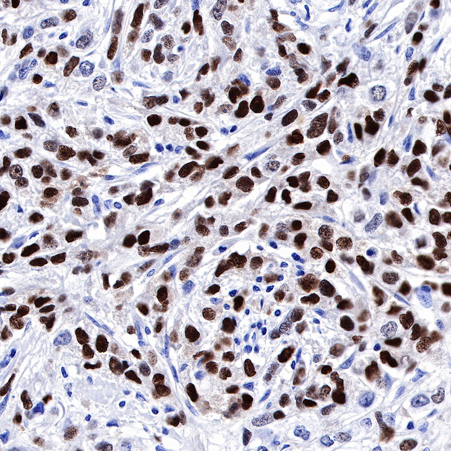
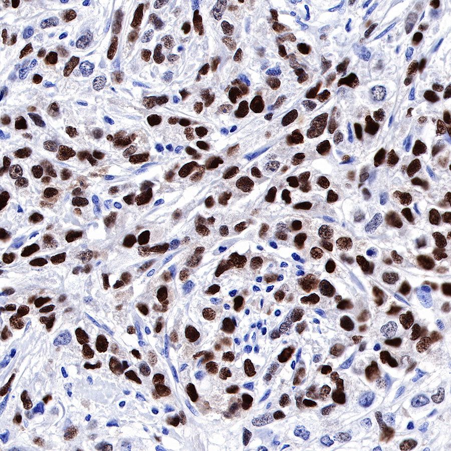
IHC shows positive staining in paraffin-embedded human breast cancer. Anti-Progesterone Receptor antibody was used at 1/500 dilution, followed by a HRP Polymer for Mouse & Rabbit IgG (ready to use). Counterstained with hematoxylin. Heat mediated antigen retrieval with Tris/EDTA buffer pH9.0 was performed before commencing with IHC staining protocol.
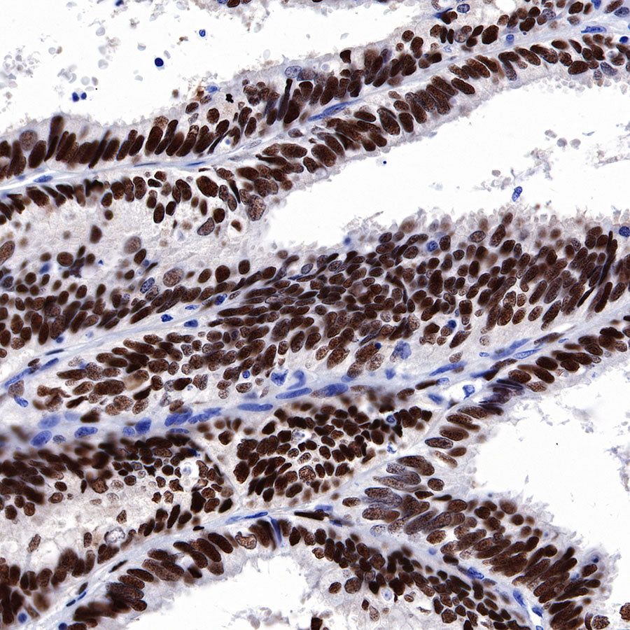
IHC shows positive staining in paraffin-embedded human endometrial cancer. Anti-Progesterone Receptor antibody was used at 1/500 dilution, followed by a HRP Polymer for Mouse & Rabbit IgG (ready to use). Counterstained with hematoxylin. Heat mediated antigen retrieval with Tris/EDTA buffer pH9.0 was performed before commencing with IHC staining protocol.
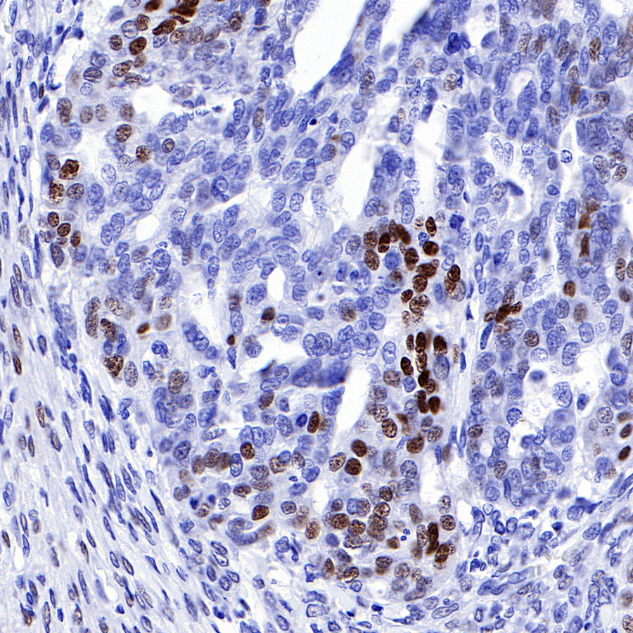
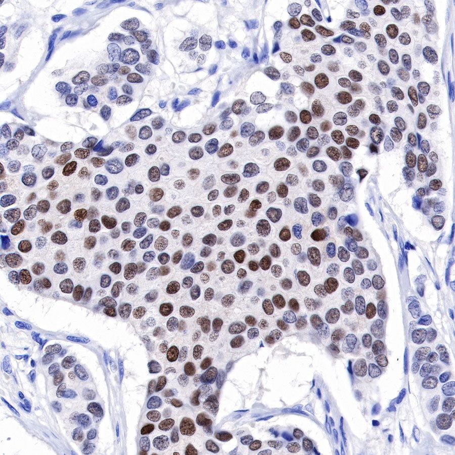
IHC shows positive staining in paraffin-embedded human ovarian cancer. Anti-Progesterone Receptor antibody was used at 1/500 dilution, followed by a HRP Polymer for Mouse & Rabbit IgG (ready to use). Counterstained with hematoxylin. Heat mediated antigen retrieval with Tris/EDTA buffer pH9.0 was performed before commencing with IHC staining protocol.

IHC shows positive staining in paraffin-embedded human breast cancer. Anti-Progesterone Receptor antibody was used at 1/500 dilution, followed by a HRP Polymer for Mouse & Rabbit IgG (ready to use). Counterstained with hematoxylin. Heat mediated antigen retrieval with Tris/EDTA buffer pH9.0 was performed before commencing with IHC staining protocol.

Negative control: IHC shows negative staining in paraffin-embedded human tonsil. Anti-Progesterone Receptor antibody was used at 1/500 dilution, followed by a HRP Polymer for Mouse & Rabbit IgG (ready to use). Counterstained with hematoxylin. Heat mediated antigen retrieval with Tris/EDTA buffer pH9.0 was performed before commencing with IHC staining protocol.
Immunocytochemistry
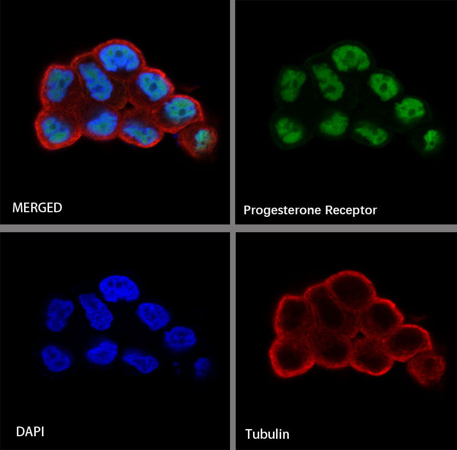
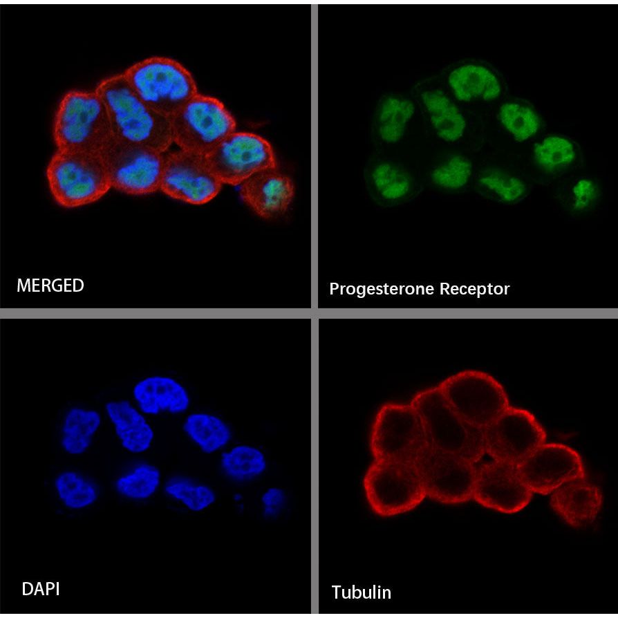
ICC shows positive staining in T47-D cells. Anti-Progesterone Receptor antibody was used at 1/500 dilution (Green) and incubated overnight at 4°C. Goat polyclonal Antibody to Rabbit IgG - H&L (Alexa Fluor® 488) was used as secondary antibody at 1/1000 dilution. The cells were fixed with 4% PFA and permeabilized with 0.1% PBS-Triton X-100. Nuclei were counterstained with DAPI (Blue). Counterstain with tubulin (red).
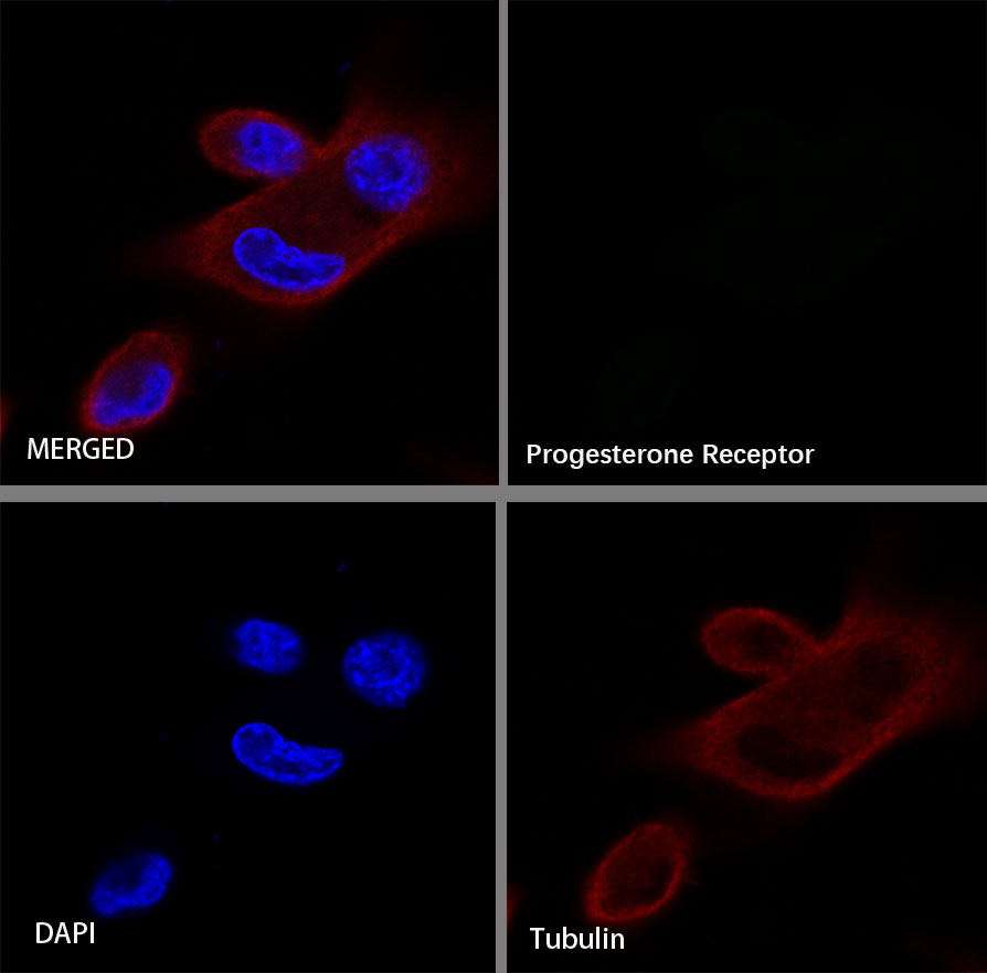
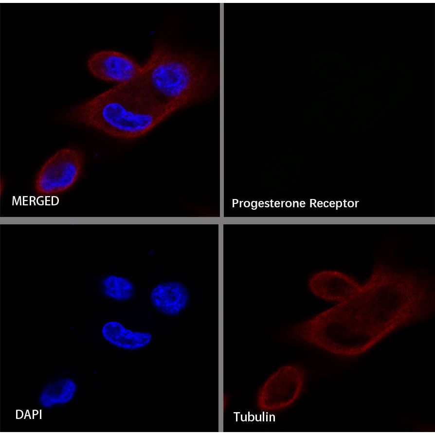
Negative control:ICC shows negative staining in MDA-MB-231 cells. Anti-Progesterone Receptor antibody was used at 1/500 dilution and incubated overnight at 4°C. Goat polyclonal Antibody to Rabbit IgG - H&L (Alexa Fluor® 488) was used as secondary antibody at 1/1000 dilution. The cells were fixed with 4% PFA and permeabilized with 0.1% PBS-Triton X-100. Nuclei were counterstained with DAPI (Blue). Counterstain with tubulin (red).