WB result of PDCD6 Rabbit mAb
Primary antibody: PDCD6 Rabbit mAb at 1/1000 dilution
Lane 1: Jurkat whole cell lysate 20 µg
Lane 2: 293T whole cell lysate 20 µg
Lane 3: HeLa whole cell lysate 20 µg
Lane 4: HepG2 whole cell lysate 20 µg
Lane 5: HCT 116 whole cell lysate 20 µg
Secondary antibody: Goat Anti-Rabbit IgG, (H+L), HRP conjugated at 1/10000 dilution
Predicted MW: 22 kDa
Observed MW: 22 kDa
(This blot was developed with high sensitivity substrate)
PDCD6 Recombinant Rabbit mAb (S-578-98)
PDCD6 Recombinant Rabbit mAb (S-578-98)
Price:
Regular price
$100 USD
Regular price
Sale price
$100 USD
Unit price
per
For shipping services or bulk orders, you may request a quotation.
Secure checkout with

View full details
Product Details
Product Details
Product Specification
| Host | Rabbit |
| Antigen | PDCD6 |
| Synonyms | Programmed cell death protein 6, Apoptosis-linked gene 2 protein homolog, ALG2 |
| Immunogen | Synthetic Peptide |
| Location | Cytoplasm, Nucleus |
| Accession | O75340 |
| Clone Number | S-578-98 |
| Antibody Type | Recombinant mAb |
| Isotype | IgG |
| Application | WB, IHC-P, ICC, IP, ICFCM |
| Reactivity | Hu, Ms, Rt |
| Purification | Protein A |
| Concentration | 0.5 mg/ml |
| Conjugation | Unconjugated |
| Physical Appearance | Liquid |
| Storage Buffer | PBS, 40% Glycerol, 0.05%BSA, 0.03% Proclin 300 |
| Stability & Storage | 12 months from date of receipt / reconstitution, -20 °C as supplied |
Dilution
| application | dilution | species |
| WB | 1:1000 | |
| IHC | 1:200 | |
| ICFCM | 1:50 | |
| ICC | 1:500 | |
| IP | 1:50 |
Background
PDCD6 is a calcium-binding protein belonging to the penta-EF-hand protein family. Calcium binding is important for homodimerization and for conformational changes required for binding to other protein partners. It participates in T cell receptor-, Fas-, and glucocorticoid-induced programmed cell death.
Picture
Picture
Western Blot
WB result of PDCD6 Rabbit mAb
Primary antibody: PDCD6 Rabbit mAb at 1/1000 dilution
Lane 1: mouse spleen lysate 20 µg
Secondary antibody: Goat Anti-Rabbit IgG, (H+L), HRP conjugated at 1/10000 dilution
Predicted MW: 22 kDa
Observed MW: 22 kDa
(This blot was developed with high sensitivity substrate)
WB result of PDCD6 Rabbit mAb
Primary antibody: PDCD6 Rabbit mAb at 1/1000 dilution
Lane 1: rat spleen lysate 20 µg
Secondary antibody: Goat Anti-Rabbit IgG, (H+L), HRP conjugated at 1/10000 dilution
Predicted MW: 22 kDa
Observed MW: 22 kDa
(This blot was developed with high sensitivity substrate)
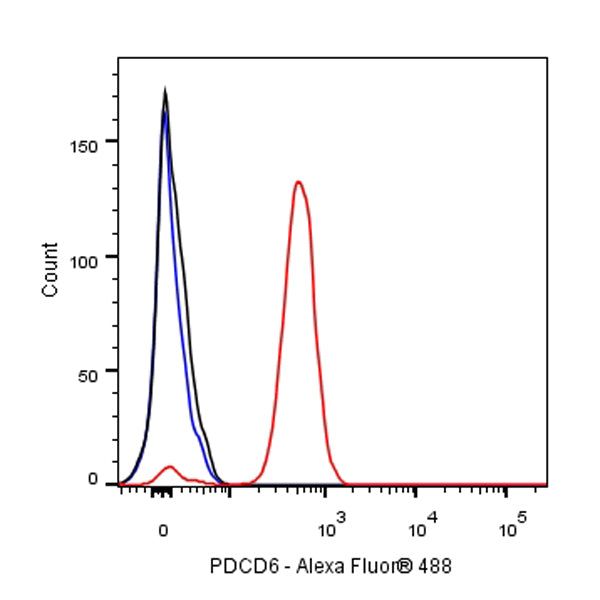
FC
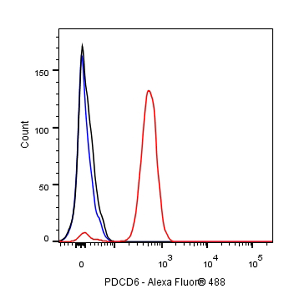
Flow cytometric analysis of 4% PFA fixed 90% methanol permeabilized HCT116 (Human colorectal carcinoma epithelial cell) cells labelling PDCD6 antibody at 1/50 dilution (1 μg)/ (Red) compared with a Rabbit monoclonal IgG (Black) isotype control and an unlabelled control (cells without incubation with primary antibody and secondary antibody) (Blue). Goat Anti - Rabbit IgG Alexa Fluor® 488 was used as the secondary antibody.
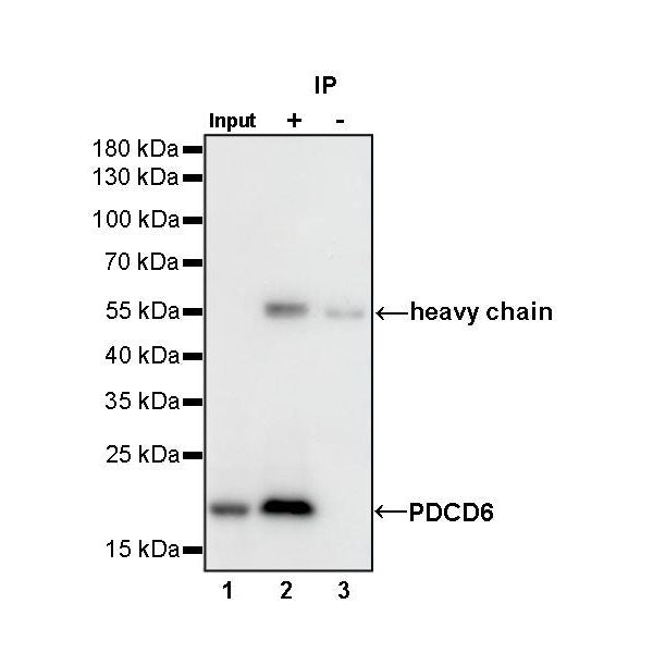
IP
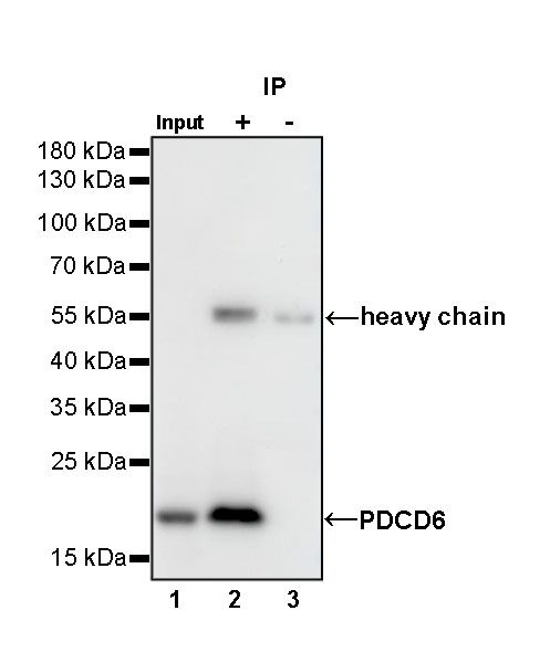
PDCD6 Rabbit mAb at 1/50 dilution (1 µg) immunoprecipitating PDCD6 in 0.4 mg HeLa whole cell lysate.
Western blot was performed on the immunoprecipitate using PDCD6 Rabbit mAb at 1/1000 dilution.
Secondary antibody (HRP) for IP was used at 1/400 dilution.
Lane 1: HeLa whole cell lysate 20 µg (Input)
Lane 2: PDCD6 Rabbit mAb IP in HeLa whole cell lysate
Lane 3: Rabbit monoclonal IgG IP in HeLa whole cell lysate
Predicted MW: 22 kDa
Observed MW: 22 kDa
(This blot was developed with high sensitivity substrate)
Immunohistochemistry
IHC shows positive staining in paraffin-embedded human breast. Anti-PDCD6 antibody was used at 1/200 dilution, followed by a HRP Polymer for Mouse & Rabbit IgG (ready to use). Counterstained with hematoxylin. Heat mediated antigen retrieval with Tris/EDTA buffer pH9.0 was performed before commencing with IHC staining protocol.
IHC shows positive staining in paraffin-embedded human colon. Anti-PDCD6 antibody was used at 1/200 dilution, followed by a HRP Polymer for Mouse & Rabbit IgG (ready to use). Counterstained with hematoxylin. Heat mediated antigen retrieval with Tris/EDTA buffer pH9.0 was performed before commencing with IHC staining protocol.
IHC shows positive staining in paraffin-embedded human breast cancer. Anti-PDCD6 antibody was used at 1/200 dilution, followed by a HRP Polymer for Mouse & Rabbit IgG (ready to use). Counterstained with hematoxylin. Heat mediated antigen retrieval with Tris/EDTA buffer pH9.0 was performed before commencing with IHC staining protocol.
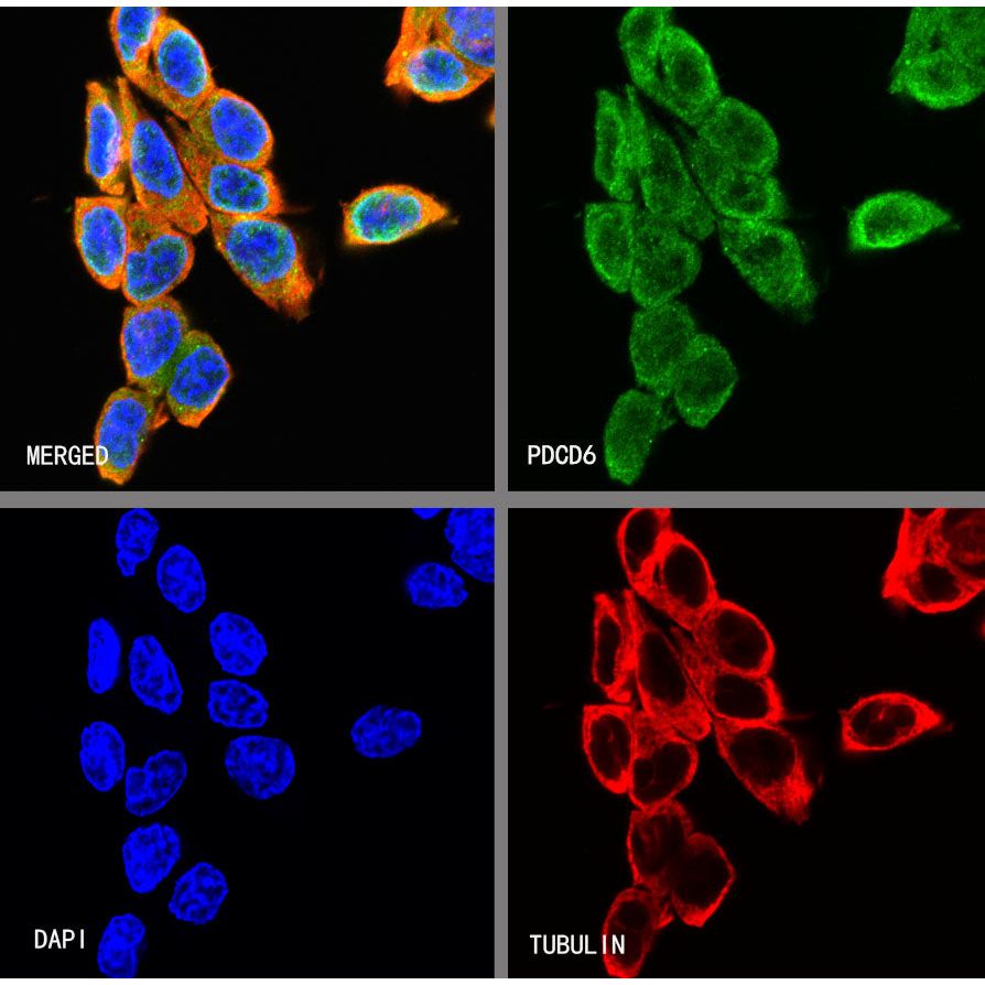
Immunocytochemistry
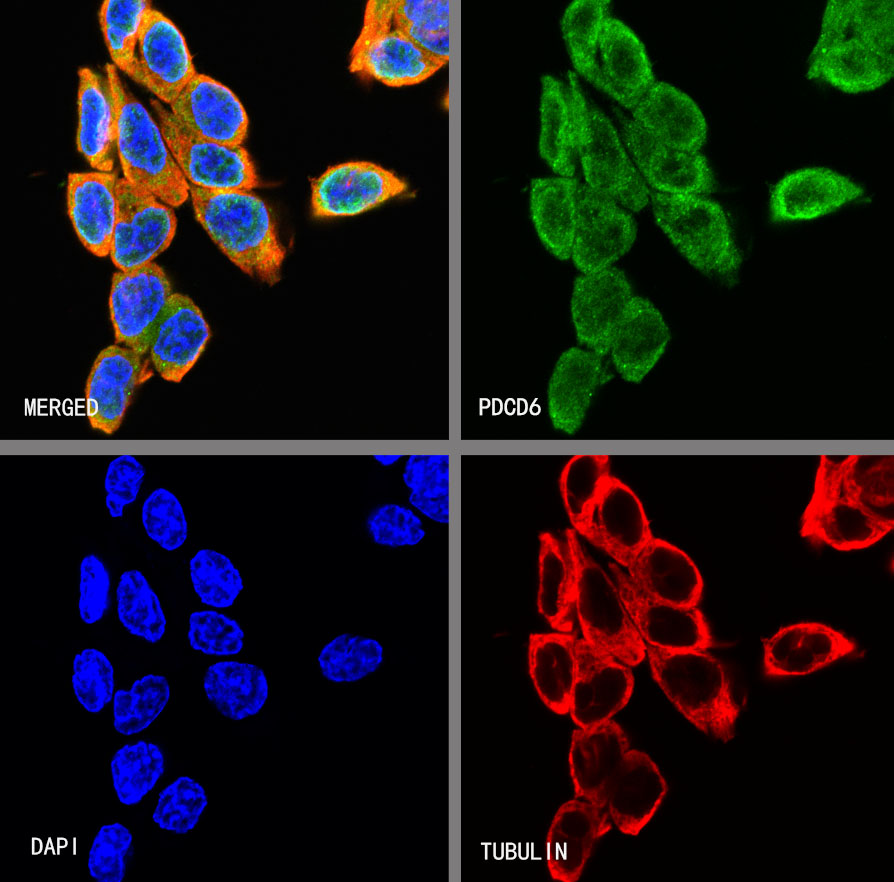
ICC shows positive staining in HCT116 cells. Anti- PDCD6 antibody was used at 1/500 dilution (Green) and incubated overnight at 4°C. Goat polyclonal Antibody to Rabbit IgG - H&L (Alexa Fluor® 488) was used as secondary antibody at 1/1000 dilution. The cells were fixed with 4% PFA and permeabilized with 0.1% PBS-Triton X-100. Nuclei were counterstained with DAPI (Blue). Counterstain with tubulin (Red).