Product Specification
| Host |
Rabbit |
| Antigen |
OCT3/4 |
| Synonyms |
POU5F1, OTF-3 |
| Immunogen |
Synthetic Peptide |
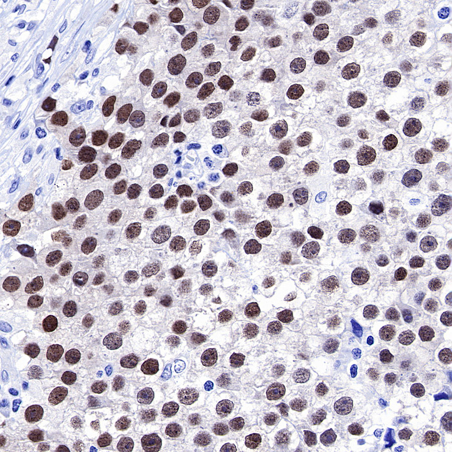
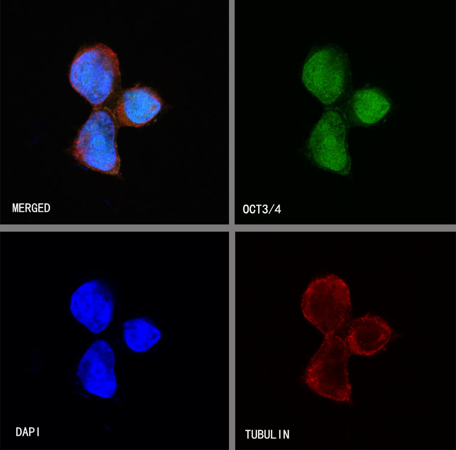
| Location |
Nucleus |
| Accession |
Q01860 |
| Clone Number |
SDT-165-76 |
| Antibody Type |
Rabbit mAb |
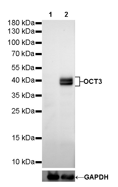
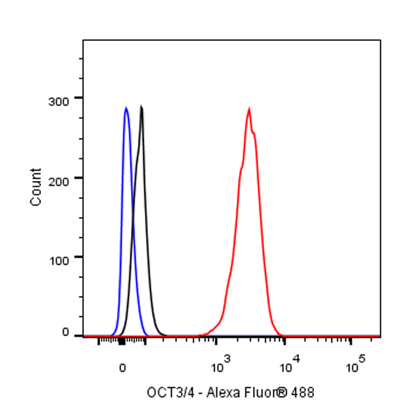
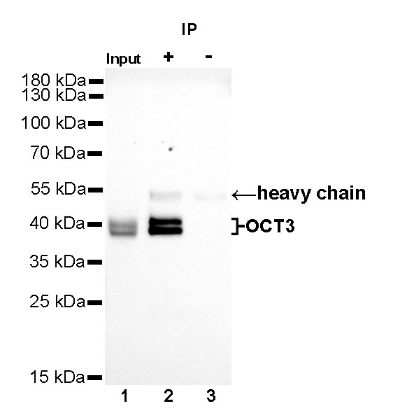
| Application |
WB, IHC-P, ICC, ICFCM, IP |
| Reactivity |
Hu |
| Predicted Reactivity |
Cz, Pg, Ms |
| Purification |
Protein A |
| Concentration |
0.25 mg/ml |
| Physical Appearance |
Liquid |
| Storage Buffer |
PBS, 40% Glycerol, 0.05% BSA, 0.03% Proclin 300 |
| Stability & Storage |
12 months from date of receipt / reconstitution, -20 °C as supplied |
Dilution
| application |
dilution |
species |
| WB |
1:500 |
|
| IHC-P |
1:1000 |
|
| IP |
1:25 |
|
| ICC |
1:50 |
|
| ICFCM |
1:250 |
|
Background
Oct-4, a transcription factor also known as Oct-3, Oct-3/4, Otf3 or NF-A3, is encoded by the Pou5f1 gene (located on chromosome 6 in human and 17 in mouse) and belongs to the POU (Pit, Oct, Unc) family of DNA binding-proteins. These proteins regulate the expression of target genes by binding to the octamer motif ATGCAAAT within their promoter or enhancer regions. Oct4, whose expression is associated with pluripotent properties of stem cells, is an essential factor controlling early stages of mammalian embryogenesis.