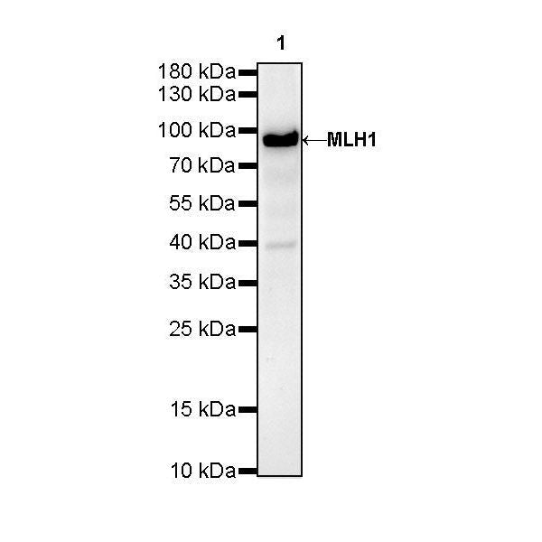
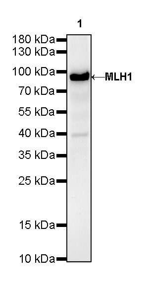
WB result of MLH1 Rabbit mAb
Primary antibody: MLH1 Rabbit mAb at 1/1000 dilution
Lane 1: rat testis lysate 20 µg
Secondary antibody: Goat Anti-Rabbit IgG, (H+L), HRP conjugated at 1/10000 dilution
Predicted MW: 84 kDa
Observed MW: 84 kDa
(This blot was developed with high sensitivity substrate)
MLH1 Recombinant Rabbit mAb (SDT-112-50-2)
MLH1 Recombinant Rabbit mAb (SDT-112-50-2)
Price:
Regular price
$100 USD
Regular price
Sale price
$100 USD
Unit price
per
For shipping services or bulk orders, you may request a quotation.
Secure checkout with

View full details
Product Details
Product Details
Product Specification
| Host | Rabbit |
| Antigen | MLH1 |
| Synonyms | DNA mismatch repair protein Mlh1, MutL protein homolog 1, COCA2 |
| Immunogen | Synthetic Peptide |
| Location | Nucleus |
| Accession | P40692 |
| Clone Number | SDT-112-50-2 |
| Antibody Type | Recombinant mAb |
| Application | WB, ICC |
| Reactivity | Hu |
| Predicted Reactivity | Rt |
| Purification | Protein A |
| Concentration | 0.5 mg/ml |
| Conjugation | Unconjugated |
| Physical Appearance | Liquid |
| Storage Buffer | PBS, 40% Glycerol, 0.05% BSA, 0.03% Proclin 300 |
| Stability & Storage | 12 months from date of receipt / reconstitution, -20 °C as supplied |
Dilution
| application | dilution | species |
| WB | 1:1000 | |
| ICC | 1:50 |
Background
DNA mismatch repair protein Mlh1 or MutL protein homolog 1 is a protein that in humans is encoded by the MLH1 gene located on chromosome 3. It is a gene commonly associated with hereditary nonpolyposis colorectal cancer. MLH1 protein is one component of a system of seven DNA mismatch repair proteins that work coordinately in sequential steps to initiate repair of DNA mismatches in humans [PMID: 18543306]. Defects in mismatch repair, found in about 13% of colorectal cancers, are much more frequently due to deficiency of MLH1 than deficiencies of other DNA mismatch repair proteins [PMID: 15887099]. The seven DNA mismatch repair proteins in humans are MLH1, MLH3, MSH2, MSH3, MSH6, PMS1 and PMS2 [ PMID: 18543306].
Picture
Picture
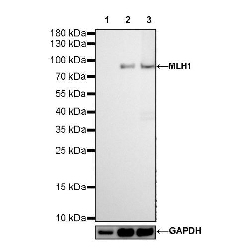
Western Blot
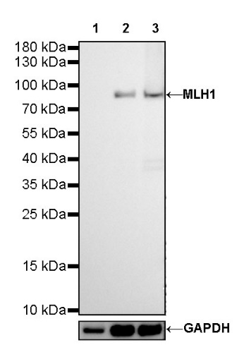
WB result of MLH1 Rabbit mAb
Primary antibody: MLH1 Rabbit mAb at 1/1000 dilution
Lane 1: HCT 116 whole cell lysate 20 µg
Lane 2: HeLa whole cell lysate 20 µg
Lane 3: Jurkat whole cell lysate 20 µg
Negative control: HCT 116 whole cell lysate
Secondary antibody: Goat Anti-Rabbit IgG, (H+L), HRP conjugated at 1/10000 dilution
Predicted MW: 84 kDa
Observed MW: 84 kDa
Immunocytochemistry
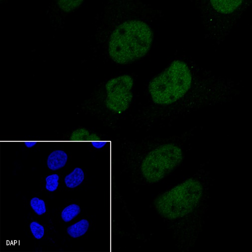
ICC shows positive staining in HeLa cells. Anti-MLH1 antibody was used at 1/50 dilution (Green) and incubated overnight at 4°C. Goat polyclonal Antibody to Rabbit IgG - H&L (Alexa Fluor® 488) was used as secondary antibody at 1/1000 dilution. The cells were fixed with 4% PFA and permeabilized with 0.1% PBS-Triton X-100. Nuclei were counterstained with DAPI (Blue).

Negative control: ICC shows negative staining in HCT-116 cells. Anti-MLH1 antibody was used at 1/50 dilution (Green) and incubated overnight at 4°C. Goat polyclonal Antibody to Rabbit IgG - H&L (Alexa Fluor® 488) was used as secondary antibody at 1/1000 dilution. The cells were fixed with 4% PFA and permeabilized with 0.1% PBS-Triton X-100. Nuclei were counterstained with DAPI (Blue).