WB result of MGMT Rabbit mAb
Primary antibody: MGMT Rabbit mAb at 1/1000 dilution
Lane 1: MDA-MB-231 whole cell lysate 20 µg
Lane 2: MCF7 whole cell lysate 20 µg
Lane 3: Jurkat whole cell lysate 20 µg
Lane 4: HT-29 whole cell lysate 20 µg
Negative control: MDA-MB-231 whole cell lysate
Secondary antibody: Goat Anti-Rabbit IgG, (H+L), HRP conjugated at 1/10000 dilution
Predicted MW: 22 kDa
Observed MW: 21 kDa
(This blot was developed with high sensitivity substrate)
MGMT Recombinant Rabbit mAb (SDT-522-71)
MGMT Recombinant Rabbit mAb (SDT-522-71)
Price:
Regular price
$100 USD
Regular price
Sale price
$100 USD
Unit price
per
For shipping services or bulk orders, you may request a quotation.
Secure checkout with

View full details
Product Details
Product Details
Product Specification
| Host | Rabbit |
| Antigen | MGMT |
| Synonyms | Methylated-DNA--protein-cysteine methyltransferase, 6-O-methylguanine-DNA methyltransferase, O-6-methylguanine-DNA-alkyltransferase |
| Immunogen | Synthetic Peptide |
| Location | Nucleus |
| Accession | P16455 |
| Clone Number | SDT-522-71 |
| Antibody Type | Recombinant mAb |
| Isotype | IgG |
| Application | WB, IHC-P, ICC, ICFCM, IP |
| Reactivity | Hu |
| Purification | Protein A |
| Concentration | 0.5 mg/ml |
| Conjugation | Unconjugated |
| Physical Appearance | Liquid |
| Storage Buffer | PBS, 40% Glycerol, 0.05%BSA, 0.03% Proclin 300 |
| Stability & Storage | 12 months from date of receipt / reconstitution, -20 °C as supplied |
Dilution
| application | dilution | species |
| WB | 1:1000 | |
| IHC | 1:100 | |
| ICC | 1:50 | |
| IP | 1:50 | |
| ICFCM | 1:500 |
Background
O6-Methylguanine-DNA Methyltransferase (MGMT) is an important DNA repair protein involved in tumor cell resistance to the cytostatic activity of chemotherapeutic alkylating agents. This protein is also effective in protecting normal cells against the genotoxic and carcinogenic effects of DNA alkylation. The alkylating drug resistance is caused by MGMT's ability to remove DNA alkyl groups introduced in the O6 position of guanine. MGMT is expressed in highly variable amounts, depending upon the cell and tissue type, species, and cellular growth characteristics. In addition, MGMT activity varies among groups of tumors and within a particular type of tumor.
Picture
Picture
Western Blot
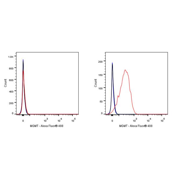
FC
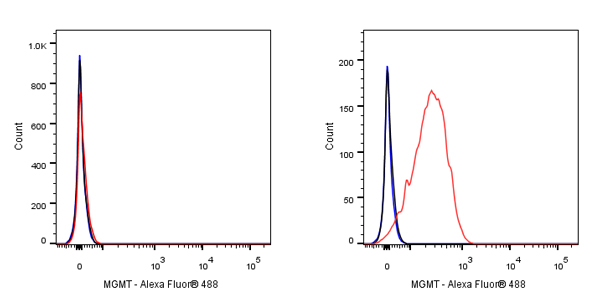
Flow cytometric analysis of 4% PFA fixed 90% methanol permeabilized MDA-MB-231 (Human breast adenocarcinoma epithelial cell, left) / MCF7(Human breast adenocarcinoma epithelial cell, Right) labelling MGMT antibody at 1/500 dilution (0.1 μg) / (Red) compared with a Rabbit monoclonal IgG (Black) isotype control and an unlabelled control (cells without incubation with primary antibody and secondary antibody) (Blue). Goat Anti - Rabbit IgG Alexa Fluor® 488 was used as the secondary antibody.
Negative control: MDA-MB-231
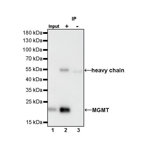
IP
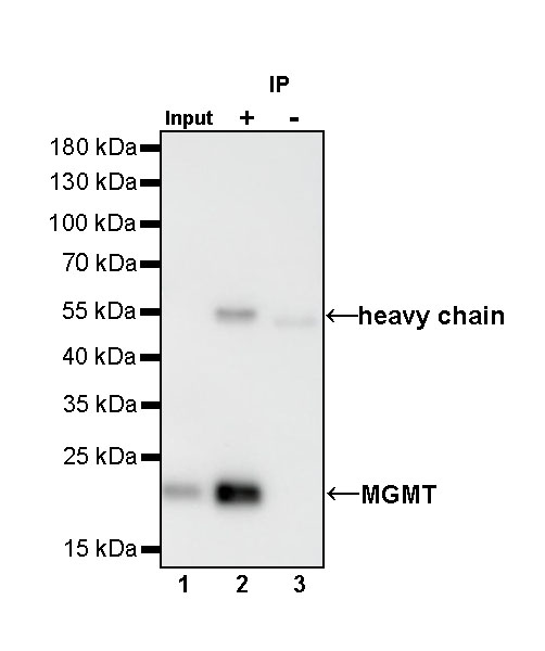
MGMT Rabbit mAb at 1/50 dilution (1 µg) immunoprecipitating MGMT in 0.4 mg MCF7 whole cell lysate.
Western blot was performed on the immunoprecipitate using MGMT Rabbit mAb at 1/1000 dilution.
Secondary antibody (HRP) for IP was used at 1/400 dilution.
Lane 1: MCF7 whole cell lysate 20 µg (Input)
Lane 2: MGMT Rabbit mAb IP in MCF7 whole cell lysate
Lane 3: Rabbit monoclonal IgG IP in MCF7 whole cell lysate
Predicted MW: 22 kDa
Observed MW: 21 kDa
Immunohistochemistry
IHC shows positive staining in paraffin-embedded human testis. Anti-MGMT antibody was used at 1/100 dilution, followed by a HRP Polymer for Mouse & Rabbit IgG (ready to use). Counterstained with hematoxylin. Heat mediated antigen retrieval with Tris/EDTA buffer pH9.0 was performed before commencing with IHC staining protocol.
IHC shows positive staining in paraffin-embedded human stomach. Anti-MGMT antibody was used at 1/100 dilution, followed by a HRP Polymer for Mouse & Rabbit IgG (ready to use). Counterstained with hematoxylin. Heat mediated antigen retrieval with Tris/EDTA buffer pH9.0 was performed before commencing with IHC staining protocol.
IHC shows positive staining in paraffin-embedded human diffuse large B-cell lymphoma. Anti-MGMT antibody was used at 1/100 dilution, followed by a HRP Polymer for Mouse & Rabbit IgG (ready to use). Counterstained with hematoxylin. Heat mediated antigen retrieval with Tris/EDTA buffer pH9.0 was performed before commencing with IHC staining protocol.
IHC shows positive staining in paraffin-embedded human lung squamous cell carcinoma. Anti-MGMT antibody was used at 1/100 dilution, followed by a HRP Polymer for Mouse & Rabbit IgG (ready to use). Counterstained with hematoxylin. Heat mediated antigen retrieval with Tris/EDTA buffer pH9.0 was performed before commencing with IHC staining protocol.
IHC shows positive staining in paraffin-embedded human ovarian carcinoma. Anti-MGMT antibody was used at 1/100 dilution, followed by a HRP Polymer for Mouse & Rabbit IgG (ready to use). Counterstained with hematoxylin. Heat mediated antigen retrieval with Tris/EDTA buffer pH9.0 was performed before commencing with IHC staining protocol.
IHC shows positive staining in paraffin-embedded human oligodendroglioma. Anti-MGMT antibody was used at 1/100 dilution, followed by a HRP Polymer for Mouse & Rabbit IgG (ready to use). Counterstained with hematoxylin. Heat mediated antigen retrieval with Tris/EDTA buffer pH9.0 was performed before commencing with IHC staining protocol.
Immunocytochemistry
ICC shows positive staining in MCF7 cells. Anti-MGMT antibody was used at 1/50 dilution (Green) and incubated overnight at 4°C. Goat polyclonal Antibody to Rabbit IgG - H&L (Alexa Fluor® 488) was used as secondary antibody at 1/1000 dilution. The cells were fixed with 4% PFA and permeabilized with 0.1% PBS-Triton X-100. Nuclei were counterstained with DAPI (Blue). Counterstain with tubulin (red).