WB result of LYPD3/C4.4A Rabbit mAb
Primary antibody: LYPD3/C4.4A Rabbit mAb at 1/1000 dilution
Lane 1: HaCaT whole cell lysate 20 µg
Lane 2: BxPC-3 whole cell lysate 20 µg
Lane 3: MCF7 whole cell lysate 20 µg
Secondary antibody: Goat Anti-Rabbit IgG, (H+L), HRP conjugated at 1/10000 dilution
Predicted MW: 36 kDa
Observed MW: 76 kDa
(This blot was developed with high sensitivity substrate)
LYPD3/C4.4A Recombinant Rabbit mAb (S-R222)
LYPD3/C4.4A Recombinant Rabbit mAb (S-R222)
Price:
Regular price
$100 USD
Regular price
Sale price
$100 USD
Unit price
per
For shipping services or bulk orders, you may request a quotation.
Secure checkout with

View full details
Product Details
Product Details
Product Specification
| Host | Rabbit |
| Antigen | LYPD3/C4.4A |
| Synonyms | Ly6/PLAUR domain-containing protein 3, GPI-anchored metastasis-associated protein C4.4A homolog, Matrigel-induced gene C4 protein (MIG-C4) |
| Location | Cell membrane |
| Accession | O95274 |
| Clone Number | S-R222 |
| Antibody Type | Recombinant mAb |
| Application | WB, IHC-P, FCM, IP |
| Reactivity | Hu |
| Purification | Protein A |
| Concentration | 0.5 mg/ml |
| Conjugation | Unconjugated |
| Physical Appearance | Liquid |
| Storage Buffer | PBS, 40% Glycerol, 0.05%BSA, 0.03% Proclin 300 |
| Stability & Storage | 12 months from date of receipt / reconstitution, -20 °C as supplied |
Dilution
| application | dilution | species |
| WB | 1:1000 | null |
| IP | 1:50 | null |
| IHC | 1:2000 | null |
| FCM | 1:500 | null |
Background
C4.4A (Ly6/PLAUR domain-containing protein 3, LYPD3), first reported in 1998, is a tumorigenic and high-glycosylated cell surface protein that has been proven to be linked with the carcinogenic effects in different solid tumors. The elevated expression of LYPD3 is not only demonstrated to be associated with lung adenocarcinoma carcinogenesis and poor prognosis but also there is evidence that LYPD3 can lead to the initiation and development of cancers and the chemoresistance of metastatic cancers by impacting the proliferation and apoptosis of the tumor, which are involved in many important regulatory mechanisms of cancers.
Picture
Picture
Western Blot
FC
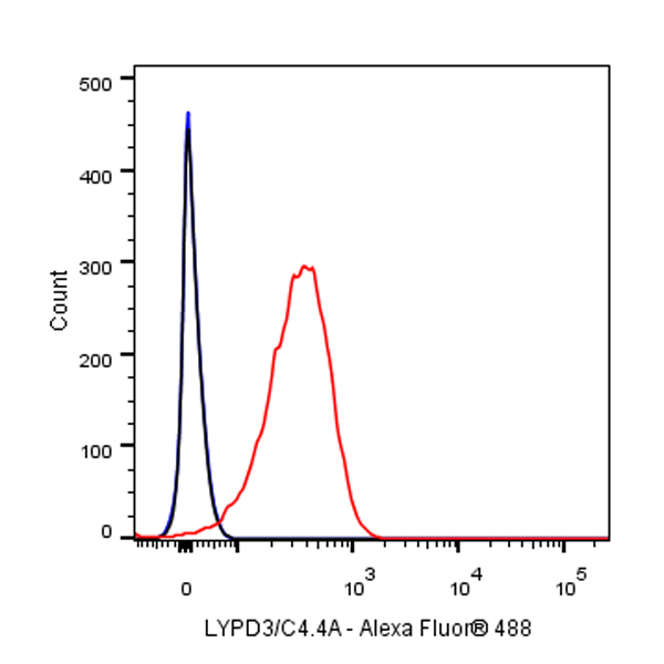
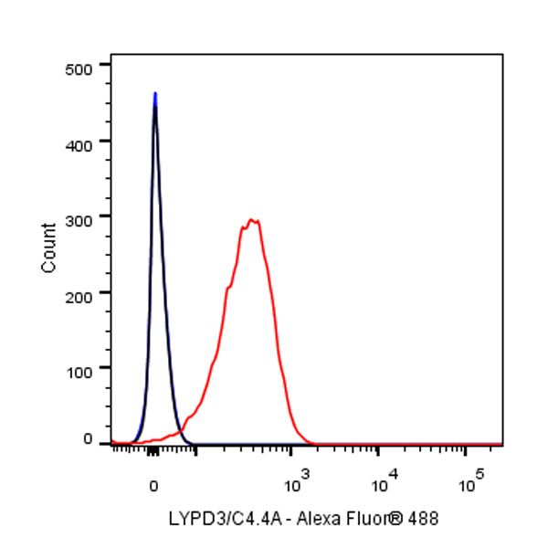
Flow cytometric analysis of HACAT (Human skin keratinocyte) cells labelling LYPD3/C4.4A antibody at 1/500 (0.1 μg) dilution / (red) compared with a Rabbit monoclonal IgG (Black) isotype control and an unlabelled control (cells without incubation with primary antibody and secondary antibody) (Blue). Goat Anti - Rabbit IgG Alexa Fluor® 488 was used as the secondary antibody.
IP
LYPD3/C4.4A Rabbit mAb at 1/50 dilution (1 µg) immunoprecipitating LYPD3/C4.4A in 0.4 mg HaCaT whole cell lysate.
Western blot was performed on the immunoprecipitate using LYPD3/C4.4A Rabbit mAb at 1/1000 dilution.
Secondary antibody (HRP) for IP was used at 1/400 dilution.
Lane 1: HaCaT whole cell lysate 20 µg (Input)
Lane 2: LYPD3/C4.4A Rabbit mAb IP in HaCaT whole cell lysate
Lane 3: Rabbit monoclonal IgG IP in HaCaT whole cell lysate
Predicted MW: 36 kDa
Observed MW: 76 kDa
Immunohistochemistry
IHC shows positive staining in paraffin-embedded human esophagus. Anti-LYPD3/C4.4A antibody was used at 1/2000 dilution, followed by a HRP Polymer for Mouse & Rabbit IgG (ready to use). Counterstained with hematoxylin. Heat mediated antigen retrieval with Tris/EDTA buffer pH9.0 was performed before commencing with IHC staining protocol.
IHC shows positive staining in paraffin-embedded human skin. Anti-LYPD3/C4.4A antibody was used at 1/2000 dilution, followed by a HRP Polymer for Mouse & Rabbit IgG (ready to use). Counterstained with hematoxylin. Heat mediated antigen retrieval with Tris/EDTA buffer pH9.0 was performed before commencing with IHC staining protocol.
IHC shows positive staining in paraffin-embedded human tonsil. Anti-LYPD3/C4.4A antibody was used at 1/2000 dilution, followed by a HRP Polymer for Mouse & Rabbit IgG (ready to use). Counterstained with hematoxylin. Heat mediated antigen retrieval with Tris/EDTA buffer pH9.0 was performed before commencing with IHC staining protocol.
IHC shows positive staining in paraffin-embedded human cervical squamous cell carcinoma. Anti-LYPD3/C4.4A antibody was used at 1/2000 dilution, followed by a HRP Polymer for Mouse & Rabbit IgG (ready to use). Counterstained with hematoxylin. Heat mediated antigen retrieval with Tris/EDTA buffer pH9.0 was performed before commencing with IHC staining protocol.
IHC shows positive staining in paraffin-embedded human transitional cell carcinoma. Anti-LYPD3/C4.4A antibody was used at 1/2000 dilution, followed by a HRP Polymer for Mouse & Rabbit IgG (ready to use). Counterstained with hematoxylin. Heat mediated antigen retrieval with Tris/EDTA buffer pH9.0 was performed before commencing with IHC staining protocol.