WB result of Lamin B1 Rabbit mAb Primary antibody: Lamin B1 Rabbit mAb at 1/1000 dilution Lane 1: HeLa whole cell lysate 20 µg Lane 2: Ramos whole cell lysate 20 µg Lane 3: MCF7 whole cell lysate 20 µg Lane 4: Jurkat whole cell lysate 20 µg Lane 5: MOLT-4 whole cell lysate 20 µg Lane 6: Caco-2 whole cell lysate 20 µg Secondary antibody: Goat Anti-Rabbit IgG, (H+L), HRP conjugated at 1/10000 dilution Predicted MW: 68 kDa Observed MW: 68 kDa
Lamin B1 Recombinant Rabbit mAb (SDT-307-108)
Lamin B1 Recombinant Rabbit mAb (SDT-307-108)
Price:
Regular price
$70 USD
Regular price
Sale price
$70 USD
Unit price
per
For shipping services or bulk orders, you may request a quotation.
Secure checkout with

View full details
Product Details
Product Details
Product Specification
| Host | Rabbit |
| Antigen | Lamin B1 |
| Synonyms | LMNB1; Lamin-B1 |
| Location | Nucleus lamina |
| Accession | P20700 |
| Clone Number | SDT-307-108 |
| Antibody Type | Recombinant mAb |
| Application | WB, IHC-P, IP, IF, ChIP |
| Reactivity | Hu, Ms, Rt |
| Purification | Protein A |
| Concentration | 0.5 mg/ml |
| Conjugation | Unconjugated |
| Physical Appearance | Liquid |
| Storage Buffer | PBS, 40% Glycerol, 0.05% BSA, 0.03% Proclin 300 |
| Stability & Storage | 12 months from date of receipt / reconstitution, -20 °C as supplied |
Dilution
| application | dilution | species |
| WB | 1:5000-1:50000 | |
| IP | 1:50 | |
| IHC | 1:2000 | |
| IF | 1:500 | |
| ChIP | 1:20-1:50 |
Background
Lamin-B1 is a protein that in humans is encoded by the LMNB1 gene. It is one of the essential members of the nuclear lamina protein family. Its main function is to maintain the integrity of nuclear skeleton, as well as to participate in the cell proliferation and aging by affecting the chromosome distribution. gene expression, and DNA damage repair. The abnormal expression of lamin B1 is related to certain diseases, including neurological diseases [e.g. neural tube defects (NDTs), adult-onset autosomal dominant leukodystrophy (ADLD)] and tumors (e.g. pancreatic cancer). It is also a potential tumor marker as well as drug target [PMID: 30499270].
Picture
Picture
Western Blot
WB result of Lamin B1 Rabbit mAb Primary antibody: Lamin B1 Rabbit mAb at 1/1000 dilution Lane 1: NIH/3T3 whole cell lysate 20 µg Lane 2: RAW 264.7 whole cell lysate 20 µg Lane 3: mouse brain lysate 20 µg Lane 4: mouse heart lysate 20 µg Lane 5: mouse kidney lysate 20 µg Lane 6: mouse spleen lysate 20 µg Secondary antibody: Goat Anti-Rabbit IgG, (H+L), HRP conjugated at 1/10000 dilution Predicted MW: 68 kDa Observed MW: 68 kDa
WB result of Lamin B1 Rabbit mAb Primary antibody: Lamin B1 Rabbit mAb at 1/1000 dilution Lane 1: PC-12 whole cell lysate 20 µg Lane 2: rat brain lysate 20 µg Lane 3: rat heart lysate 20 µg Lane 4: rat kidney lysate 20 µg Secondary antibody: Goat Anti-Rabbit IgG, (H+L), HRP conjugated at 1/10000 dilution Predicted MW: 68 kDa Observed MW: 68 kDa
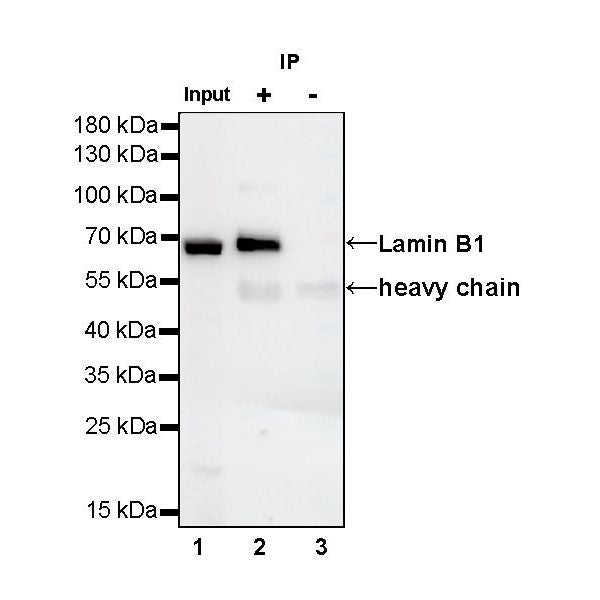
ChIP
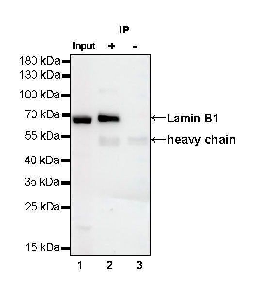
Lamin B1 Rabbit mAb at 1/50 dilution (1 µg) immunoprecipitating Lamin B1 in 0.4 mg HeLa whole cell lysate.
Western blot was performed on the immunoprecipitate using Lamin B1 Rabbit mAb at 1/1000 dilution.
Secondary antibody (HRP) for IP was used at 1/400 dilution.
Lane 1: HeLa whole cell lysate 20 µg (Input)
Lane 2: Lamin B1 Rabbit mAb IP in HeLa whole cell lysate
Lane 3: Rabbit monoclonal IgG IP in HeLa whole cell lysate
Predicted MW: 68 kDa
Observed MW: 68 kDa
Immunohistochemistry
IHC shows positive staining in paraffin-embedded human spleen. Anti-Lamin B1 antibody was used at 1/2000 dilution, followed by a Goat Anti-Rabbit IgG H&L (HRP) ready to use. Counterstained with hematoxylin. Heat mediated antigen retrieval with Tris/EDTA buffer pH9.0 was performed before commencing with IHC staining protocol.
IHC shows positive staining in paraffin-embedded human stomach. Anti-Lamin B1 antibody was used at 1/2000 dilution, followed by a Goat Anti-Rabbit IgG H&L (HRP) ready to use. Counterstained with hematoxylin. Heat mediated antigen retrieval with Tris/EDTA buffer pH9.0 was performed before commencing with IHC staining protocol.
IHC shows positive staining in paraffin-embedded human endometrial cancer. Anti-Lamin B1 antibody was used at 1/2000 dilution, followed by a Goat Anti-Rabbit IgG H&L (HRP) ready to use. Counterstained with hematoxylin. Heat mediated antigen retrieval with Tris/EDTA buffer pH9.0 was performed before commencing with IHC staining protocol.
IHC shows positive staining in paraffin-embedded human lung cancer. Anti-Lamin B1 antibody was used at 1/2000 dilution, followed by a Goat Anti-Rabbit IgG H&L (HRP) ready to use. Counterstained with hematoxylin. Heat mediated antigen retrieval with Tris/EDTA buffer pH9.0 was performed before commencing with IHC staining protocol.
IHC shows positive staining in paraffin-embedded human thyroid cancer. Anti-Lamin B1 antibody was used at 1/2000 dilution, followed by a Goat Anti-Rabbit IgG H&L (HRP) ready to use. Counterstained with hematoxylin. Heat mediated antigen retrieval with Tris/EDTA buffer pH9.0 was performed before commencing with IHC staining protocol.
IHC shows positive staining in paraffin-embedded mouse spleen. Anti-Lamin B1 antibody was used at 1/2000 dilution, followed by a Goat Anti-Rabbit IgG H&L (HRP) ready to use. Counterstained with hematoxylin. Heat mediated antigen retrieval with Tris/EDTA buffer pH9.0 was performed before commencing with IHC staining protocol.
IHC shows positive staining in paraffin-embedded mouse skeletal muscle. Anti-Lamin B1 antibody was used at 1/2000 dilution, followed by a Goat Anti-Rabbit IgG H&L (HRP) ready to use. Counterstained with hematoxylin. Heat mediated antigen retrieval with Tris/EDTA buffer pH9.0 was performed before commencing with IHC staining protocol.
IHC shows positive staining in paraffin-embedded rat cerebral cortex. Anti-Lamin B1 antibody was used at 1/2000 dilution, followed by a Goat Anti-Rabbit IgG H&L (HRP) ready to use. Counterstained with hematoxylin. Heat mediated antigen retrieval with Tris/EDTA buffer pH9.0 was performed before commencing with IHC staining protocol.
IHC shows positive staining in paraffin-embedded rat liver. Anti-Lamin B1 antibody was used at 1/2000 dilution, followed by a Goat Anti-Rabbit IgG H&L (HRP) ready to use. Counterstained with hematoxylin. Heat mediated antigen retrieval with Tris/EDTA buffer pH9.0 was performed before commencing with IHC staining protocol.
Immunofluorescence
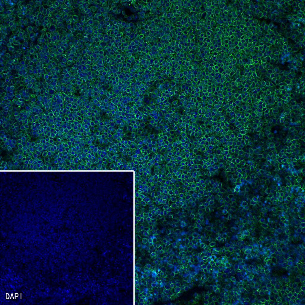
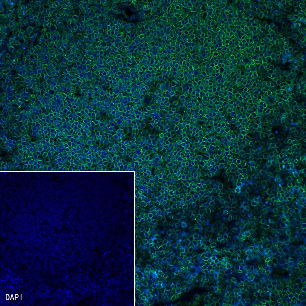
IF shows positive staining in paraffin-embedded mouse spleen. Anti-Lamin B1 antibody was used at 1/500 dilution (Green) and incubated overnight at 4°C. Goat polyclonal Antibody to Rabbit IgG - H&L (Alexa Fluor® 488) was used as secondary antibody at 1/1000 dilution. Counterstained with DAPI (Blue). Heat mediated antigen retrieval with EDTA buffer pH9.0 was performed before commencing with IF staining protocol.
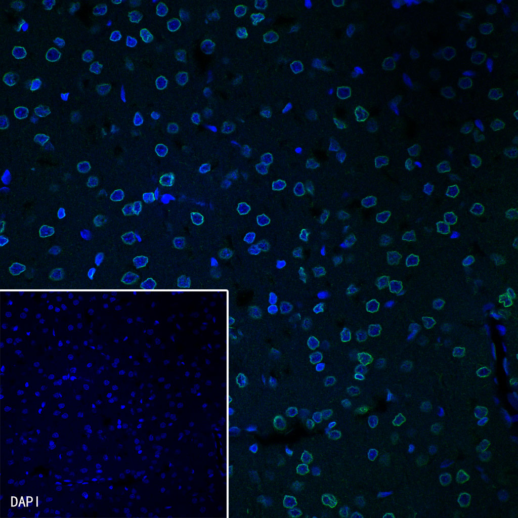
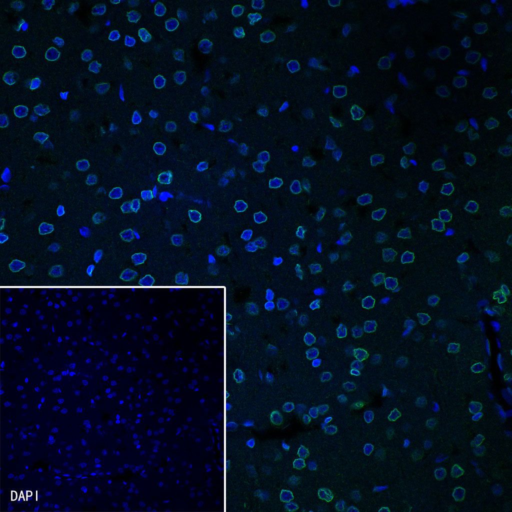
IF shows positive staining in paraffin-embedded rat brain. Anti-Lamin B1 antibody was used at 1/500 dilution (Green) and incubated overnight at 4°C. Goat polyclonal Antibody to Rabbit IgG - H&L (Alexa Fluor® 488) was used as secondary antibody at 1/1000 dilution. Counterstained with DAPI (Blue). Heat mediated antigen retrieval with EDTA buffer pH9.0 was performed before commencing with IF staining protocol.
ChIP
Chromatin immunoprecipitation (ChIP) was performed on HeLa cells cross - linked with 1% formaldehyde for 10 min, then chromatin was fragmented by sonication. Parallel reactions used Lamin B1 Recombinant Rabbit mAb (SDT-307-108) and Rabbit mAb IgG Isotype Control (SDT-R173) at 1:50 for immunoprecipitation.
Post - immunoprecipitation, both samples were washed, eluted, cross - links reversed. Purified DNA was analyzed by qPCR.
qPCR (%input: immunoprecipitated DNA/input DNA)
showed the enrichment of BRCA1, DBB1 and HBB in
Lamin B1 Recombinant Rabbit mAb (SDT-307-108)-
immunoprecipitated sample.