Product Specification
| Host |
Rabbit |
| Antigen |
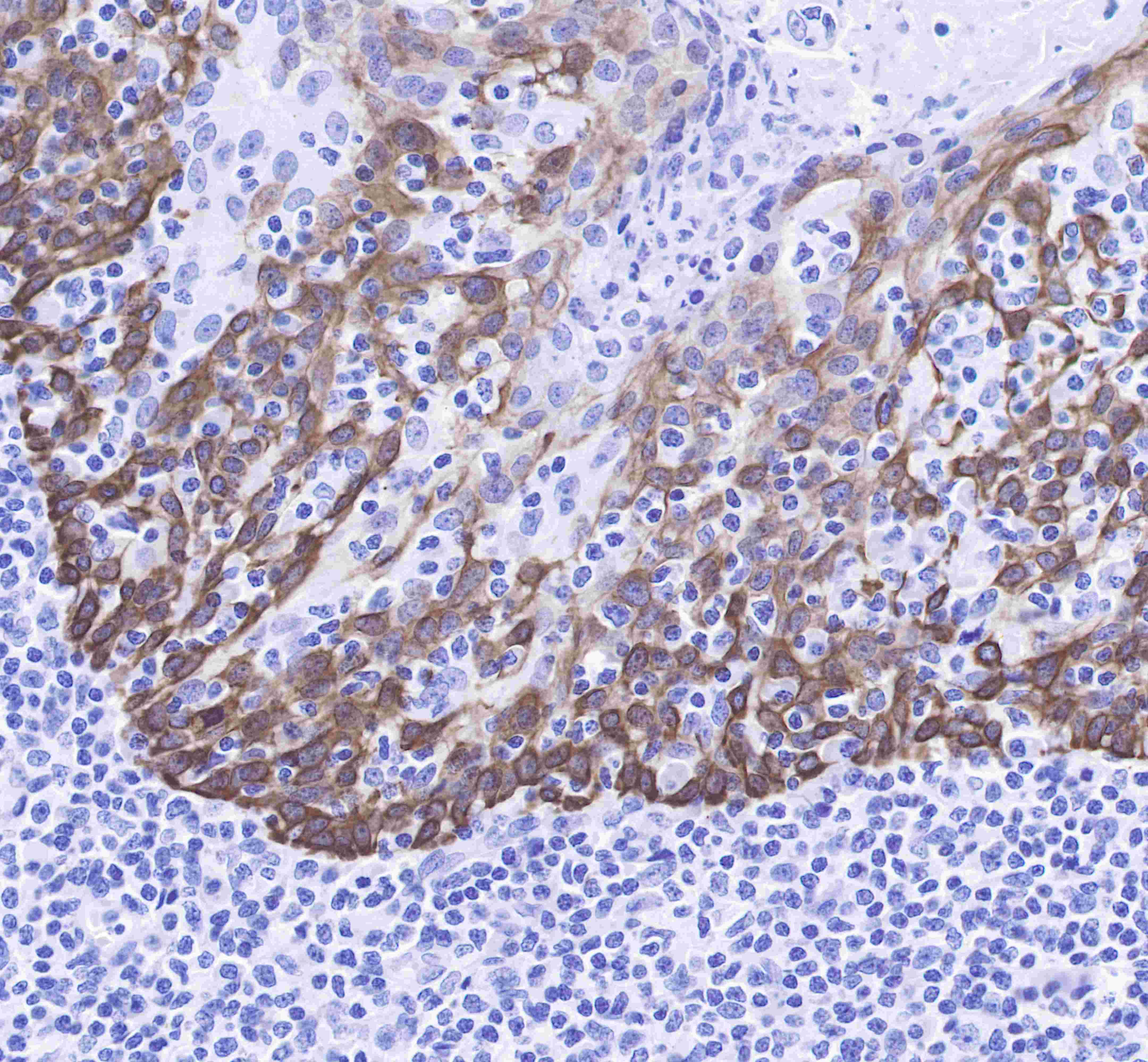
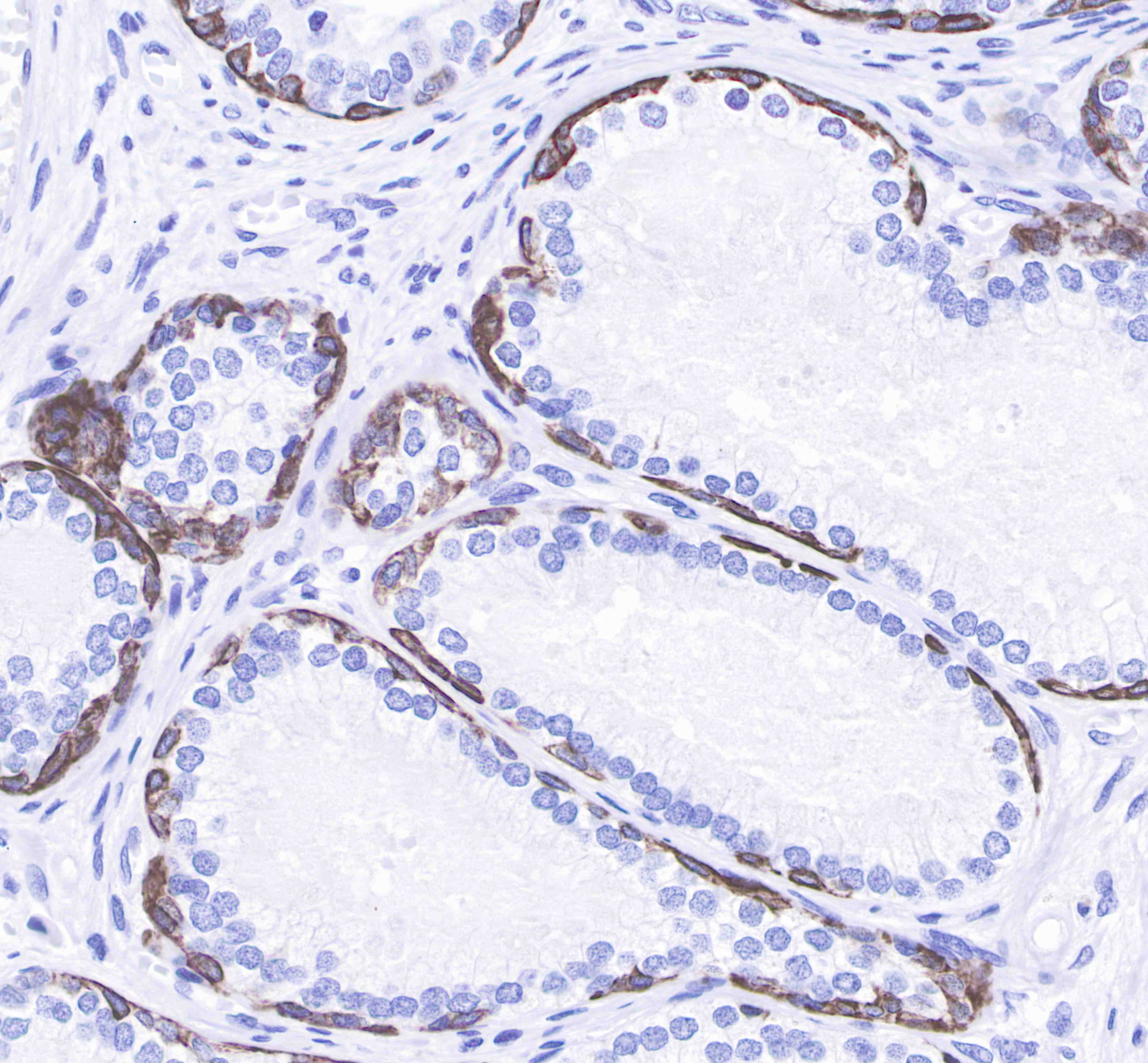
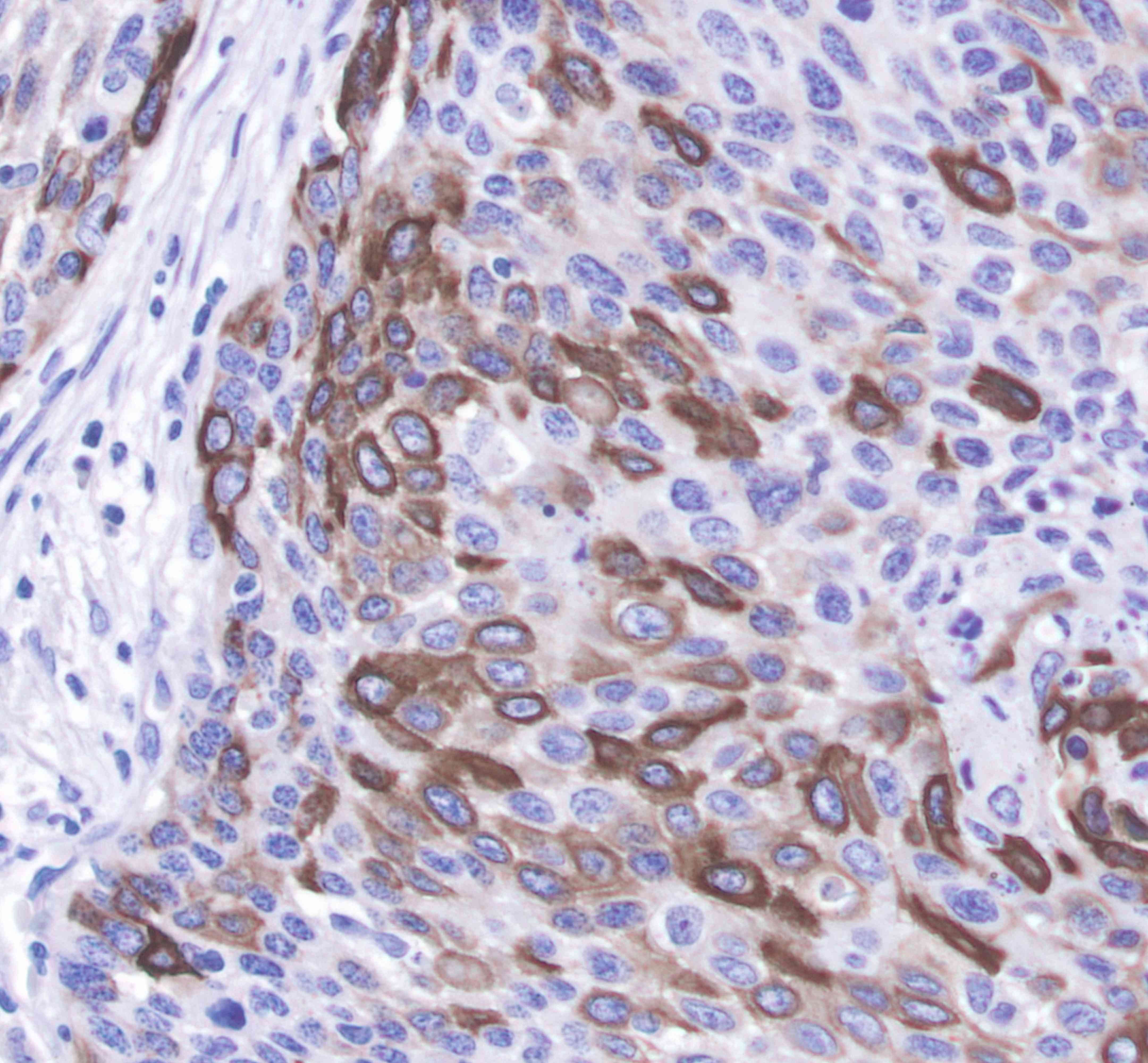
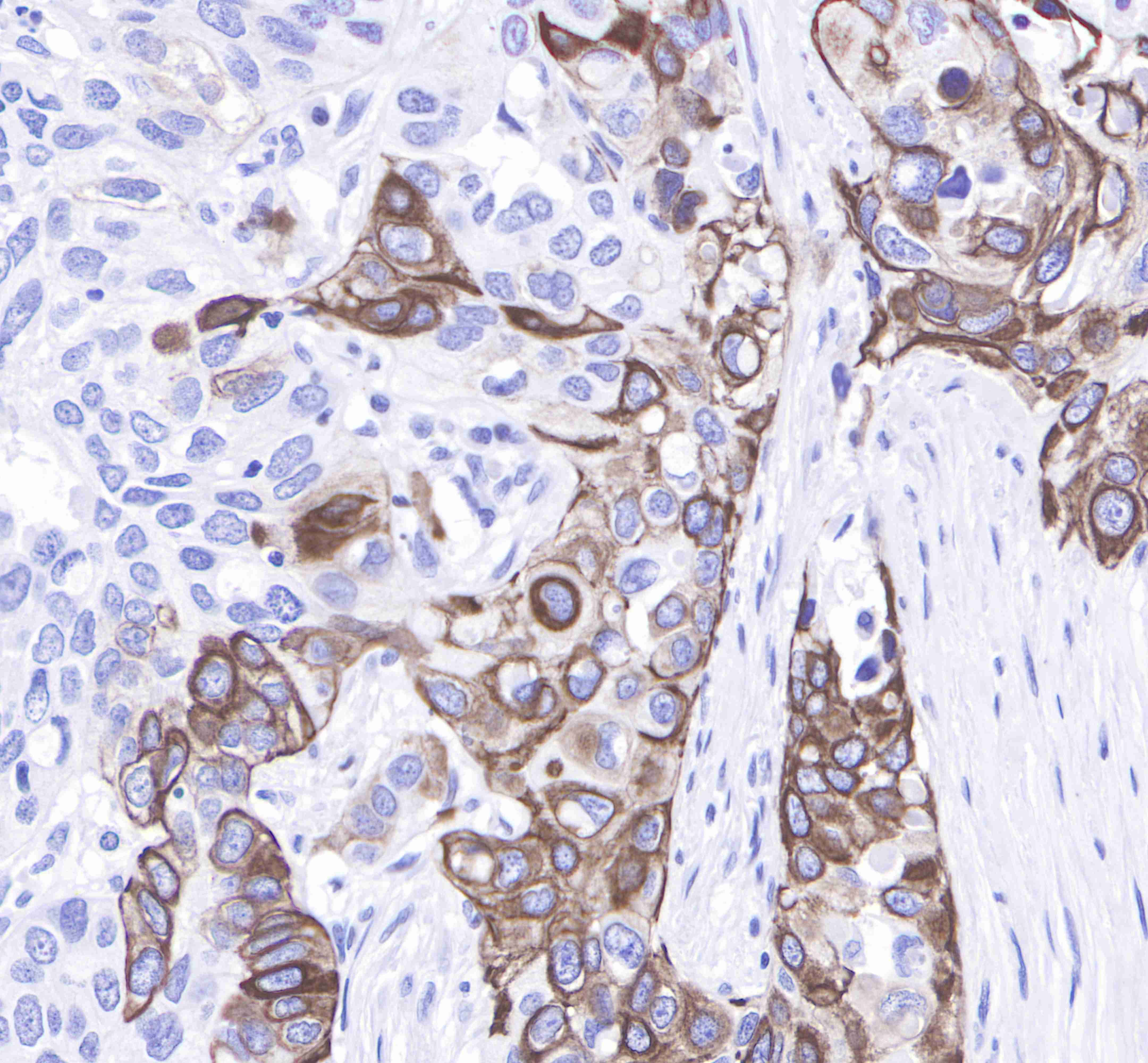
Keratin 14 |
| Synonyms |
Cytokeratin-14,CK-14, KRT14 |
| Immunogen |
Synthetic Peptide |
| Accession |
P02533 |
| Clone Number |
SDT-023-1 |
| Antibody Type |
Rabbit mAb |
| Application |
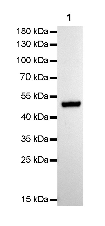
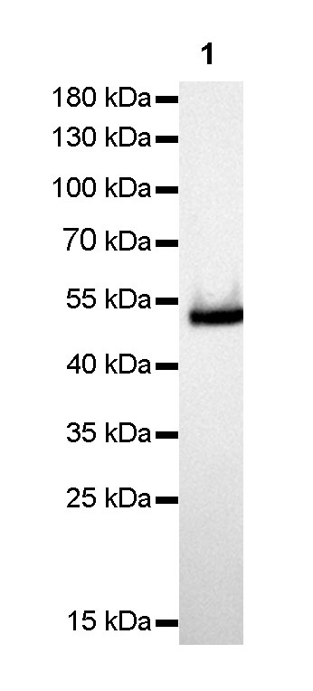
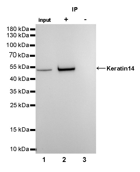
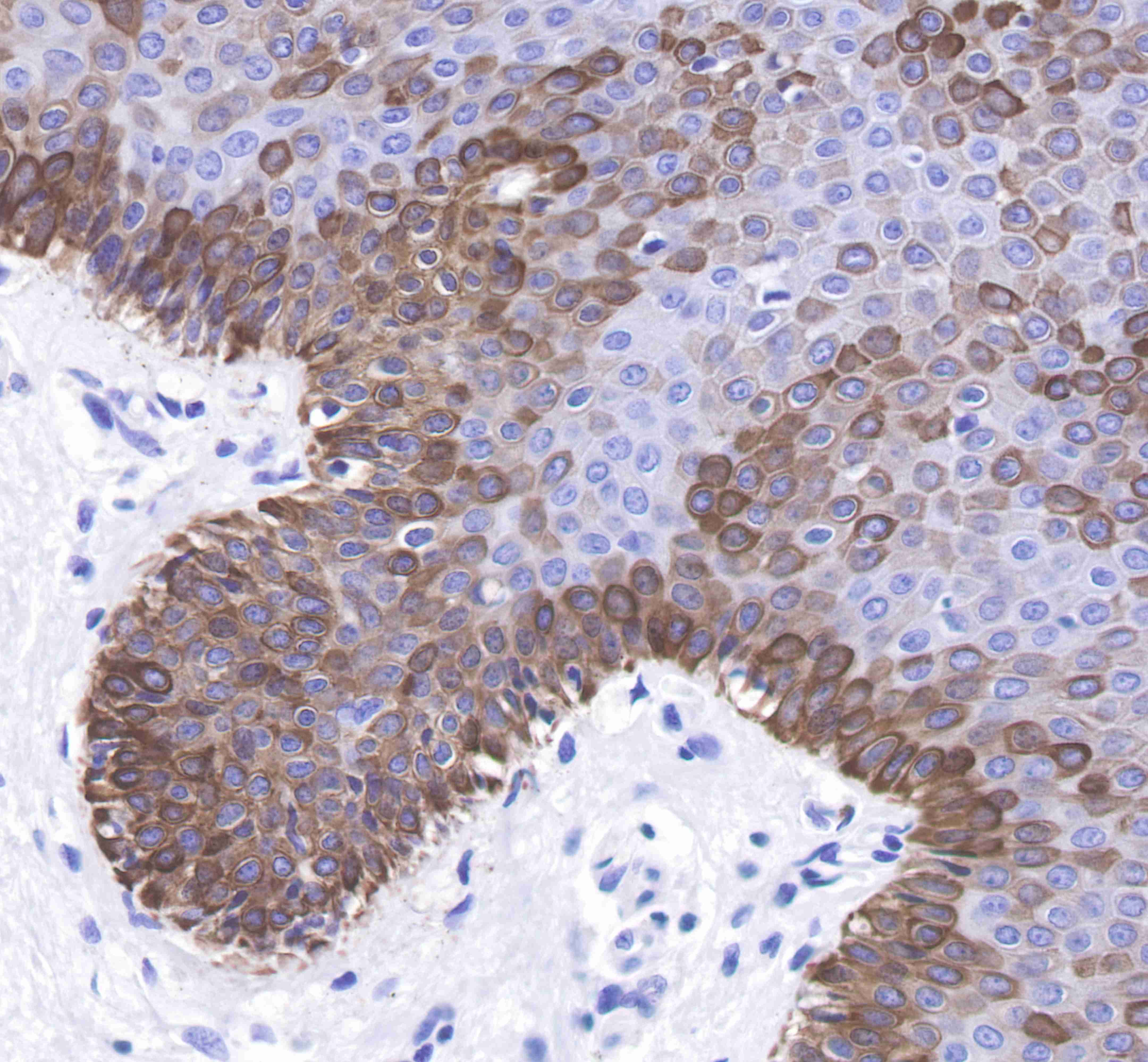
WB, IHC-P, ICC, IP |
| Reactivity |
Hu, Ms |
| Predicted Reactivity |
Rt |
| Purification |
Protein A |
| Research Area |
Signal Transduction |
| Concentration |
0.5mg/ml |
| Molecular Weight |
51kDa |
| Conjugation |
Unconjugated |
| Physical Appearance |
Liquid |
| Storage Buffer |
PBS, 40% Glycerol, 0.05%BSA, 0.03% Proclin 300 |
| Stability & Storage |
12 months from date of receipt / reconstitution, -20 °C as supplied |
Dilution
| application |
dilution |
species |
| IHC-P |
1:1000 |
|
| ICC |
1:500 |
|
| WB |
1:1000 |
|
| IP |
1:25 |
|
Background
Keratin 14 is a member of the type I keratin family of intermediate filament proteins. Keratin 14 was the first type I keratin sequence determined. Keratin 14 is also known as cytokeratin-14 (CK-14) or keratin-14 (KRT14). In humans it is encoded by the KRT14 gene. Keratin 14 is expressed in mitotically active basal layer cells, along with its partner keratin 5 (K5), and their expression is down-regulated as cells differentiate. Keratin 14 has been studied as a prognostic marker in breast cancer. Keratin 14 distinguishes stratified epithelial cells from simple epithelial cells and has been reported useful in the identification of squamous cell carcinomas.