Product Specification
| Host |
Rabbit |
| Antigen |
GLUT2 |
| Synonyms |
SLC2A2 |
| Immunogen |
N/A |
| Location |
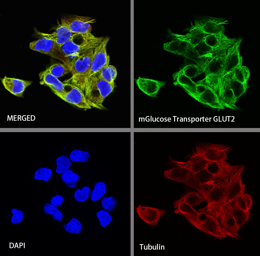
Cell membrane |
| Accession |
P11168 |
| Clone Number |
SDT-R036 |
| Antibody Type |
Rabbit mAb |
| Application |
WB, ICC |
| Reactivity |
Hu, Ms, Rt |
| Purification |
Protein A |
| Concentration |
0.5 mg/ml |
| Physical Appearance |
Liquid |
| Storage Buffer |
PBS, 40% Glycerol, 0.05%BSA, 0.03% Proclin 300 |
| Stability & Storage |
12 months from date of receipt / reconstitution, -20 °C as supplied |
Dilution
| application |
dilution |
species |
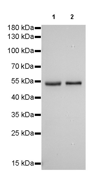
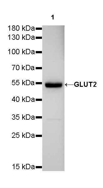
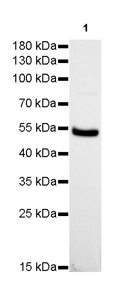
| WB |
1:1000 |
null |
| ICC |
1:100 |
null |
Background
GLUT2 transports both glucose and fructose with low affinity and plays a critical role in glucose sensing mechanisms. Alterations in the function or expression of GLUT2 are involved in the Fanconi–Bickel syndrome, diabetes, and cancer.