Product Specification
| Host |
Rabbit |
| Antigen |
FUBP1 |
| Synonyms |
FBP |
| Immunogen |
N/A |
| Location |
Nucleus |
| Accession |
Q96AE4 |
| Clone Number |
SDT-R060 |
| Antibody Type |
Rabbit mAb |
| Application |
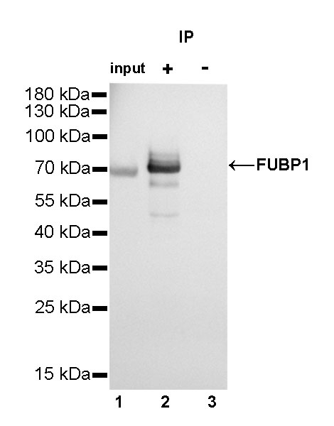
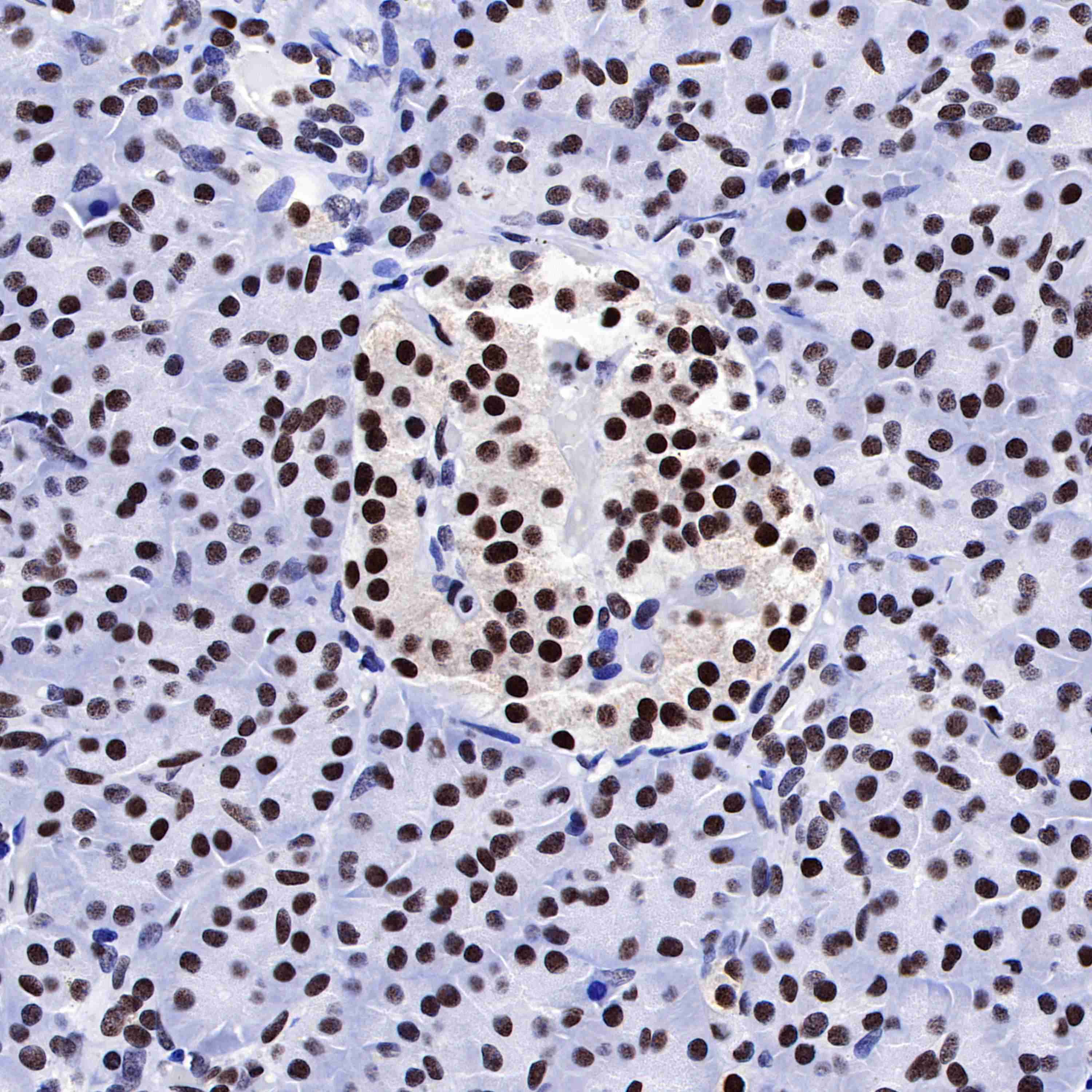
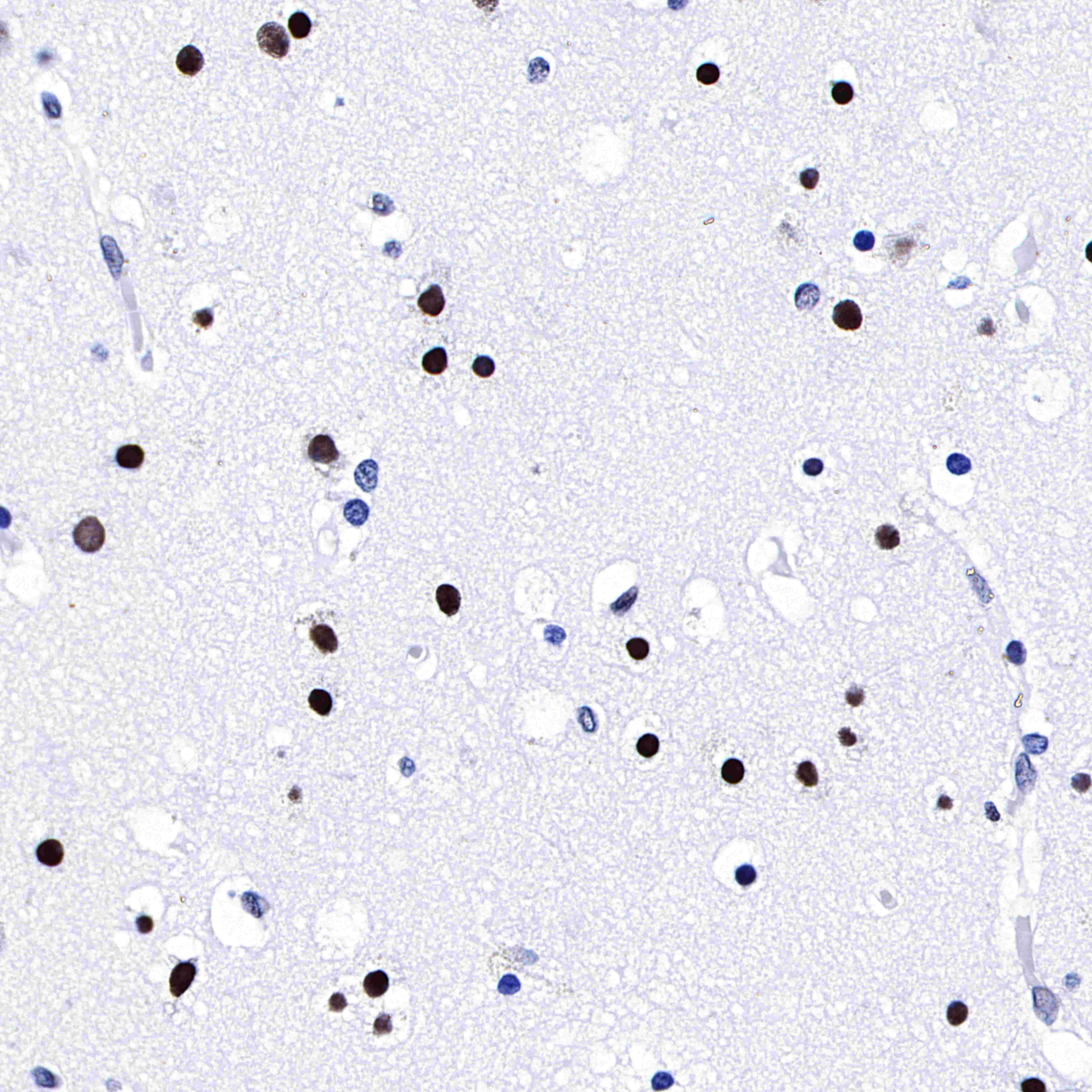
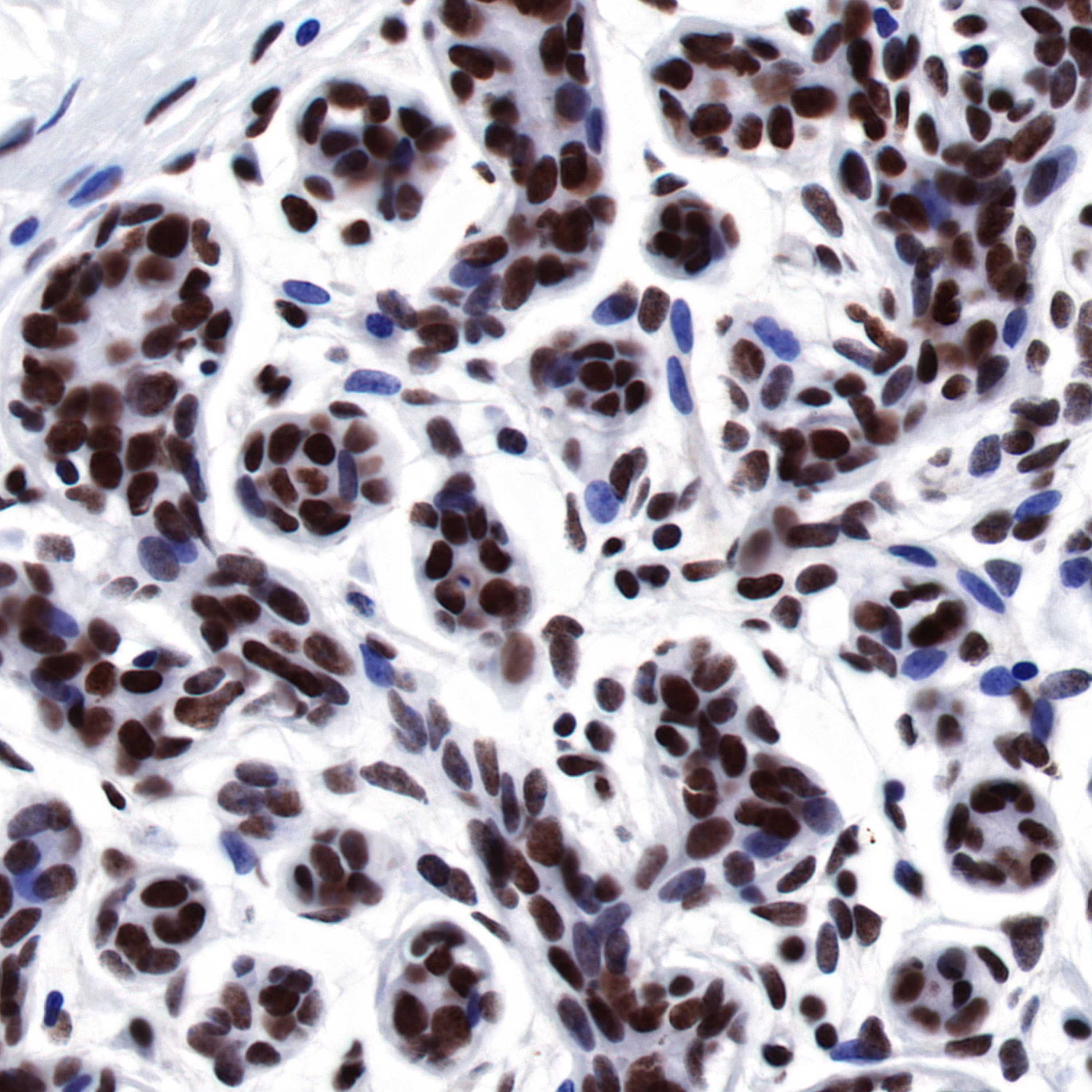
WB, IHC-P, ICC, IP, ICFCM |
| Reactivity |
Hu, Ms, Rt |
| Purification |
Protein A |
| Concentration |
0.5 mg/ml |
| Physical Appearance |
Liquid |
| Storage Buffer |
PBS, 40% Glycerol, 0.05%BSA, 0.03% Proclin 300 |
| Stability & Storage |
12 months from date of receipt / reconstitution, -20 °C as supplied |
Dilution
| application |
dilution |
species |
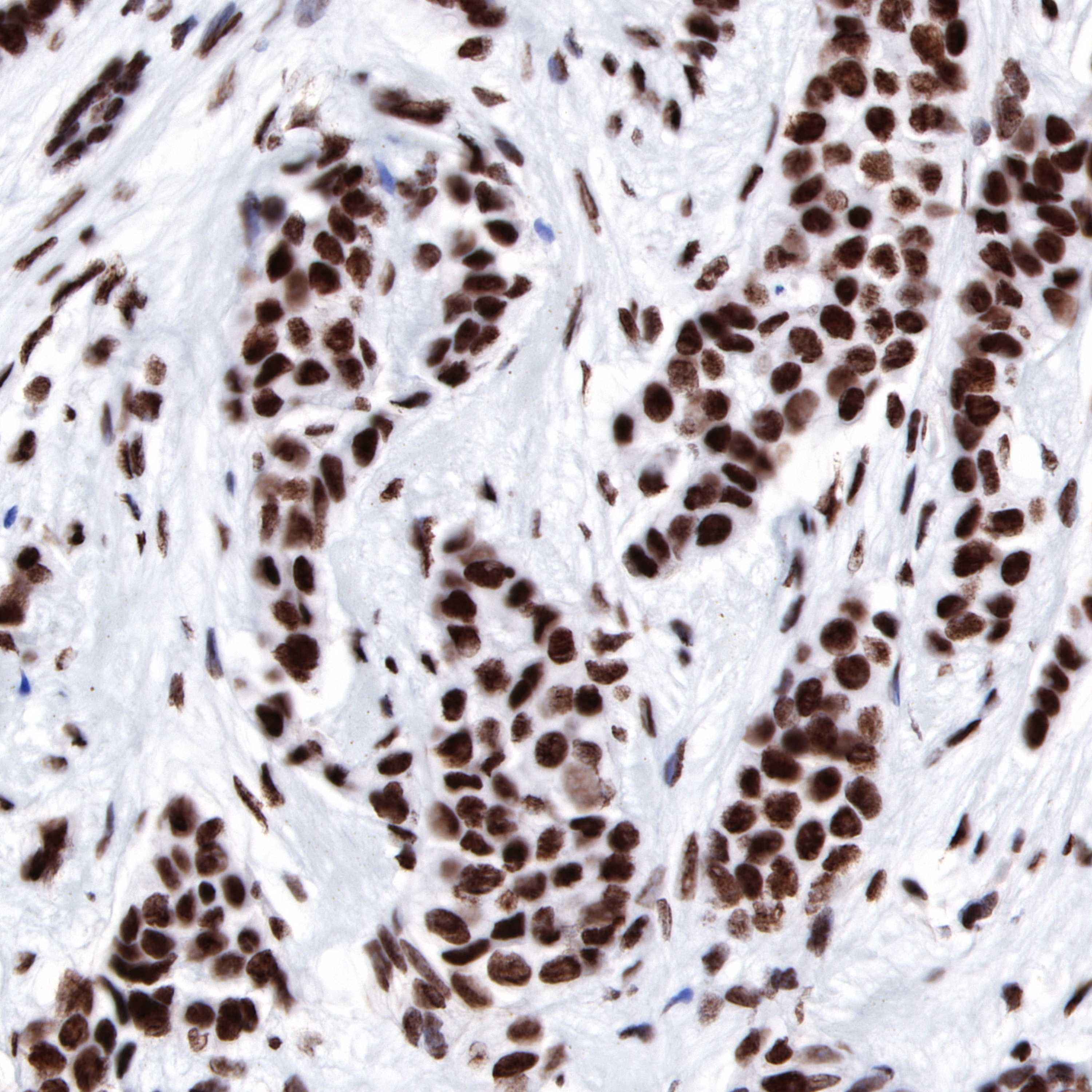
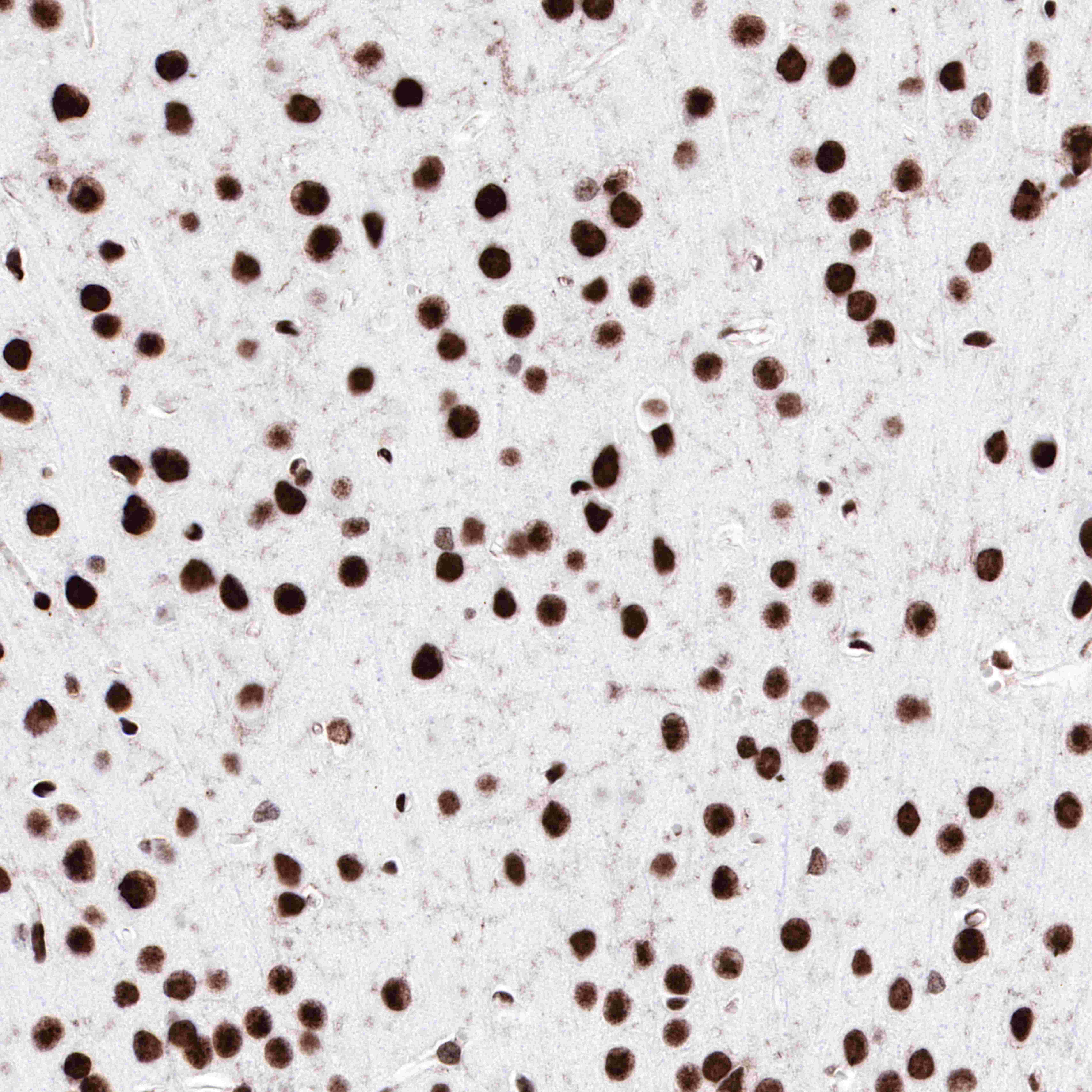
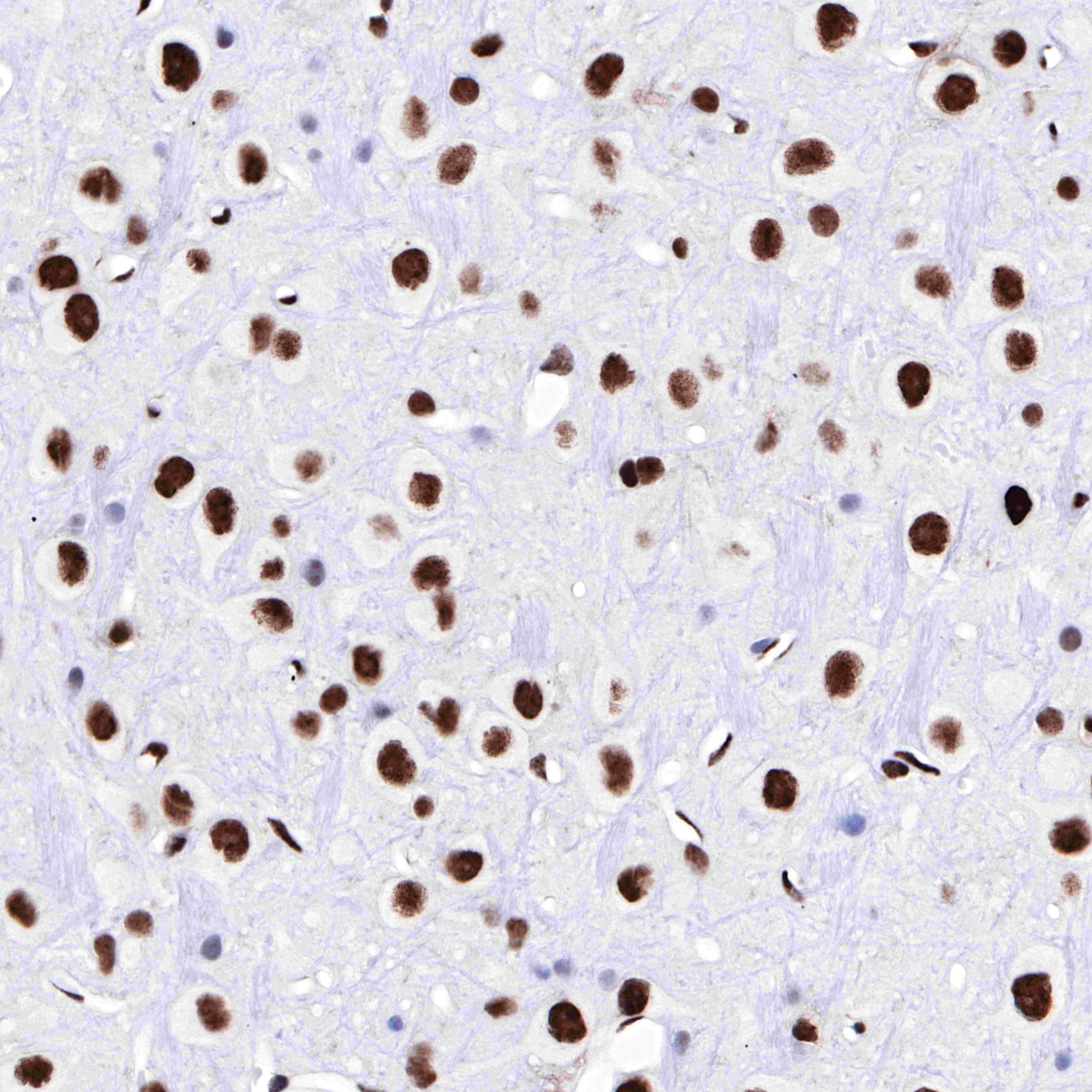
| IHC-P |
1:1000 |
|
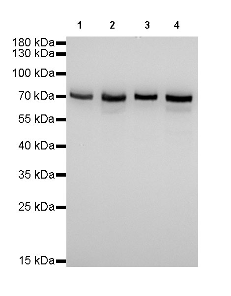
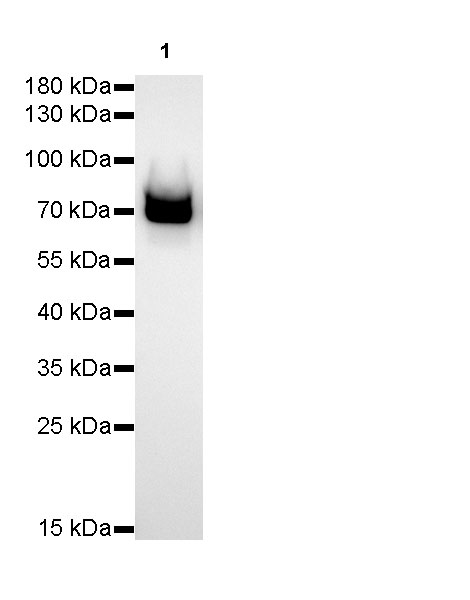
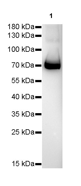
| WB |
1:1000-1:10000 |
|
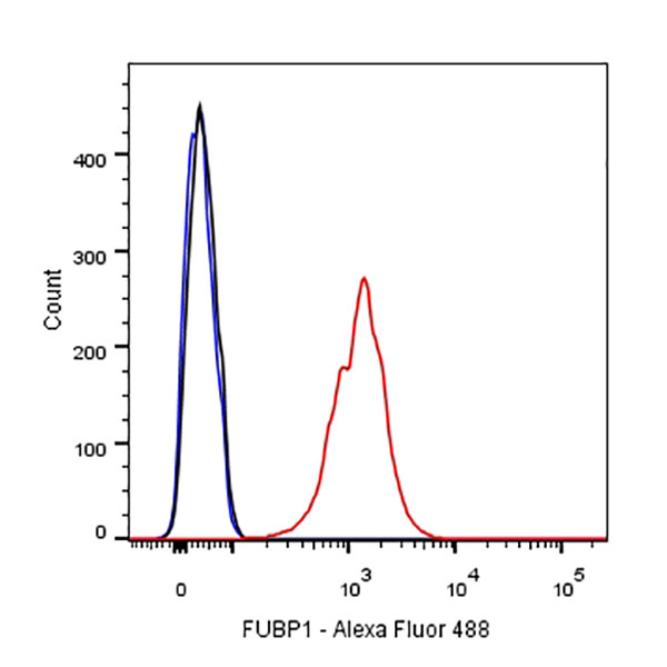
| ICFCM |
1:500 |
|
| IP |
1:25 |
|
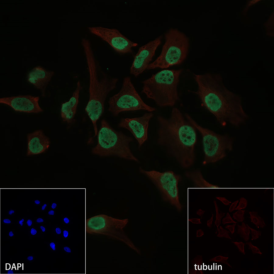
| ICC |
1:500 |
|
Background
Human distal upstream element (Fuse) binding protein 1 (FUBP1) is a transcriptional regulator of c-Myc and represents an important prognostic marker in many cancers.Regulates MYC expression by binding to a single-stranded far-upstream element (FUSE) upstream of the MYC promoter. May act both as activator and repressor of transcription.