WB result of EGFR (delE746-A750) Rabbit mAb
Primary antibody: EGFR (delE746-A750) Rabbit mAb at 1/1000 dilution
Lane 1: A431 whole cell lysate 20 µg
Lane 2: A549 whole cell lysate 20 µg
Lane 3: H1975 whole cell lysate 20 µg
Lane 4: HCC827 whole cell lysate 20 µg
Negative control: A431 whole cell lysate; A549 whole cell lysate; H1975 whole cell lysate
Secondary antibody: Goat Anti-Rabbit IgG, (H+L), HRP conjugated at 1/10000 dilution
Predicted MW: 134 kDa
Observed MW: 175 kDa
EGFR (delE746-A750) Recombinant Rabbit mAb (S-451-168)
EGFR (delE746-A750) Recombinant Rabbit mAb (S-451-168)
Price:
Regular price
$100 USD
Regular price
Sale price
$100 USD
Unit price
per
For shipping services or bulk orders, you may request a quotation.
Secure checkout with

View full details
Product Details
Product Details
Product Specification
| Host | Rabbit |
| Antigen | EGFR (delE746-A750) |
| Synonyms | Epidermal growth factor receptor, Proto-oncogene c-ErbB-1, Receptor tyrosine-protein kinase erbB-1, ERBB, ERBB1, HER1 |
| Immunogen | Synthetic Peptide |
| Location | Cell membrane |
| Accession | P00533 |
| Clone Number | S-451-168 |
| Antibody Type | Recombinant mAb |
| Isotype | IgG |
| Application | WB, IHC-P, ICC, IP |
| Reactivity | Hu |
| Purification | Protein A |
| Concentration | 0.5 mg/ml |
| Conjugation | Unconjugated |
| Physical Appearance | Liquid |
| Storage Buffer | PBS, 40% Glycerol, 0.05%BSA, 0.03% Proclin 300 |
| Stability & Storage | 12 months from date of receipt / reconstitution, -20 °C as supplied |
Dilution
| application | dilution | species |
| WB | 1:1000 | |
| IP | 1:50 | |
| IHC-P | 1:500 | |
| ICC | 1:500 |
Background
The epidermal growth factor receptor (EGFR; ErbB-1; HER1 in humans) is a transmembrane protein that is a receptor for members of the epidermal growth factor family (EGF family) of extracellular protein ligands. The EGFR is essential for ductal development of the mammary glands, and agonists of the EGFR such as amphiregulin, TGF-α, and heregulin induce both ductal and lobuloalveolar development even in the absence of estrogen and progesterone. EGFR mutations, del E746-A750 in exon 19 and L858R in exon 21 in tumor cells of NMLC represent biomarkers of response to tyrosine kinase inhibitors (TKI) therapy. Patients with tumors positive for EGFR mutations show better response and greater survival. These mutations occupy 90% of mutations in lung cancer.
Picture
Picture
Western Blot
IP
EGFR (delE746-A750) Rabbit mAb at 1/50 dilution (1 µg) immunoprecipitating EGFR (delE746-A750) in 0.4 mg HCC827 whole cell lysate.
Western blot was performed on the immunoprecipitate using EGFR (delE746-A750) Rabbit mAb at 1/1000 dilution.
Secondary antibody (HRP) for IP was used at 1/400 dilution.
Lane 1: HCC827 whole cell lysate 20 µg (Input)
Lane 2: EGFR (delE746-A750) Rabbit mAb IP in HCC827 whole cell lysate
Lane 3: Rabbit monoclonal IgG IP in HCC827 whole cell lysate
Predicted MW: 134 kDa
Observed MW: 175 kDa
Immunohistochemistry
IHC shows positive staining in paraffin-embedded HCC827 cells and negative staining in paraffin-embedded A549, H1975 and A431 cells. Anti-EGFR (delE746-A750) antibody was used at 1/500 dilution, followed by a HRP Polymer for Mouse & Rabbit IgG (ready to use). Counterstained with hematoxylin. Heat mediated antigen retrieval with Tris/EDTA buffer pH9.0 was performed before commencing with IHC staining protocol.
IHC shows positive staining in paraffin-embedded human lung adenocarcinoma. Anti-EGFR (delE746-A750) antibody was used at 1/500 dilution, followed by a HRP Polymer for Mouse & Rabbit IgG (ready to use). Counterstained with hematoxylin. Heat mediated antigen retrieval with Tris/EDTA buffer pH9.0 was performed before commencing with IHC staining protocol.
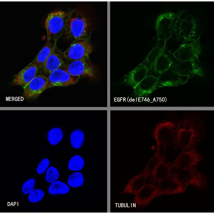
Immunocytochemistry
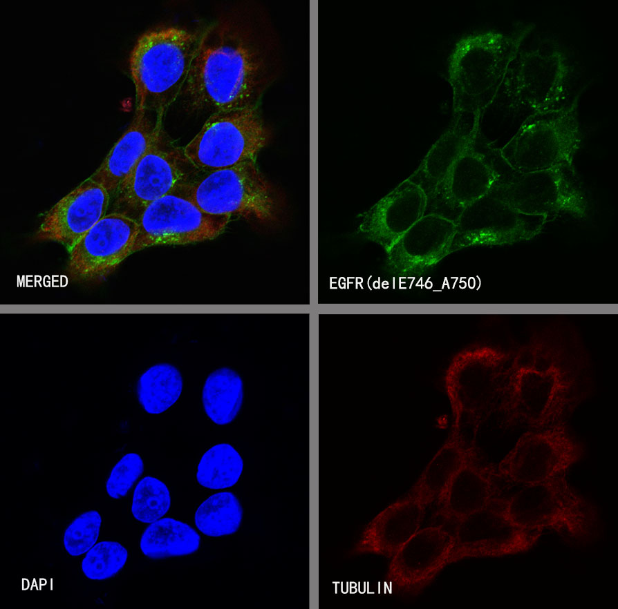
ICC shows positive staining in HCC827 cells. Anti-EGFR(delE746_A750) antibody was used at 1/500 dilution (Green) and incubated overnight at 4°C. Goat polyclonal Antibody to Rabbit IgG - H&L (Alexa Fluor® 488) was used as secondary antibody at 1/1000 dilution. The cells were fixed with 100% ice-cold methanol and permeabilized with 0.1% PBS-Triton X-100. Nuclei were counterstained with DAPI (Blue).Counterstain with tubulin (red).
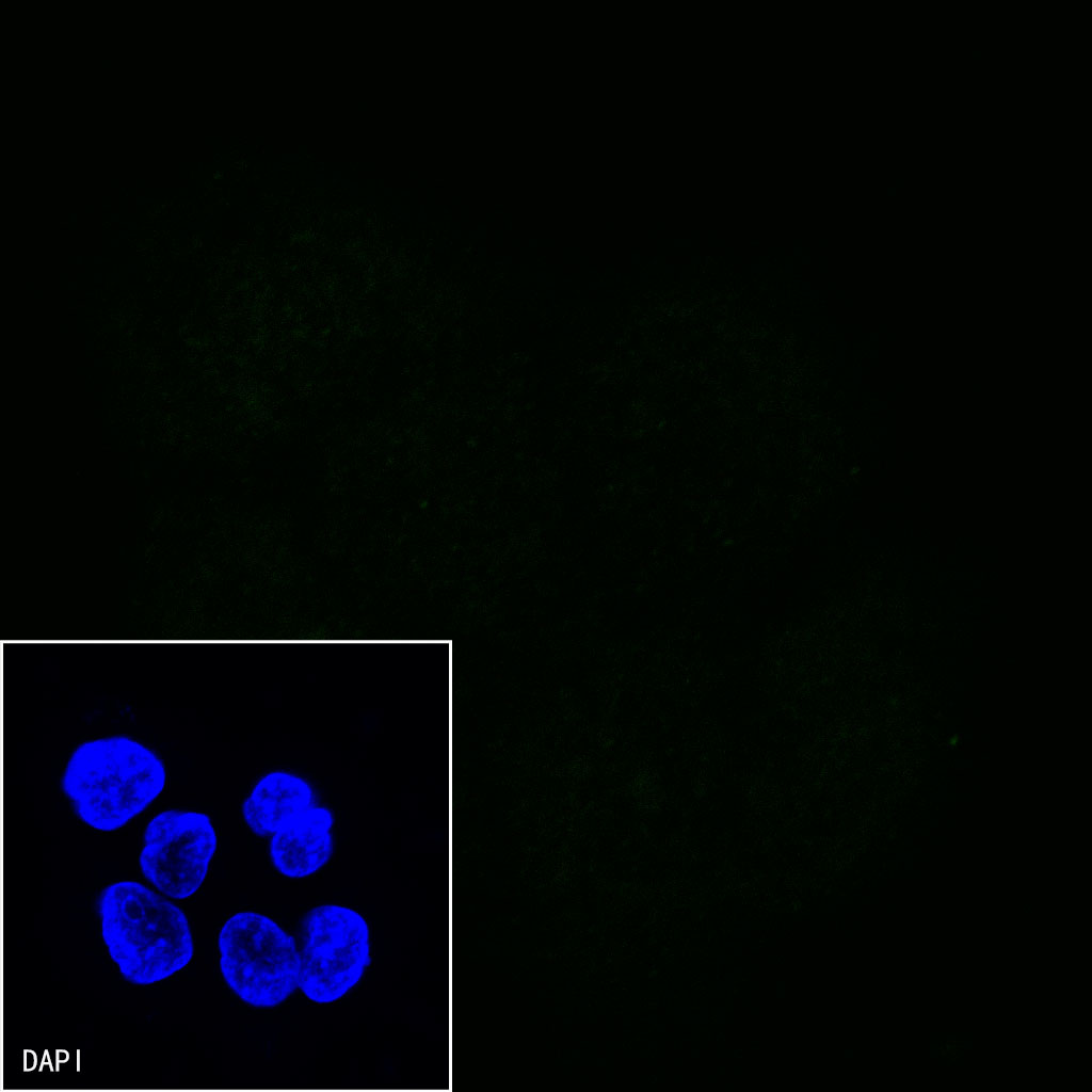
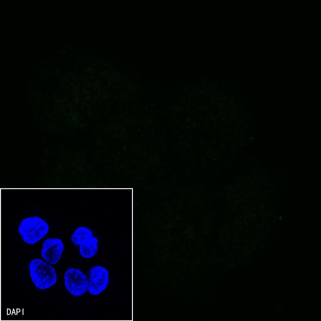
Negative control:ICC shows negative staining in A431 cells. Anti-EGFR(delE746_A750) antibody was used at 1/500 dilution and incubated overnight at 4°C. Goat polyclonal Antibody to Rabbit IgG - H&L (Alexa Fluor® 488) was used as secondary antibody at 1/1000 dilution. The cells were fixed with 100% ice-cold methanol and permeabilized with 0.1% PBS-Triton X-100. Nuclei were counterstained with DAPI (Blue).