Product Specification
| Host |
Rabbit |
| Antigen |
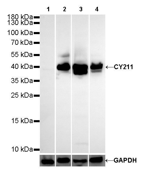
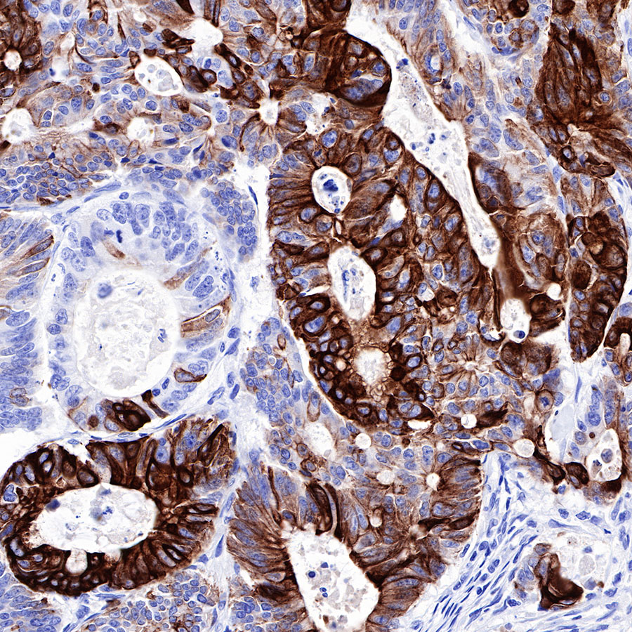
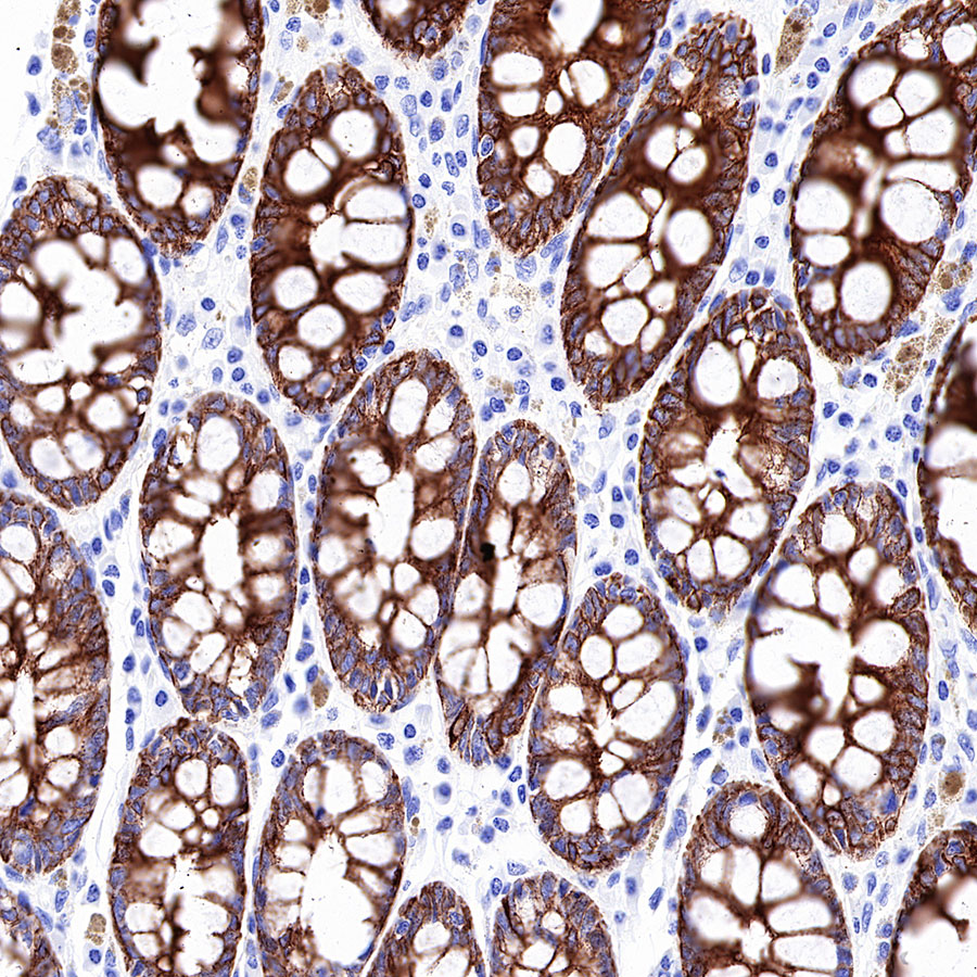
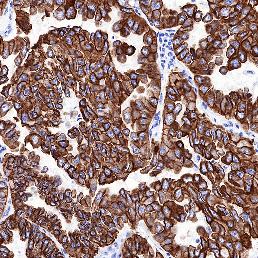
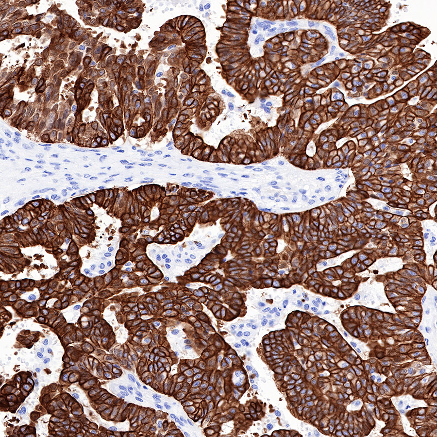
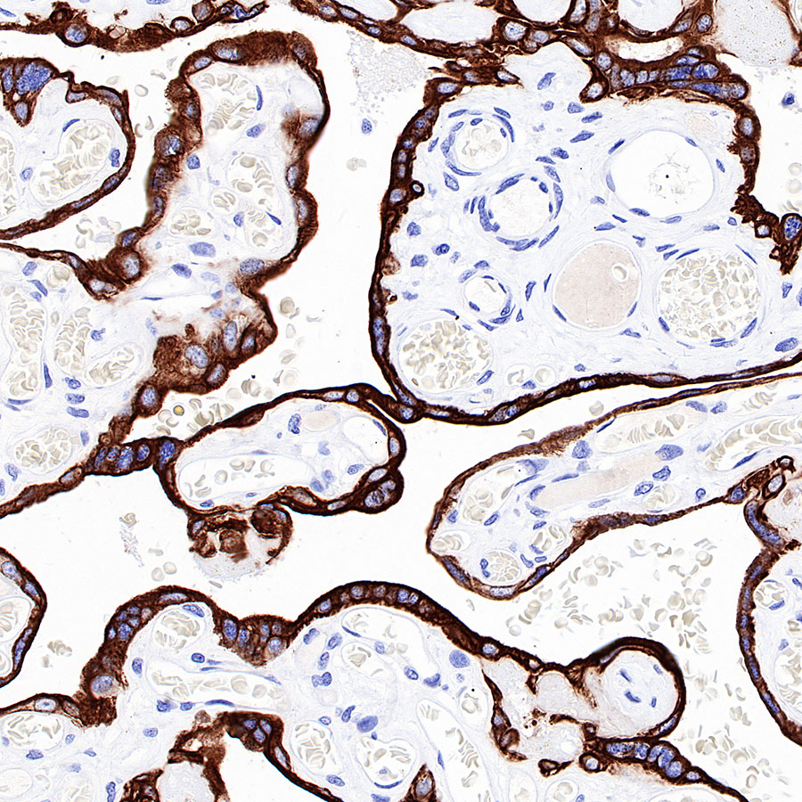
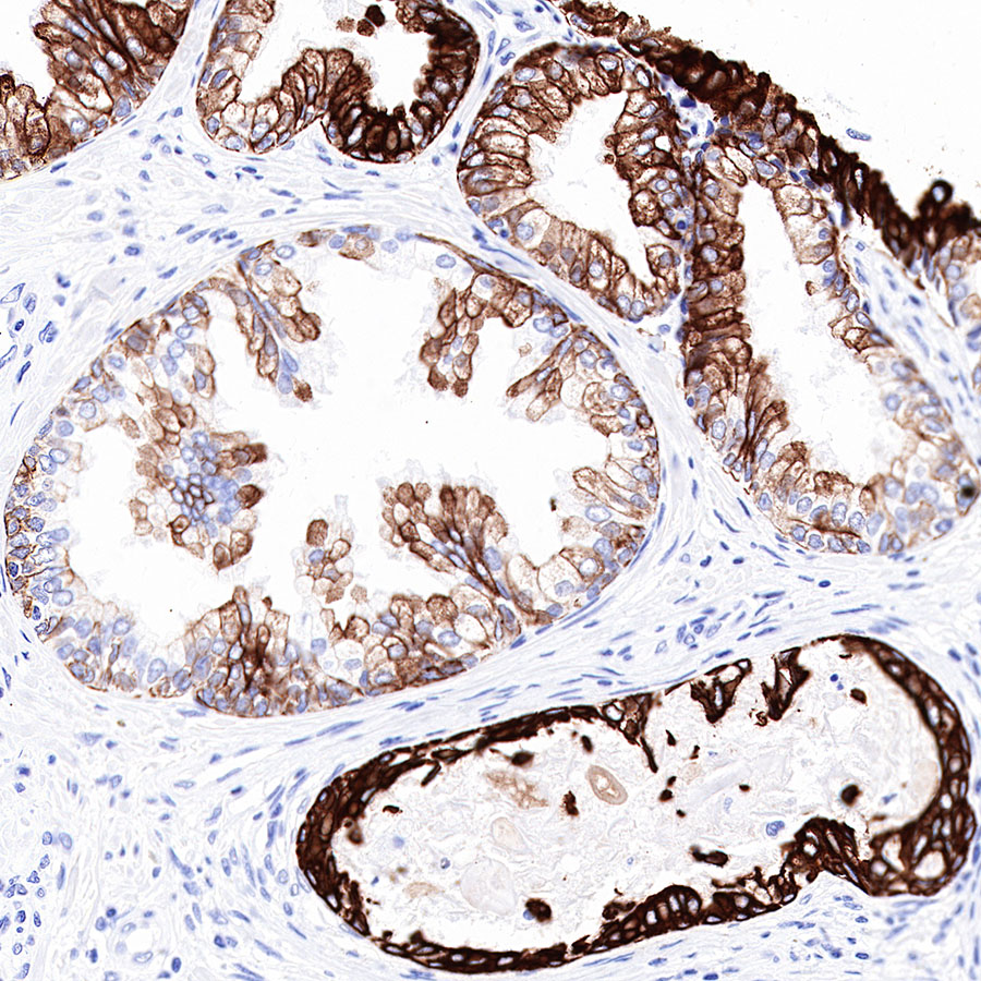
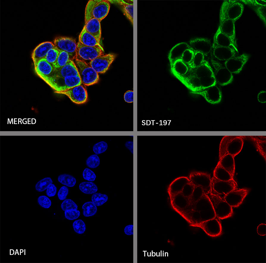
Cytokeratin 19 fragment (CY211) |
| Synonyms |
CYFRA 21-1 |
| Immunogen |
Recombinant Protein |
| Location |
Cytoplasm |
| Accession |
P08727 |
| Clone Number |
SDT-197-28 |
| Antibody Type |
Rabbit mAb |
| Application |
WB, IHC-P, ICC |
| Reactivity |
Hu |
| Purification |
Protein A |
| Concentration |
0.25 mg/ml |
| Physical Appearance |
Liquid |
| Storage Buffer |
PBS, 40% Glycerol, 0.05%BSA, 0.03% Proclin 300 |
| Stability & Storage |
12 months from date of receipt / reconstitution, -20 °C as supplied |
Dilution
| application |
dilution |
species |
| WB |
1:500 |
null |
| IHC-P |
1:1000 |
null |
| ICC |
1:250 |
null |
Background
Cytokeratin 19 fragment (CYFRA 21-1) is a fragment of cytokeratin 19 that is typically associated with epithelial cell cancers, including NSCLC, and is typically associated with the SQLC type. Since cytokeratins are structural proteins of keratin-containing intermediate filaments found in the epithelial cells, their degradation produces soluble fragments which are measurable in the blood of lung cancer patients as a tumor marker. Other examples of cytokeratin fragments include tissue polypeptide antigen and tissue polypeptide-specific antigen. CYFRA 21-1 is known to be correlated with disease response and the prognosis of lung cancer but cannot be used to differentiate cancer patients from patients with respiratory diseases.