Product Specification
| Host |
Rabbit |
| Antigen |
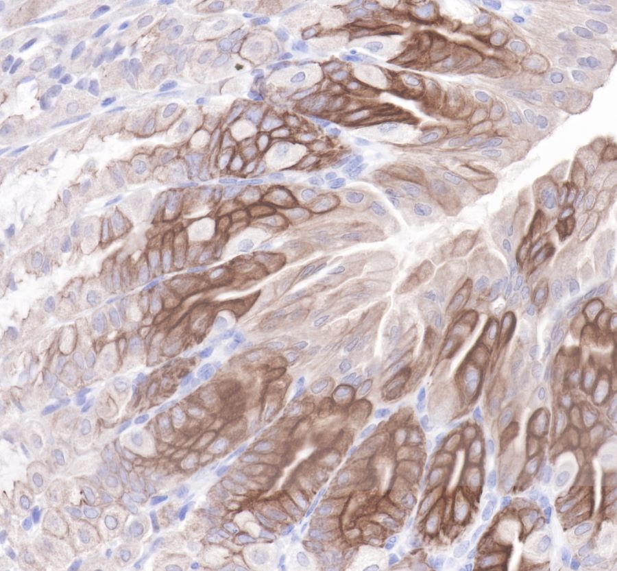
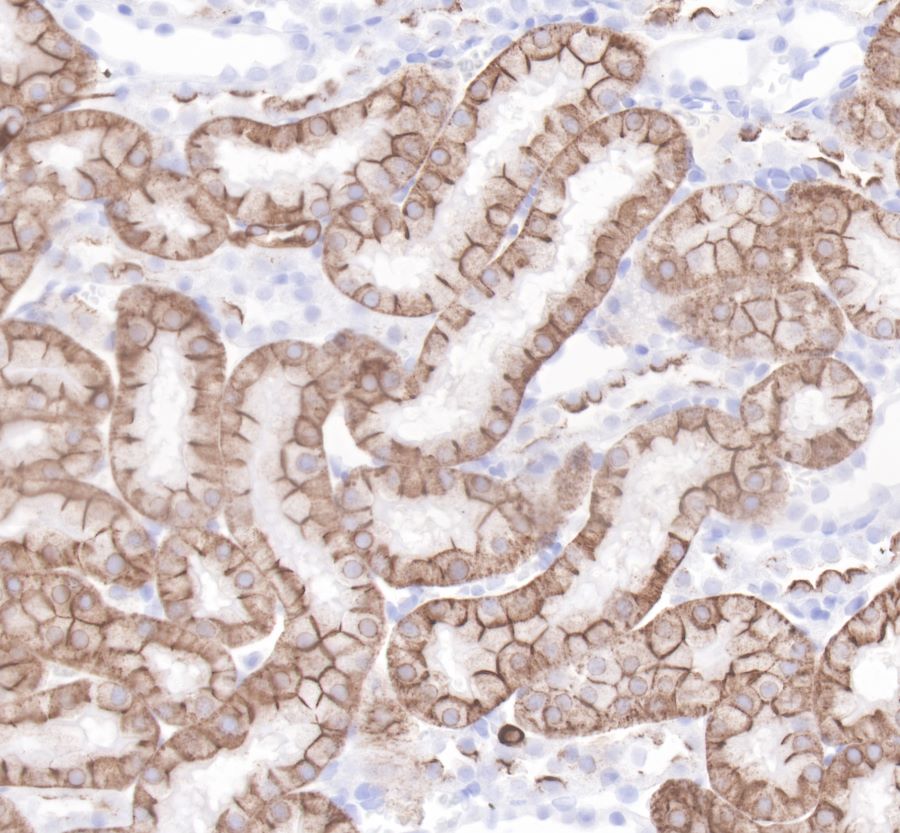
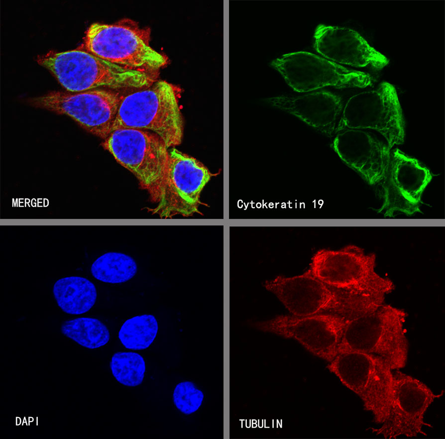
Cytokeratin 19 |
| Synonyms |
KRT19, CK-19 |
| Immunogen |
Synthetic Peptide |
| Location |
Cell membrane |
| Accession |
P08727 |
| Clone Number |
SDT-107-19 |
| Antibody Type |
Rabbit mAb |
| Application |
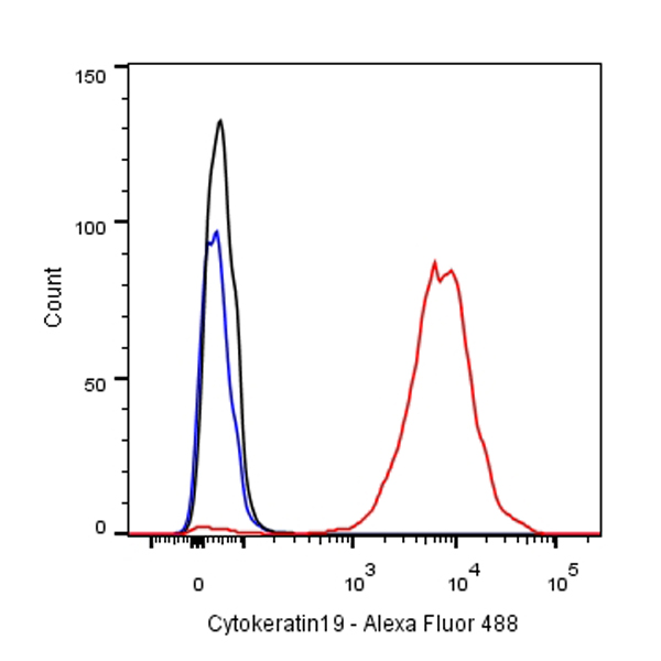
WB, IHC-P, ICC, FC |
| Reactivity |
Hu, Ms, Rt |
| Purification |
Protein A |
| Concentration |
0.25mg/ml |
| Physical Appearance |
Liquid |
| Storage Buffer |
PBS, 40% Glycerol, 0.05%BSA, 0.03% Proclin 300 |
| Stability & Storage |
12 months from date of receipt / reconstitution, -20 °C as supplied |
Dilution
| application |
dilution |
species |
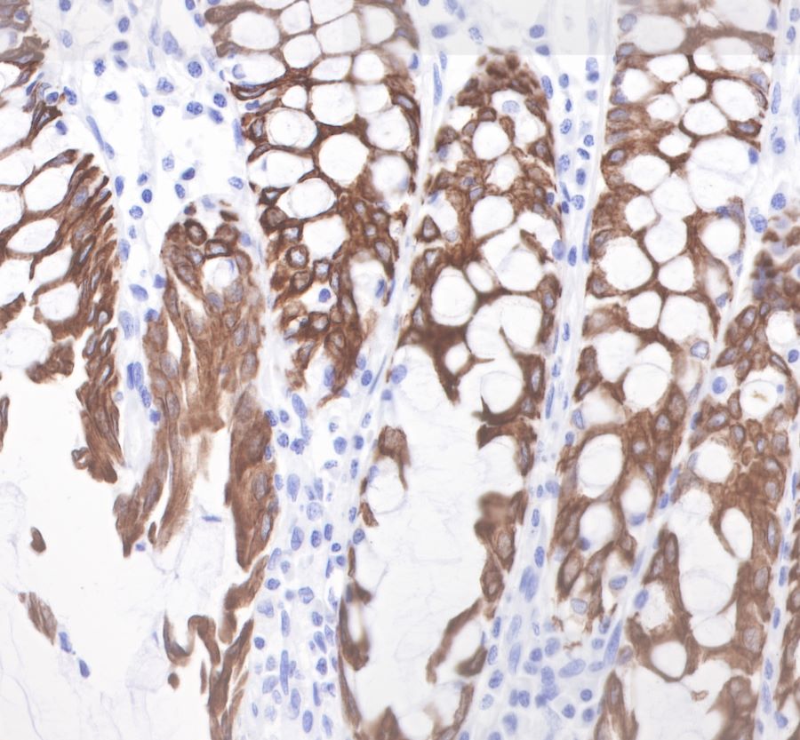
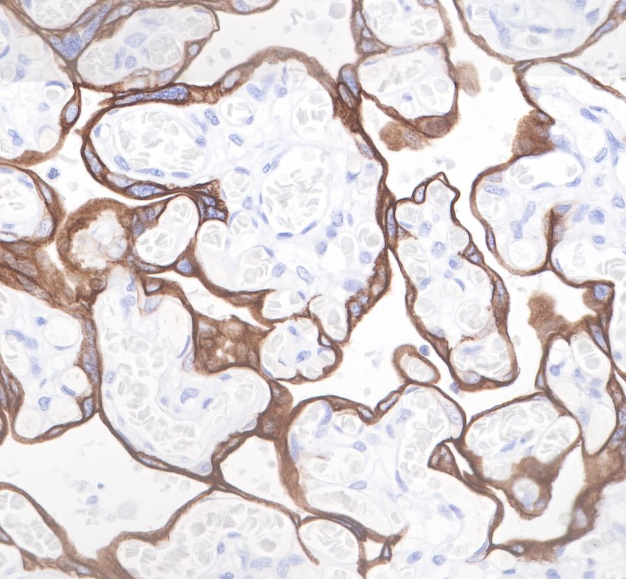
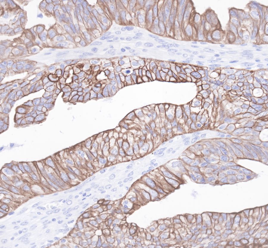
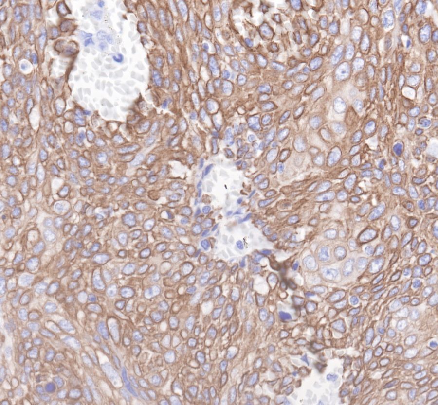
| IHC-P |
1:1000 |
|
| FC |
1:500 |
|
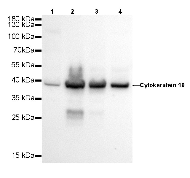
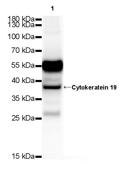
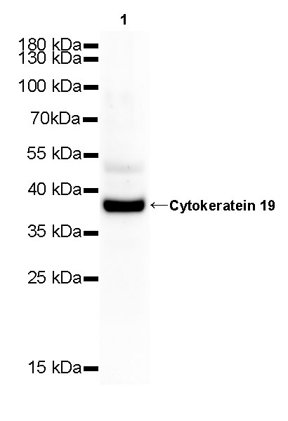
| WB |
1:500 |
|
| ICC |
1:250 |
|
Background
Cytokeratin 19 is a member of the keratin family. The keratins are intermediate filament proteins responsible for the structural integrity of epithelial cells and are subdivided into cytokeratins and hair keratins. Keratin 19 is a type I keratin. The type I cytokeratins consist of acidic proteins which are arranged in pairs of heterotypic keratin chains. Unlike its related family members, this smallest known acidic cytokeratin is not paired with a basic cytokeratin in epithelial cells. It is specifically found in the periderm, the transiently superficial layer that envelops the developing epidermis. KRT19 is also known as Cyfra 21-1.Due to its high sensitivity, KRT19 is the most used marker for the RT-PCR-mediated detection of tumor cells disseminated in lymph nodes, peripheral blood, and bone marrow of breast cancer patients.