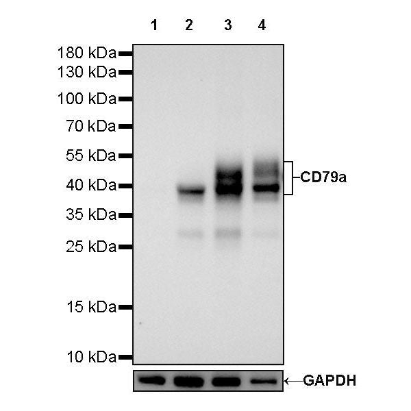
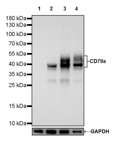
WB result of CD79a Rabbit mAb
Primary antibody: CD79a Rabbit mAb at 1/5000 dilution
Lane 1: Jurkat whole cell lysate 20 µg
Lane 2: Raji whole cell lysate 20 µg
Lane 3: Ramos whole cell lysate 20 µg
Lane 4: Daudi whole cell lysate 20 µg
Negative control: Jurkat whole cell lysate
Secondary antibody: Goat Anti-Rabbit IgG, (H+L), HRP conjugated at 1/10000 dilution
Predicted MW: 25kDa
Observed MW: 40~55kDa
CD79a/MB-1 Recombinant Rabbit mAb (SDT-030-18)
CD79a/MB-1 Recombinant Rabbit mAb (SDT-030-18)
Price:
Regular price
$100 USD
Regular price
Sale price
$100 USD
Unit price
per
For shipping services or bulk orders, you may request a quotation.
Secure checkout with

View full details
Product Details
Product Details
Product Specification
| Host | Rabbit |
| Antigen | CD79a/MB-1 |
| Synonyms | Ig-alpha, Surface IgM-associated protein, IGA |
| Immunogen | Synthetic Peptide |
| Location | Cell membrane |
| Accession | P11912 |
| Clone Number | SDT-030-18 |
| Antibody Type | Recombinant mAb |
| Application | WB, IHC-P, IF |
| Reactivity | Hu |
| Purification | Protein A |
| Concentration | 0.125 mg/ml |
| Conjugation | Unconjugated |
| Physical Appearance | Liquid |
| Storage Buffer | PBS, 40% Glycerol, 0.05% BSA, 0.03% Proclin 300 |
| Stability & Storage | 12 months from date of receipt / reconstitution, -20 °C as supplied |
Dilution
| application | dilution | species |
| WB | 1:5000 | |
| IHC-P | 1:1000 | |
| IF | 1:500 |
Background
The CD79 molecule, comprising two polypeptide chains, mb-1 (CD79a) and B29 (CD79b), is physically associated in the B-cell membrane with immunoglobulin [PMID: 7632952]. CD79a has been reported to exhibit a high degree of linage-specificity for B-cell differentiation, with a specificity of 88% and a sensitivity of 100% [PMID: 34672246].
Picture
Picture
Western Blot
Immunohistochemistry
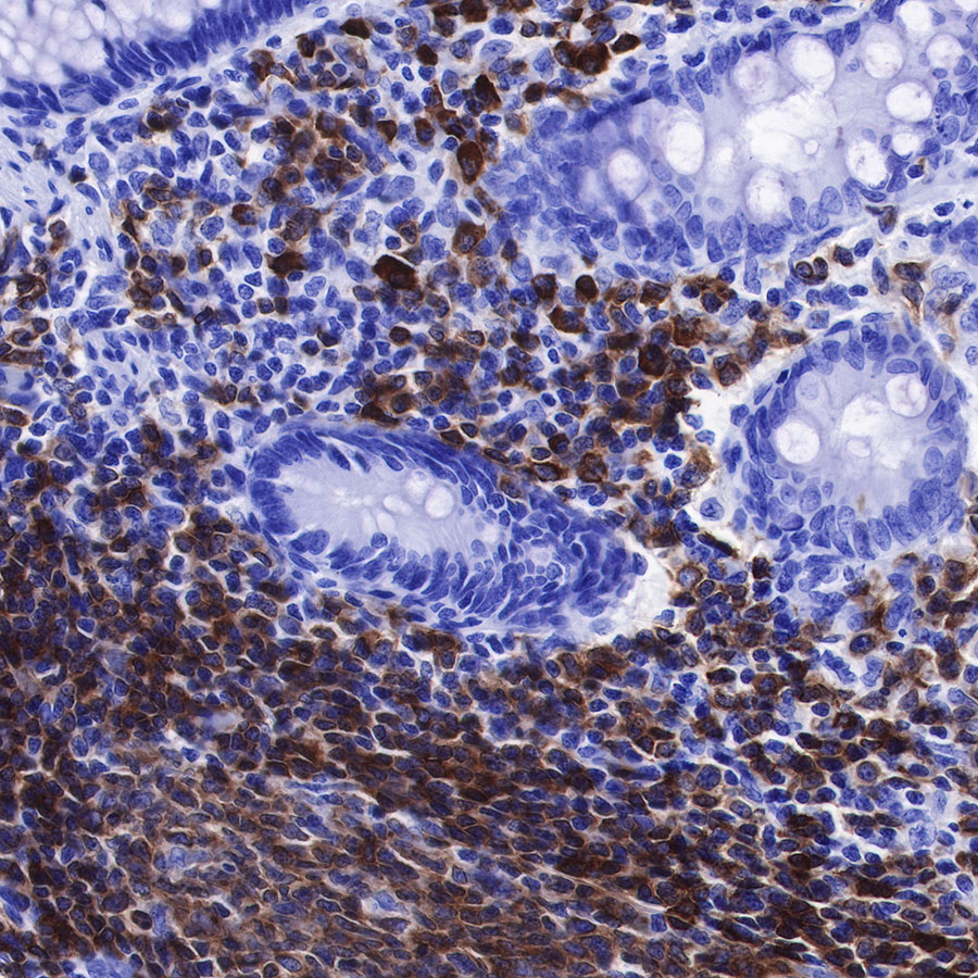
IHC shows positive staining in paraffin-embedded human colon. Anti-CD79a/MB-1 antibody was used at 1/1000 dilution, followed by a HRP Polymer for Mouse & Rabbit IgG (ready to use). Counterstained with hematoxylin. Heat mediated antigen retrieval with Tris/EDTA buffer pH9.0 was performed before commencing with IHC staining protocol.

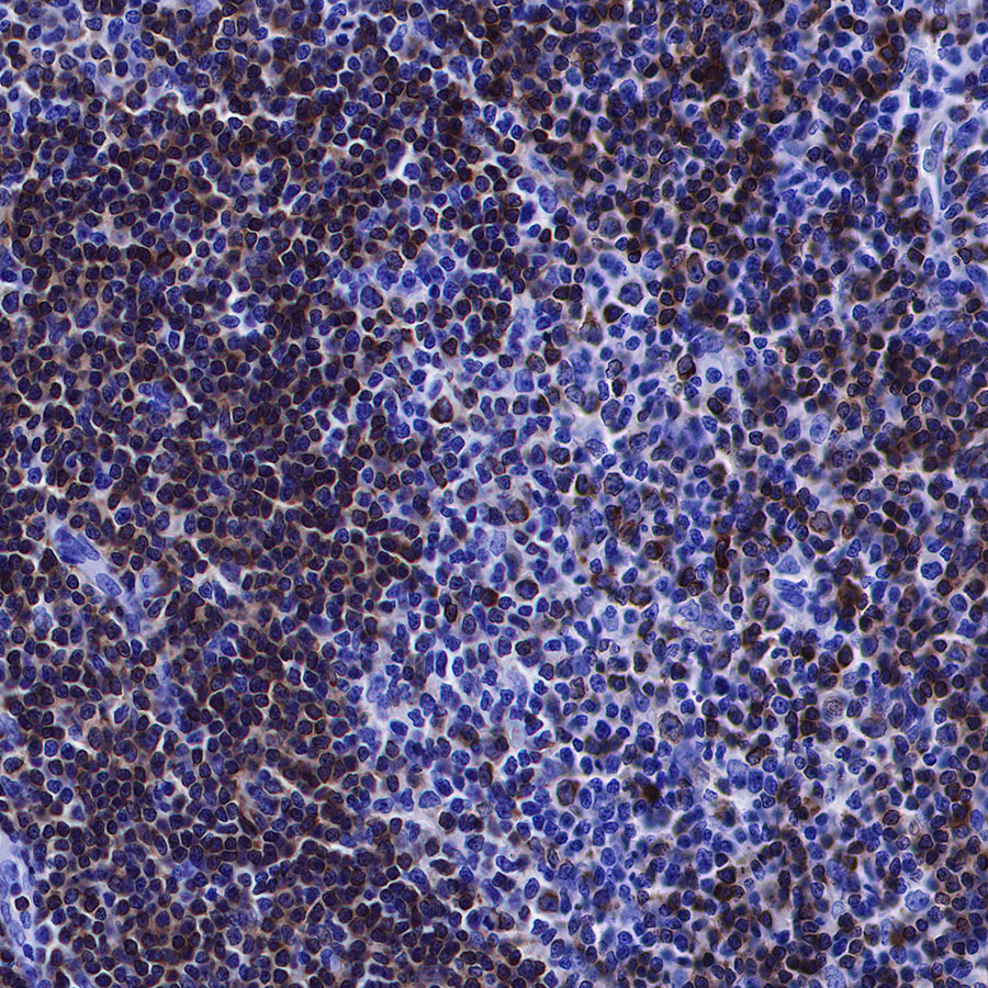
IHC shows positive staining in paraffin-embedded human spleen. Anti-CD79a/MB-1 antibody was used at 1/1000 dilution, followed by a HRP Polymer for Mouse & Rabbit IgG (ready to use). Counterstained with hematoxylin. Heat mediated antigen retrieval with Tris/EDTA buffer pH9.0 was performed before commencing with IHC staining protocol.
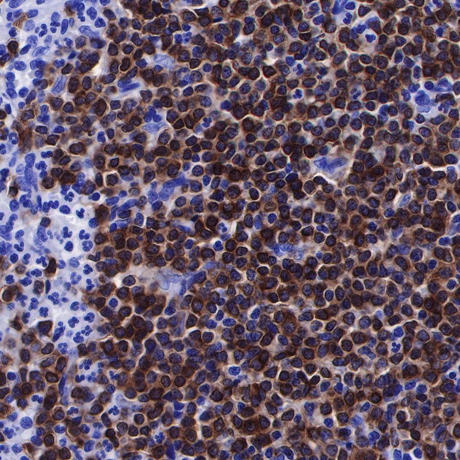
IHC shows positive staining in paraffin-embedded human tonsil. Anti-CD79a/MB-1 antibody was used at 1/1000 dilution, followed by a HRP Polymer for Mouse & Rabbit IgG (ready to use). Counterstained with hematoxylin. Heat mediated antigen retrieval with Tris/EDTA buffer pH9.0 was performed before commencing with IHC staining protocol.
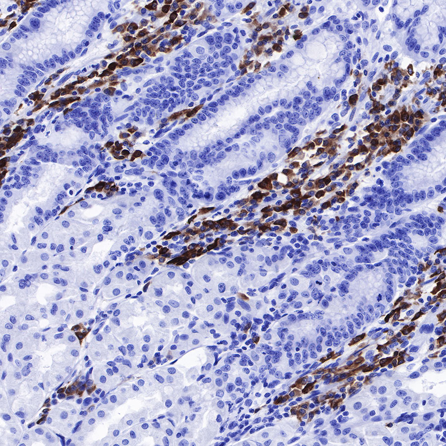
IHC shows positive staining in paraffin-embedded human stomach. Anti-CD79a/MB-1 antibody was used at 1/1000 dilution, followed by a HRP Polymer for Mouse & Rabbit IgG (ready to use). Counterstained with hematoxylin. Heat mediated antigen retrieval with Tris/EDTA buffer pH9.0 was performed before commencing with IHC staining protocol.

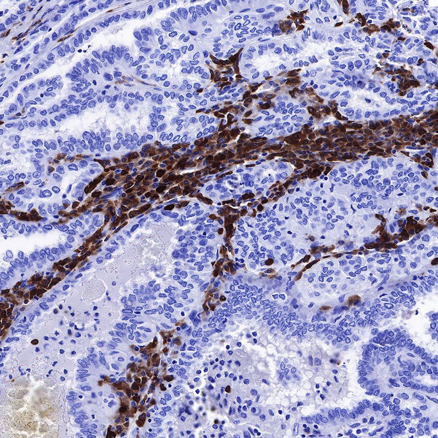
IHC shows positive staining in paraffin-embedded human thyroid cancer. Anti-CD79a/MB-1 antibody was used at 1/1000 dilution, followed by a HRP Polymer for Mouse & Rabbit IgG (ready to use). Counterstained with hematoxylin. Heat mediated antigen retrieval with Tris/EDTA buffer pH9.0 was performed before commencing with IHC staining protocol.
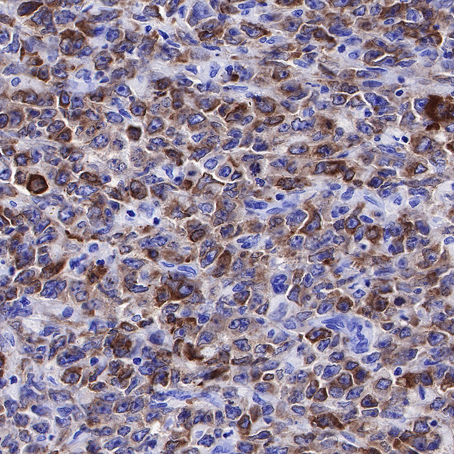
IHC shows positive staining in paraffin-embedded human diffuse large B-cell lymphoma. Anti-CD79a/MB-1 antibody was used at 1/1000 dilution, followed by a HRP Polymer for Mouse & Rabbit IgG (ready to use). Counterstained with hematoxylin. Heat mediated antigen retrieval with Tris/EDTA buffer pH9.0 was performed before commencing with IHC staining protocol.
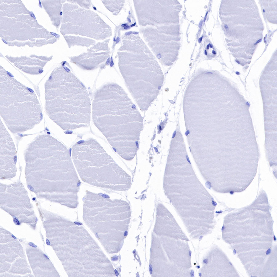
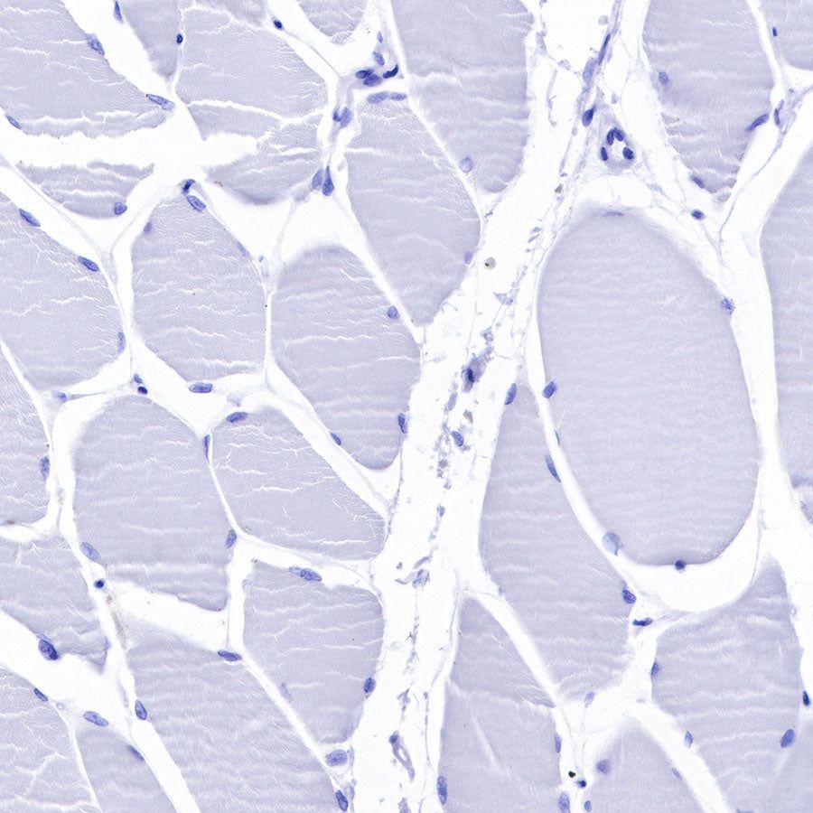
Negative control: IHC shows negative staining in paraffin-embedded human skeletal muscle. Anti-CD79a/MB-1 antibody was used at 1/1000 dilution, followed by a HRP Polymer for Mouse & Rabbit IgG (ready to use). Counterstained with hematoxylin. Heat mediated antigen retrieval with Tris/EDTA buffer pH9.0 was performed before commencing with IHC staining protocol.
Immunofluorescence

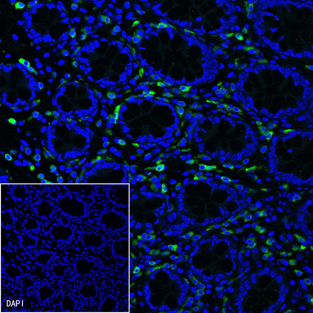
IF shows positive staining in paraffin-embedded human colon. Anti-CD79a/MB-1 antibody was used at 1/500 dilution (Green) and incubated overnight at 4°C. Goat polyclonal Antibody to Rabbit IgG - H&L (Alexa Fluor® 488) was used as secondary antibody at 1/1000 dilution. Counterstained with DAPI (Blue). Heat mediated antigen retrieval with EDTA buffer pH9.0 was performed before commencing with IF staining protocol.

IF shows positive staining in paraffin-embedded human spleen. Anti-CD79a/MB-1 antibody was used at 1/500 dilution (Green) and incubated overnight at 4°C. Goat polyclonal Antibody to Rabbit IgG - H&L (Alexa Fluor® 488) was used as secondary antibody at 1/1000 dilution. Counterstained with DAPI (Blue). Heat mediated antigen retrieval with EDTA buffer pH9.0 was performed before commencing with IF staining protocol.
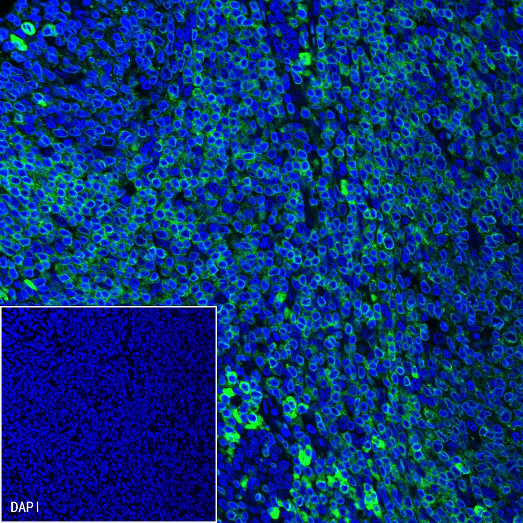
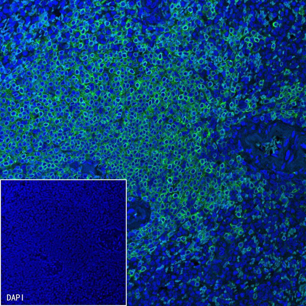
IF shows positive staining in paraffin-embedded human tonsil. Anti-CD79a/MB-1 antibody was used at 1/500 dilution (Green) and incubated overnight at 4°C. Goat polyclonal Antibody to Rabbit IgG - H&L (Alexa Fluor® 488) was used as secondary antibody at 1/1000 dilution. Counterstained with DAPI (Blue). Heat mediated antigen retrieval with EDTA buffer pH9.0 was performed before commencing with IF staining protocol.

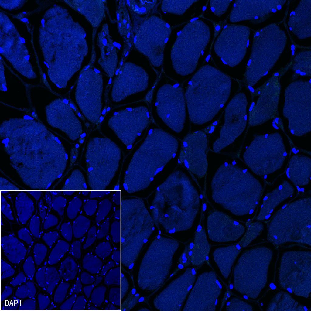
Negative control: IF shows negative staining in paraffin-embedded human skeletal muscle. Anti-CD79a/MB-1 antibody was used at 1/500 dilution and incubated overnight at 4°C. Goat polyclonal Antibody to Rabbit IgG - H&L (Alexa Fluor® 488) was used as secondary antibody at 1/1000 dilution. Counterstained with DAPI (Blue). Heat mediated antigen retrieval with EDTA buffer pH9.0 was performed before commencing with IF staining protocol.