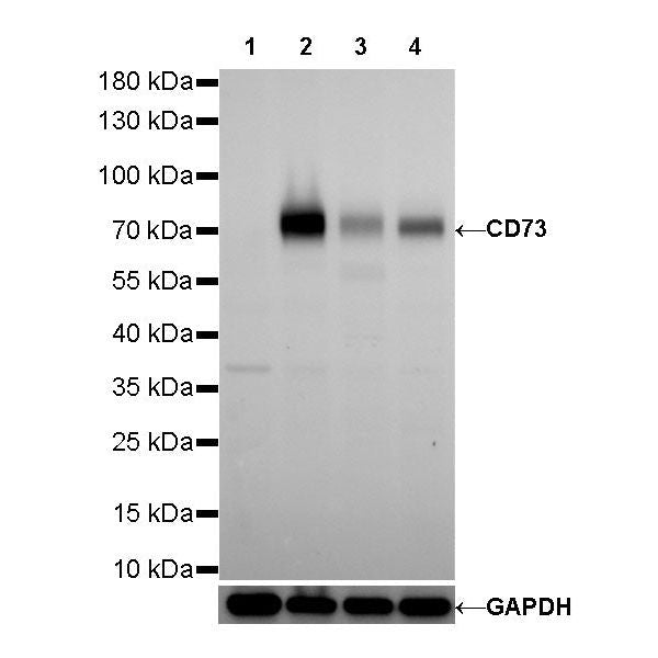
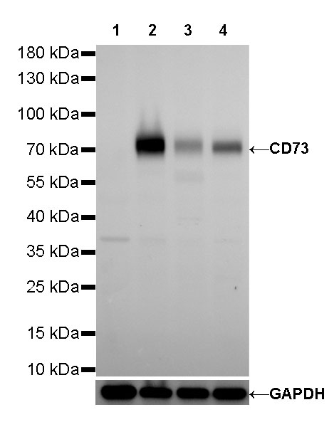
WB result of CD73 Rabbit mAb
Primary antibody: CD73 Rabbit mAb at 1/1000 dilution
Lane 1: Raji whole cell lysate 20 µg
Lane 2: HT-29 whole cell lysate 20 µg
Lane 3: A431 whole cell lysate 20 µg
Lane 4: HepG2 whole cell lysate 20 µg
Negative control: Raji whole cell lysate
Secondary antibody: Goat Anti-Rabbit IgG, (H+L), HRP conjugated at 1/10000 dilution
Predicted MW: 63 kDa
Observed MW: 70 kDa
Exposure time: 120s
CD73 Recombinant Rabbit mAb (SDT-218-52)
CD73 Recombinant Rabbit mAb (SDT-218-52)
Price:
Regular price
$100 USD
Regular price
Sale price
$100 USD
Unit price
per
For shipping services or bulk orders, you may request a quotation.
Secure checkout with

View full details
Product Details
Product Details
Product Specification
| Host | Rabbit |
| Antigen | CD73 |
| Synonyms | 5'-nucleotidase, 5'-NT, 5'-deoxynucleotidase |
| Immunogen | Synthetic Peptide |
| Location | Cell membrane |
| Accession | P21589 |
| Clone Number | SDT-218-52 |
| Antibody Type | Rabbit mAb |
| Application | WB, IHC-P |
| Reactivity | Hu, Ms, Rt |
| Purification | Protein A |
| Concentration | 0.5 mg/ml |
| Physical Appearance | Liquid |
| Storage Buffer | PBS, 40% Glycerol, 0.05% BSA, 0.03% Proclin 300 |
| Stability & Storage | 12 months from date of receipt / reconstitution, -20 °C as supplied |
Dilution
| application | dilution | species |
| IHC-P | 1:500-1:1000 | |
| WB | 1:1000 |
Background
CD73, also known as ecto-5′-nucleotidase (ecto-5′-NT, EC 3.1.3.5) is a glycosylphosphatidyl inositol (GPI)-anchored cell surface protein that is encoded by NT5E gene. CD73 is widely expressed on different tissues [PubMed: 9553767, PubMed: 18404475] and cell types including, but not limited to the subsets of T cells and B cells [PubMed: 2550543], endothelial cells [PubMed: 9015312, PubMed: 15358667] and epithelial cells [PubMed: 9169488].
Picture
Picture
Western Blot
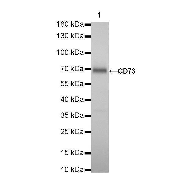
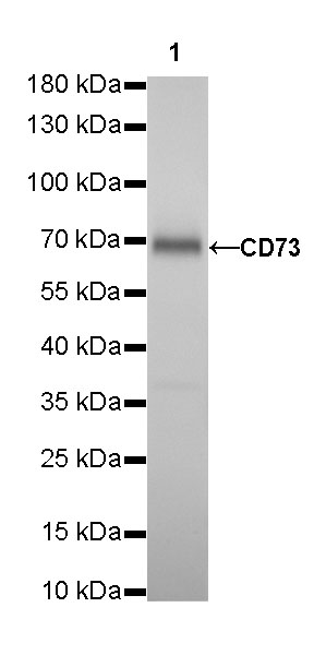
WB result of CD73 Rabbit mAb
Primary antibody: CD73 Rabbit mAb at 1/1000 dilution
Lane 1: mouse brain lysate 20 µg
Secondary antibody: Goat Anti-Rabbit IgG, (H+L), HRP conjugated at 1/10000 dilution
Predicted MW: 63 kDa
Observed MW: 68 kDa
Exposure time: 180s
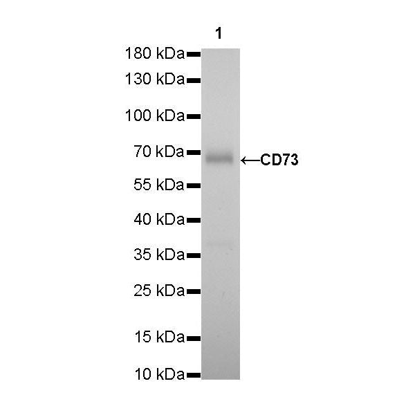
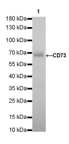
WB result of CD73 Rabbit mAb
Primary antibody: CD73 Rabbit mAb at 1/1000 dilution
Lane 1: rat brain lysate 20 µg
Secondary antibody: Goat Anti-Rabbit IgG, (H+L), HRP conjugated at 1/10000 dilution
Predicted MW: 63 kDa
Observed MW: 70 kDa
Exposure time: 180s
Immunohistochemistry
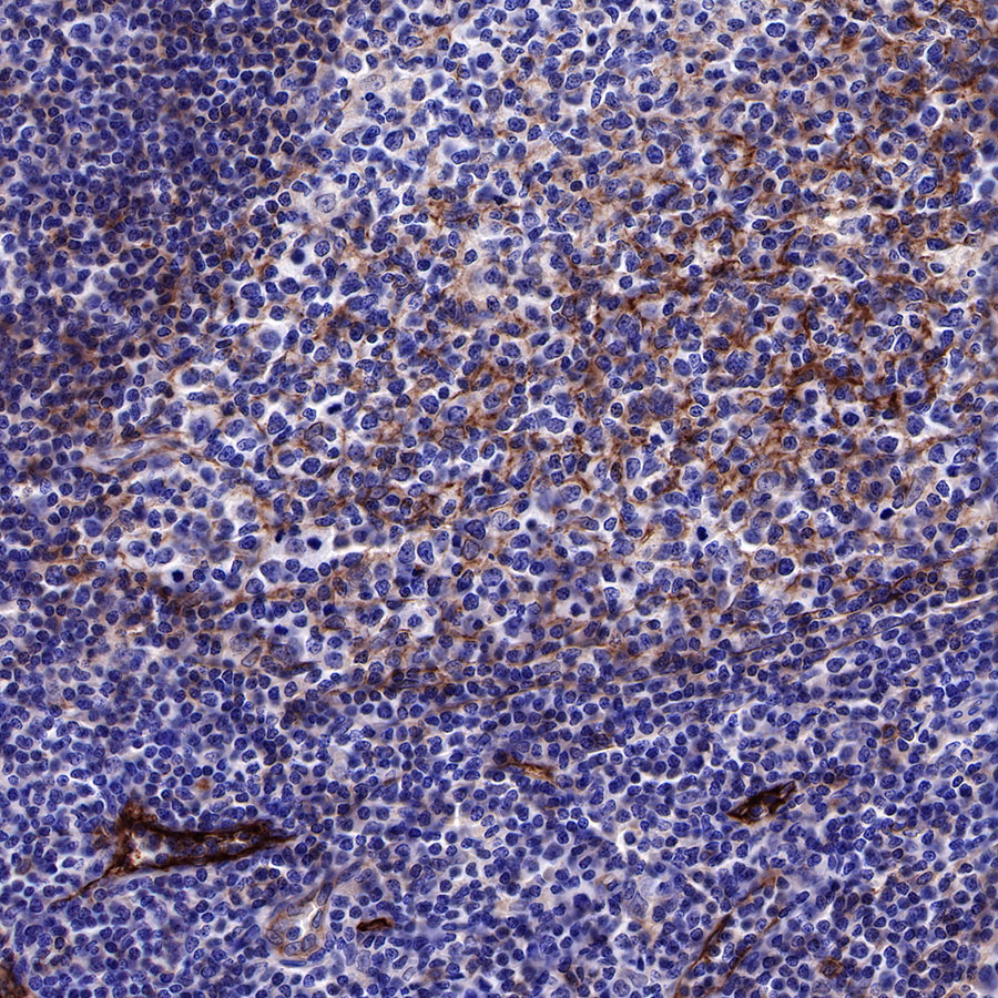
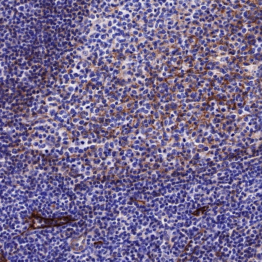
IHC shows positive staining in paraffin-embedded human tonsil. Anti-CD73 antibody was used at 1/500 dilution, followed by a HRP Polymer for Mouse & Rabbit IgG (ready to use). Counterstained with hematoxylin. Heat mediated antigen retrieval with Tris/EDTA buffer pH9.0 was performed before commencing with IHC staining protocol.

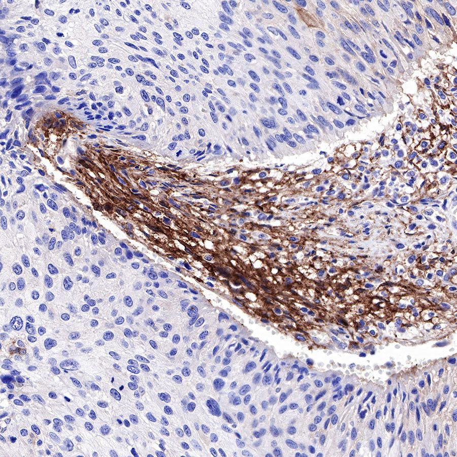
IHC shows positive staining in paraffin-embedded human cervical squamous cell carcinoma. Anti-CD73 antibody was used at 1/500 dilution, followed by a HRP Polymer for Mouse & Rabbit IgG (ready to use). Counterstained with hematoxylin. Heat mediated antigen retrieval with Tris/EDTA buffer pH9.0 was performed before commencing with IHC staining protocol.
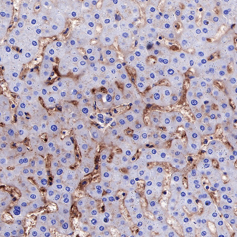
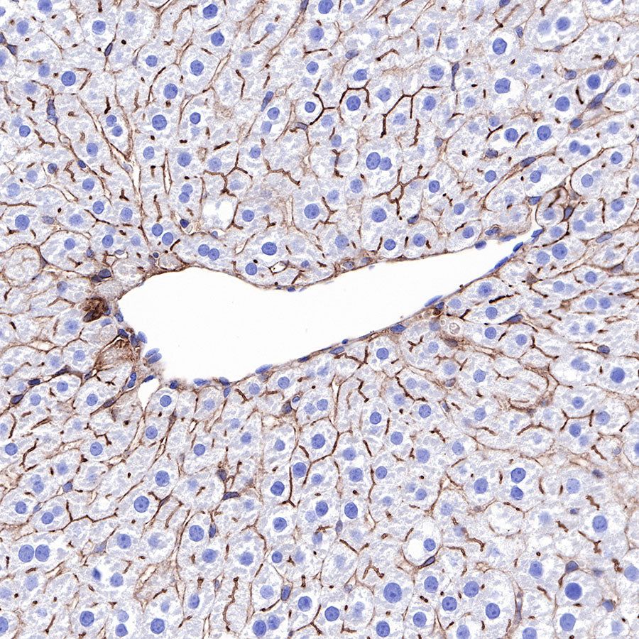
IHC shows positive staining in paraffin-embedded human liver. Anti-CD73 antibody was used at 1/1000 dilution, followed by a HRP Polymer for Mouse & Rabbit IgG (ready to use). Counterstained with hematoxylin. Heat mediated antigen retrieval with Tris/EDTA buffer pH9.0 was performed before commencing with IHC staining protocol.

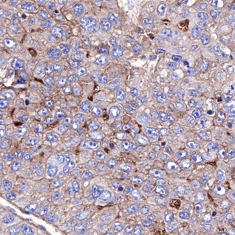
IHC shows positive staining in paraffin-embedded human hepatocellular carcinoma. Anti-CD73 antibody was used at 1/500 dilution, followed by a HRP Polymer for Mouse & Rabbit IgG (ready to use). Counterstained with hematoxylin. Heat mediated antigen retrieval with Tris/EDTA buffer pH9.0 was performed before commencing with IHC staining protocol.
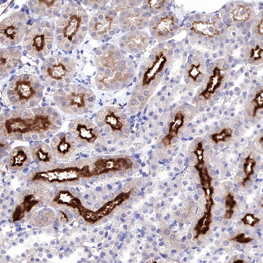
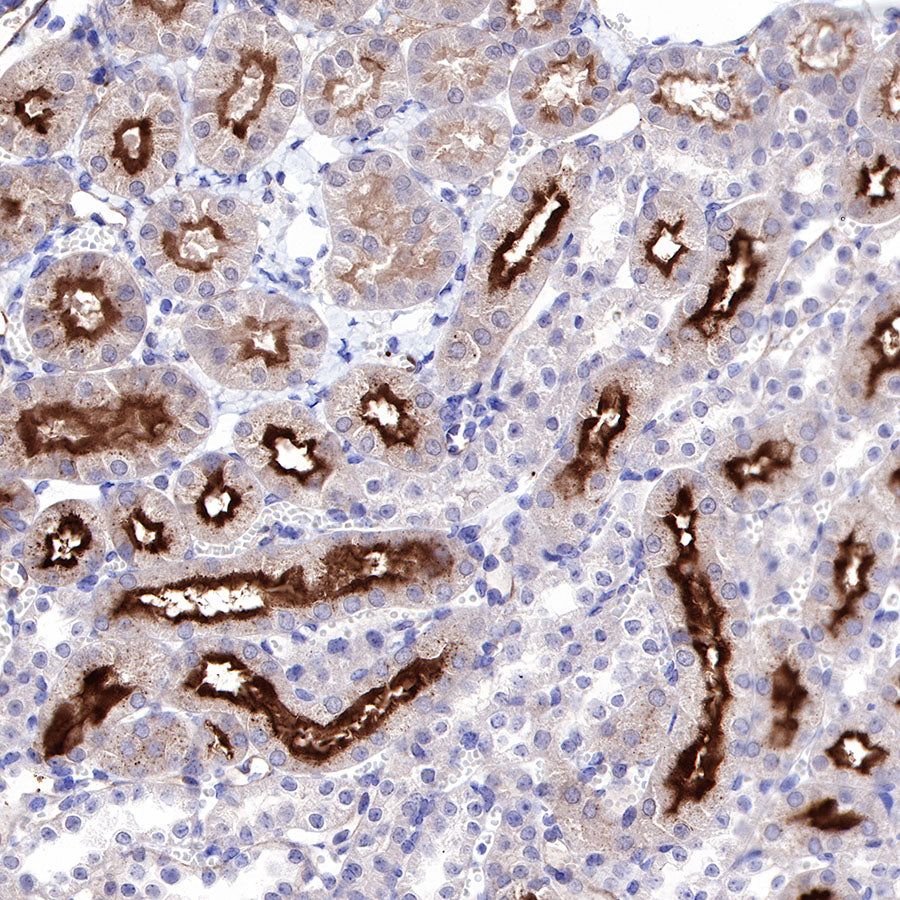
IHC shows positive staining in paraffin-embedded mouse kidney. Anti-CD73 antibody was used at 1/500 dilution, followed by a HRP Polymer for Mouse & Rabbit IgG (ready to use). Counterstained with hematoxylin. Heat mediated antigen retrieval with Tris/EDTA buffer pH9.0 was performed before commencing with IHC staining protocol.
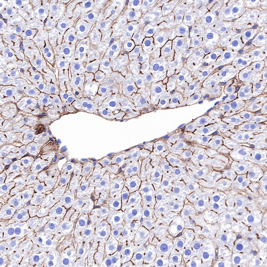
IHC shows positive staining in paraffin-embedded rat liver. Anti-CD73 antibody was used at 1/1000 dilution, followed by a HRP Polymer for Mouse & Rabbit IgG (ready to use). Counterstained with hematoxylin. Heat mediated antigen retrieval with Tris/EDTA buffer pH9.0 was performed before commencing with IHC staining protocol.