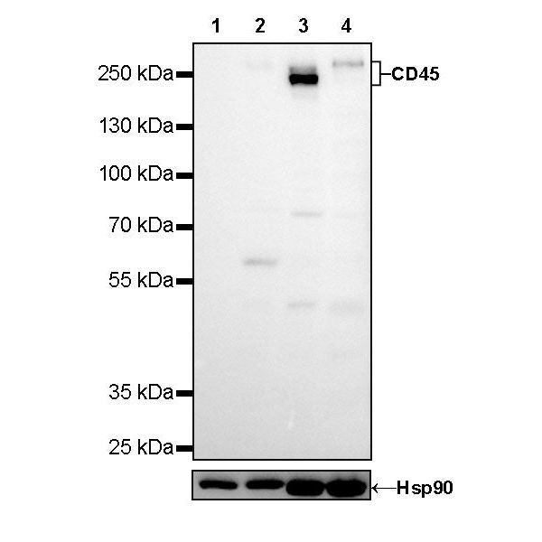
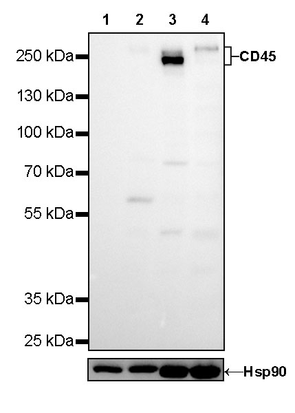
WB result of CD45 Mouse mAb
Primary antibody: CD45 Mouse mAb at 1/1250 dilution
Lane 1: MCF7 whole cell lysate 20 µg
Lane 2: THP-1 whole cell lysate 20 µg
Lane 3: Jurkat whole cell lysate 20 µg
Lane 4: Raji whole cell lysate 20 µg
Negative control: MCF7 whole cell lysate
Low expression control: THP-1 whole cell lysate
Secondary antibody: Goat Anti-Mouse IgG, (H+L), HRP conjugated at 1/10000 dilution
Predicted MW: 147kDa
Observed MW: 240~260kDa
CD45 Mouse mAb (429-1)
CD45 Mouse mAb (429-1)
Price:
Regular price
$100 USD
Regular price
Sale price
$100 USD
Unit price
per
For shipping services or bulk orders, you may request a quotation.
Secure checkout with

View full details
Product Details
Product Details
Product Specification
| Host | Mouse |
| Antigen | CD45 |
| Synonyms | Leukocyte common antigen (L-CA) |
| Immunogen | Synthetic Peptide |
| Location | Cell membrane |
| Accession | P08575 |
| Clone Number | 429-1 |
| Antibody Type | Mouse mAb |
| Isotype | IgG1,k |
| Application | WB, IHC-P, ICC, ICFCM |
| Reactivity | Hu |
| Purification | Protein G |
| Concentration | 0.5 mg/ml |
| Conjugation | Unconjugated |
| Physical Appearance | Liquid |
| Storage Buffer | PBS, 40% Glycerol, 0.05% BSA, 0.03% Proclin 300 |
| Stability & Storage | 12 months from date of receipt / reconstitution, -20 °C as supplied |
Dilution
| application | dilution | species |
| WB | 1:1250 | |
| IHC-P | 1:1000 | |
| ICC | 1:125 | |
| ICFCM | 1:500 |
Background
CD45 is a type I transmembrane protein that is present in various isoforms on all differentiated hematopoietic cells (except erythrocytes and plasma cells). CD45 has been shown to be an essential regulator of T- and B-cell antigen receptor signaling. It functions through either direct interaction with components of the antigen receptor complexes via its extracellular domain (a form of co-stimulation), or by activating various Src family kinases required for the antigen receptor signaling via its cytoplasmic domain. CD45 also suppresses JAK kinases, and so functions as a negative regulator of cytokine receptor signaling.
Picture
Picture
Western Blot
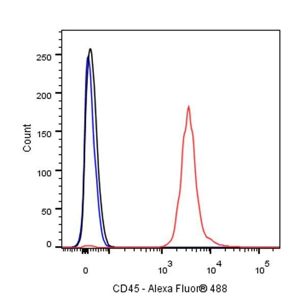
FC
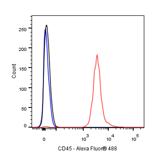
Flow cytometric analysis of 4% PFA fixed 90% methanol permeabilized Jurkat (Human T cell leukemia T lymphocyte) cells labelling Amyloid Precursor Protein antibody at 1/500 dilution (0.1 μg)/ (Red) compared with a Mouse monoclonal IgG (Black) isotype control and an unlabelled control (cells without incubation with primary antibody and secondary antibody) (Blue). Goat Anti - Mouse IgG Alexa Fluor® 488 was used as the secondary antibody.
Immunohistochemistry

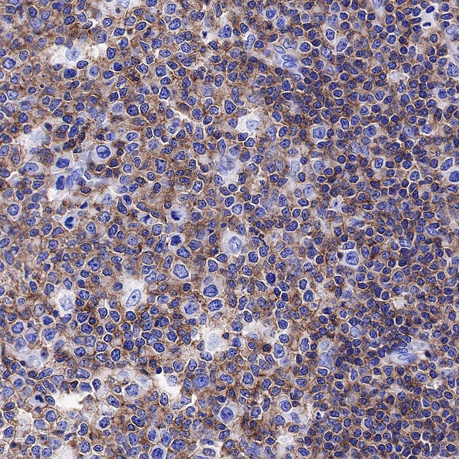
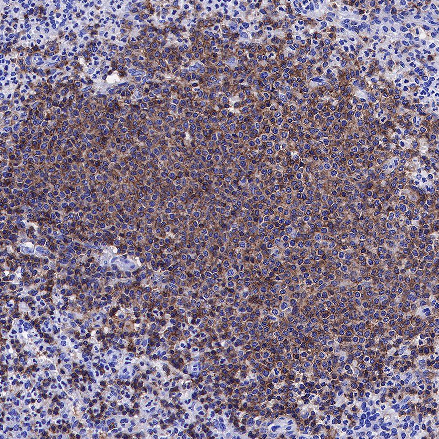
IHC shows positive staining in paraffin-embedded human tonsil. Anti-CD45 antibody was used at 1/1000 dilution, followed by a HRP Polymer for Mouse & Rabbit IgG (ready to use). Counterstained with hematoxylin. Heat mediated antigen retrieval with Tris/EDTA buffer pH9.0 was performed before commencing with IHC staining protocol.

IHC shows positive staining in paraffin-embedded human spleen. Anti-CD45 antibody was used at 1/1000 dilution, followed by a HRP Polymer for Mouse & Rabbit IgG (ready to use). Counterstained with hematoxylin. Heat mediated antigen retrieval with Tris/EDTA buffer pH9.0 was performed before commencing with IHC staining protocol.
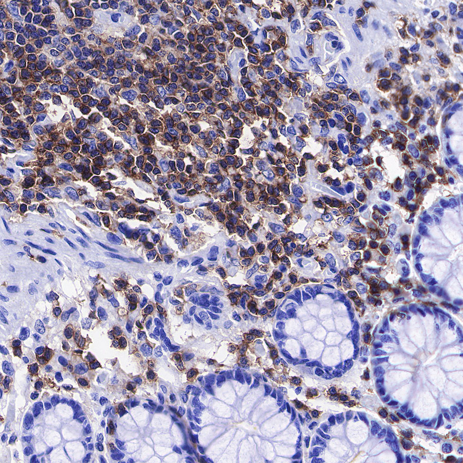
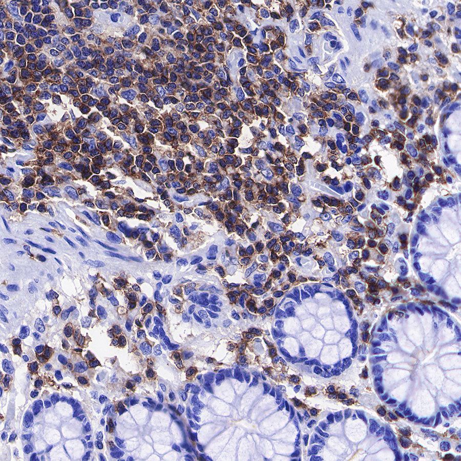
IHC shows positive staining in paraffin-embedded human colon. Anti-CD45 antibody was used at 1/1000 dilution, followed by a HRP Polymer for Mouse & Rabbit IgG (ready to use). Counterstained with hematoxylin. Heat mediated antigen retrieval with Tris/EDTA buffer pH9.0 was performed before commencing with IHC staining protocol.
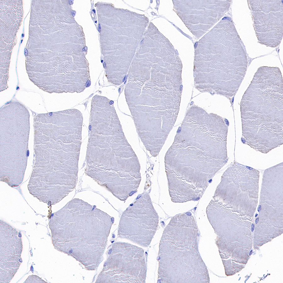
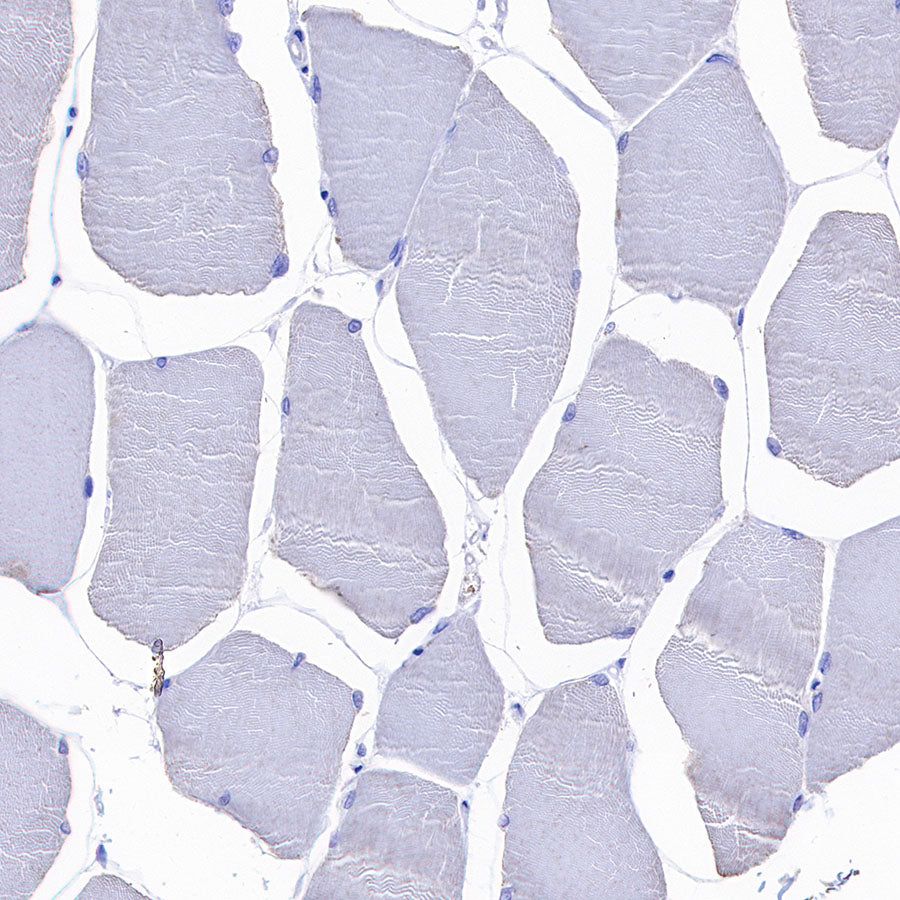
Negative control: IHC shows negative staining in paraffin-embedded human skeletal muscle. Anti-CD45 antibody was used at 1/1000 dilution, followed by a HRP Polymer for Mouse & Rabbit IgG (ready to use). Counterstained with hematoxylin. Heat mediated antigen retrieval with Tris/EDTA buffer pH9.0 was performed before commencing with IHC staining protocol.
Immunocytochemistry

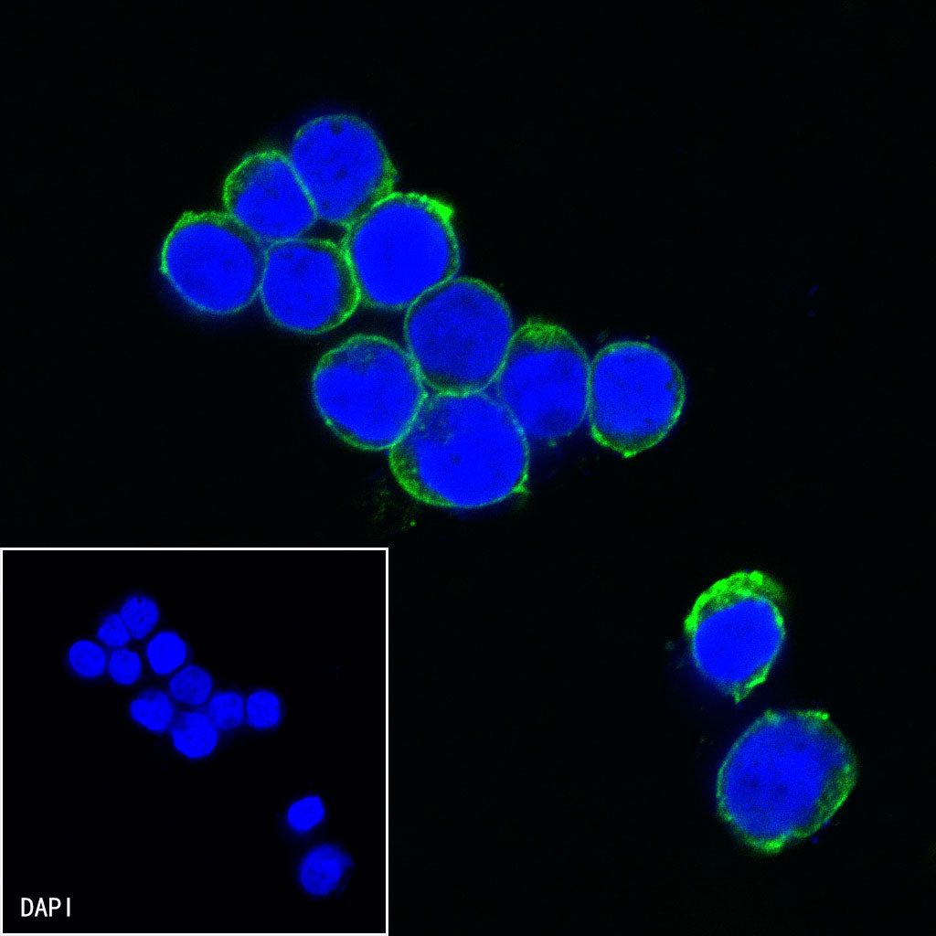
ICC shows positive staining in Jurkat cells. Anti-CD45 antibody was used at 1/125 dilution (Green) and incubated overnight at 4°C. Goat polyclonal Antibody to mouse IgG - H&L (Alexa Fluor® 488) was used as secondary antibody at 1/1000 dilution. The cells were fixed with 100% ice-cold methanol and permeabilized with 0.1% PBS-Triton X-100. Nuclei were counterstained with DAPI (Blue).
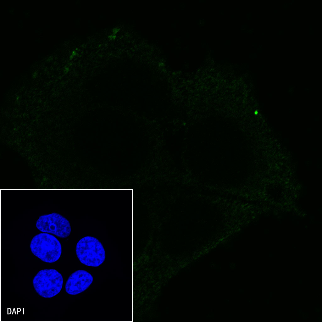
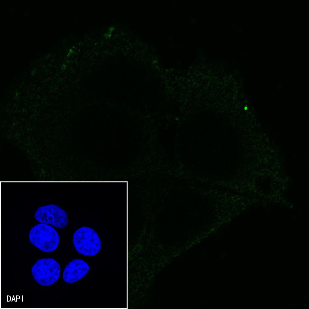
Negative control:ICC shows negative staining in MCF7 cells.Anti-CD45 antibody was used at 1/125 dilution and incubated overnight at 4°C. Goat polyclonal Antibody to mouse IgG - H&L (Alexa Fluor® 488) was used as secondary antibody at 1/1000 dilution. The cells were fixed with 100% ice-cold methanol and permeabilized with 0.1% PBS-Triton X-100. Nuclei were counterstained with DAPI (Blue).