Product Specification
| Host |
Rabbit |
| Antigen |
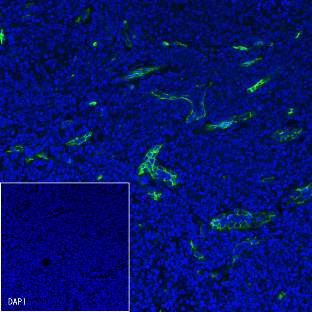
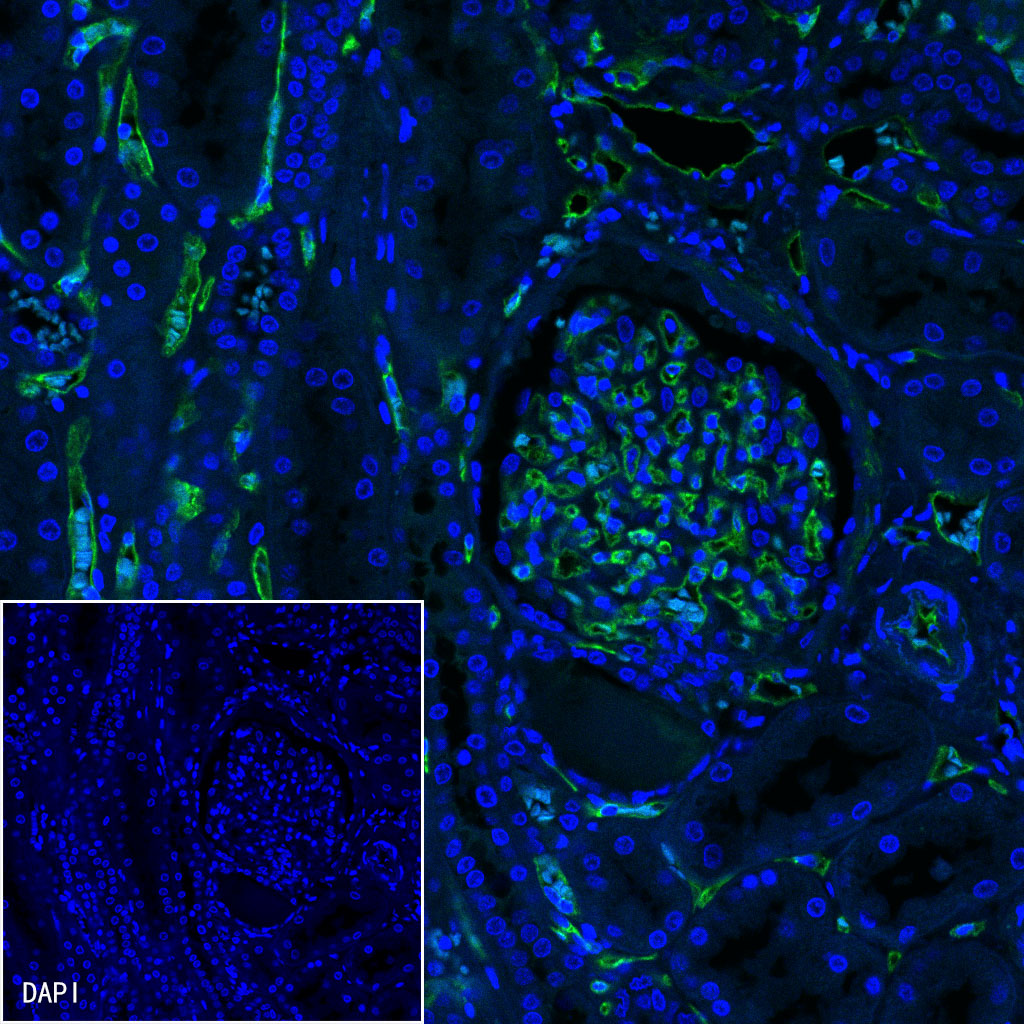
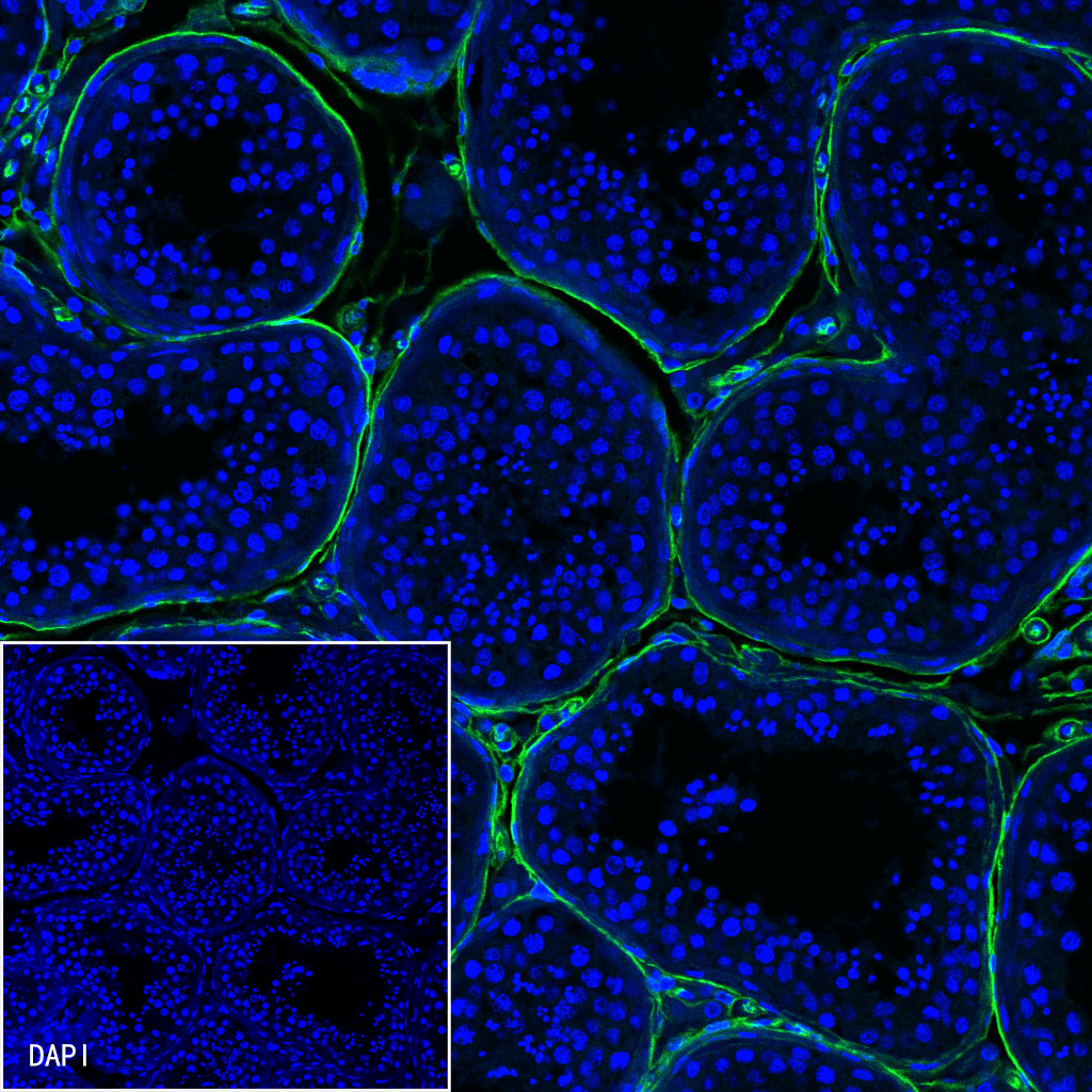
CD34 |
| Synonyms |
Hematopoietic progenitor cell antigen CD34 |
| Immunogen |
Synthetic Peptide |
| Accession |
P28906 |
| Clone Number |
SDT-051-68 |
| Antibody Type |
Rabbit mAb |
| Application |
WB, IHC-P, ICC, IP, IF |
| Reactivity |
Hu, Ms, Rt |
| Purification |
Protein A |
| Concentration |
0.5mg/ml |
| Conjugation |
Unconjugated |
| Physical Appearance |
Liquid |
| Storage Buffer |
PBS, 40% Glycerol, 0.05%BSA, 0.03% Proclin 300 |
| Stability & Storage |
12 months from date of receipt / reconstitution, -20 °C as supplied |
Dilution
| application |
dilution |
species |
| ICC |
1:500 |
|
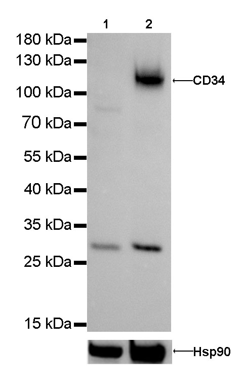
| WB |
1:1000 |
|
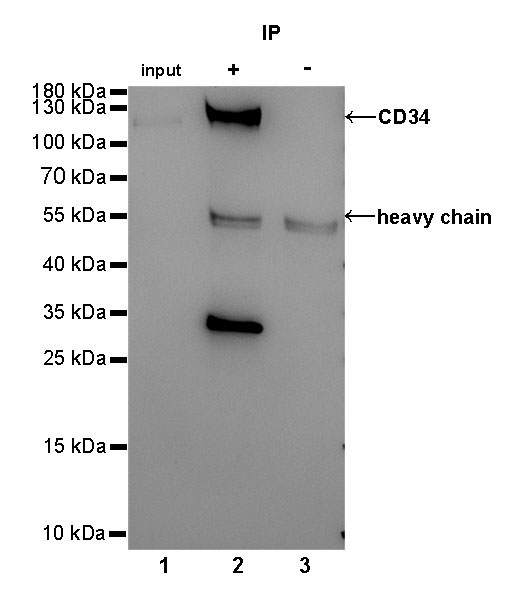
| IP |
1:25 |
|
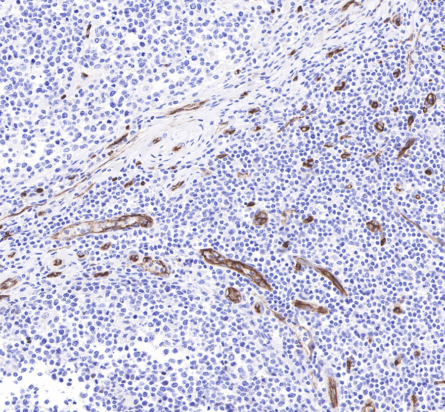
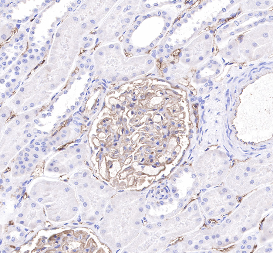
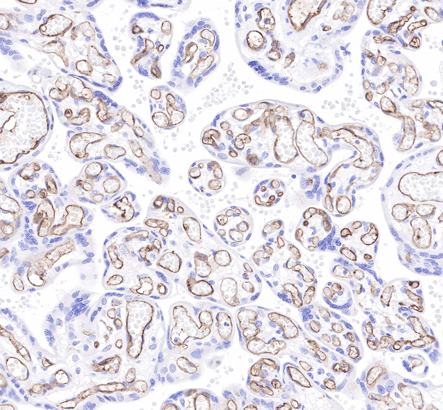
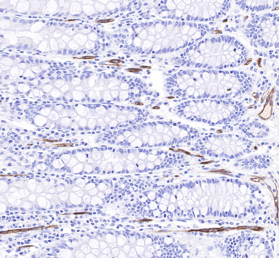
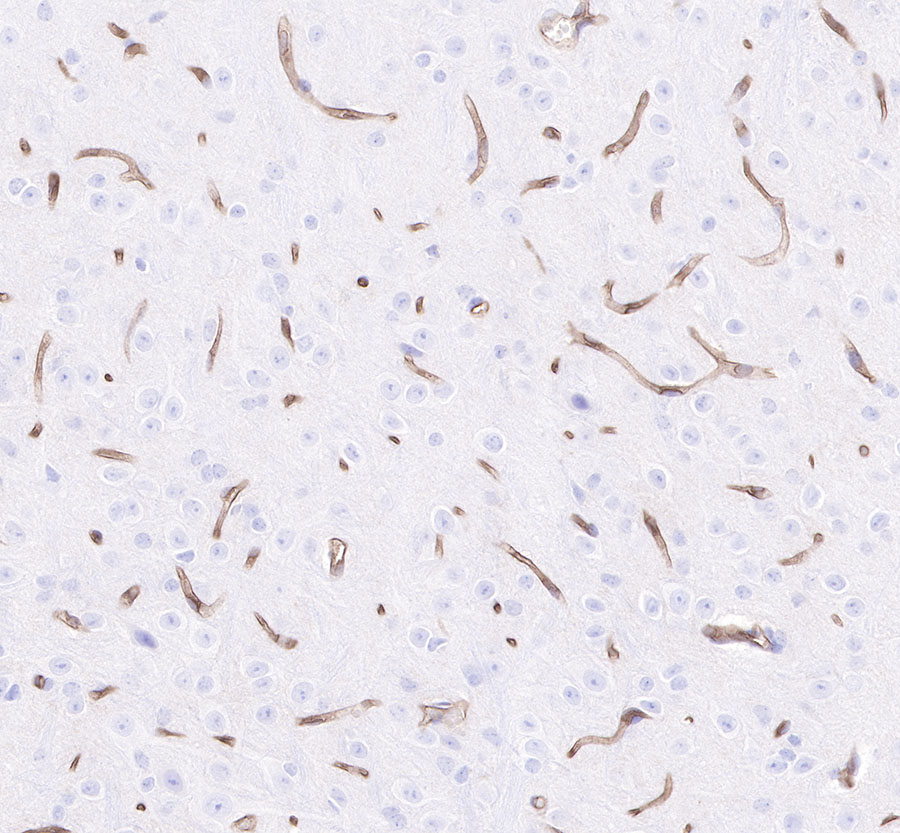
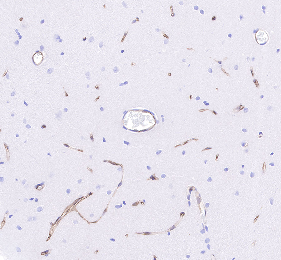
| IHC-P |
1:500-1:1000 |
|
| IF |
1:500 |
|
Background
CD34 is a transmembrane phosphoglycoprotein protein encoded by the CD34 gene. CD34 derives its name from the cluster of differentiation protocol that identifies cell surface antigens. CD34 was first described on hematopoietic stem cells independently by Civin et al. and Tindle et al, as a cell surface glycoprotein and functions as a cell-cell adhesion factor. It may also mediate the attachment of hematopoietic stem cells to bone marrow extracellular matrix or directly to stromal cells. Clinically, it is associated with the selection and enrichment of hematopoietic stem cells for bone marrow transplants. Due to these historical and clinical associations, CD34 expression is almost ubiquitously related to hematopoietic cells.