

Flow cytometric analysis of HeLa (Human cervix adenocarcinoma epithelial cell, left) / Romas (Human Burkitt's lymphoma B lymphocyte, right) cells labelling CD27 antibody at 1/2000 dilution (0.1 μg) / (red) compared with a Mouse monoclonal IgG (Black) isotype control and an unlabelled control (cells without incubation with primary antibody and secondary antibody) (Blue). Goat Anti - Mouse IgG Alexa Fluor® 488 was used as the secondary antibody.
Negative control: HeLa
CD27 Mouse mAb (S-568-2)
CD27 Mouse mAb (S-568-2)
Price:
Regular price
$45 USD
Regular price
Sale price
$45 USD
Unit price
per
For shipping services or bulk orders, you may request a quotation.
Secure checkout with

View full details
Product Details
Product Details
Product Specification
| Host | Mouse |
| Antigen | CD27 |
| Synonyms | CD27L receptor, T-cell activation antigen CD27, T14, Tumor necrosis factor receptor superfamily member 7, TNFRSF7 |
| Immunogen | Recombinant Protein |
| Location | Membrane |
| Accession | P26842 |
| Clone Number | S-568-2 |
| Antibody Type | Mouse mAb |
| Isotype | IgG1,k |
| Application | ICC, FCM |
| Reactivity | Hu |
| Purification | Protein G |
| Concentration | 2 mg/ml |
| Conjugation | Unconjugated |
| Physical Appearance | Liquid |
| Storage Buffer | PBS, 40% Glycerol, 0.05%BSA, 0.03% Proclin 300 |
| Stability & Storage | 12 months from date of receipt / reconstitution, -20 °C as supplied |
Dilution
| application | dilution | species |
| FCM | 1:2000 | |
| ICC | 1:500 |
Background
CD27 is a member of the TNF-receptor superfamily. This receptor is required for generation and long-term maintenance of T cell immunity. It binds to ligand CD70, and plays a key role in regulating B-cell activation and immunoglobulin synthesis. When CD27 binds CD70, a signaling cascade leads to the differentiation and clonal expansion of T cells. The cascade also results in improved survival and memory of cytotoxic T cells and increased production of certain cytokines. It is currently of interest to immunologists as a co-stimulatory immune checkpoint molecule, and is the target of an anti-cancer drug in clinical trials.
Picture
Picture
FC
Immunocytochemistry
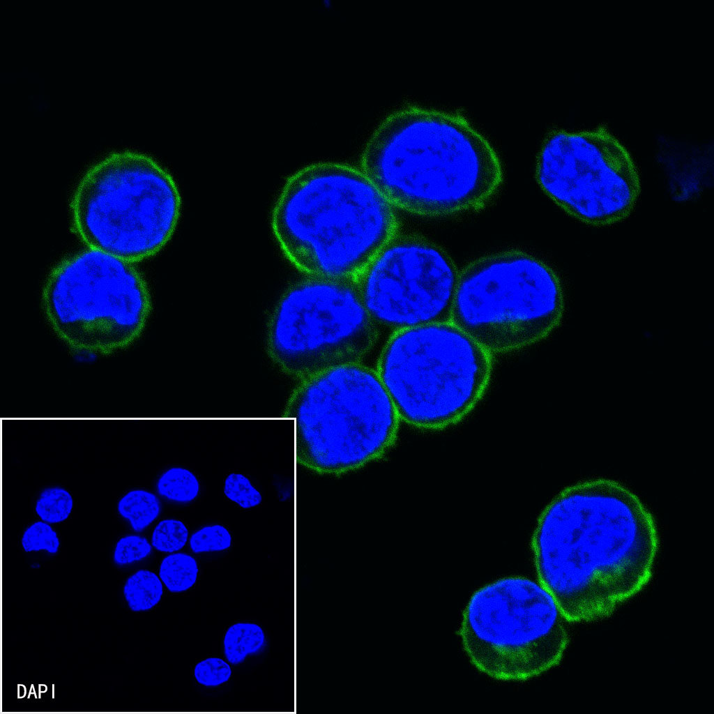
ICC shows positive staining in Ramos cells. Anti-CD27 antibody was used at 1/500 dilution (Green) and incubated overnight at 4°C. Goat polyclonal Antibody to mouse IgG - H&L (Alexa Fluor® 488) was used as secondary antibody at 1/1000 dilution. The cells were fixed with 4% PFA and permeabilized with 0.1% PBS-Triton X-100. Nuclei were counterstained with DAPI (Blue).
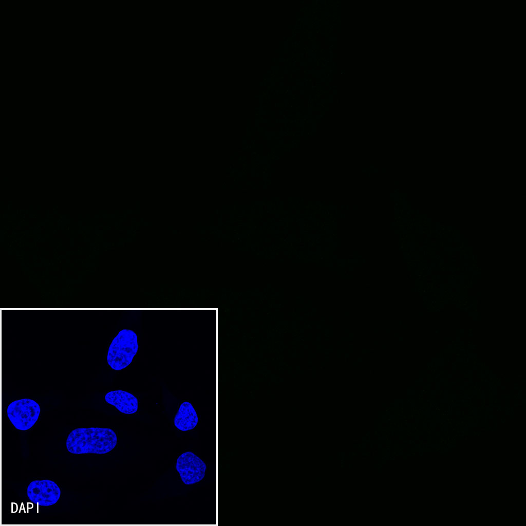
Negative control:ICC shows negative staining in HeLa cells.Anti-CD27 antibody was used at 1/500 dilution and incubated overnight at 4°C. Goat polyclonal Antibody to mouse IgG - H&L (Alexa Fluor® 488) was used as secondary antibody at 1/1000 dilution. The cells were fixed with 4% PFA and permeabilized with 0.1% PBS-Triton X-100. Nuclei were counterstained with DAPI (Blue).

