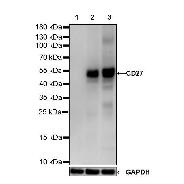
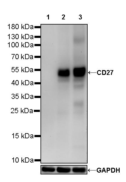
WB result of CD27 Mouse mAb
Primary antibody: CD27 Mouse mAb at 1/1000 dilution
Lane 1: HepG2 whole cell lysate 20 µg
Lane 2: Raji whole cell lysate 20 µg
Lane 3: Ramos whole cell lysate 20 µg
Negative control: HepG2 whole cell lysate
Secondary antibody: Goat Anti-Mouse IgG, (H+L), HRP conjugated at 1/10000 dilution
Predicted MW: 29 kDa
Observed MW: 50kDa
(This blot was developed with high sensitivity substrate)
CD27 Mouse mAb (S-568-1)
CD27 Mouse mAb (S-568-1)
Price:
Regular price
$100 USD
Regular price
Sale price
$100 USD
Unit price
per
For shipping services or bulk orders, you may request a quotation.
Secure checkout with

View full details
Product Details
Product Details
Product Specification
| Host | Mouse |
| Antigen | CD27 |
| Synonyms | CD27L receptor, T-cell activation antigen CD27, T14, Tumor necrosis factor receptor superfamily member 7, TNFRSF7 |
| Immunogen | Recombinant Protein |
| Location | Membrane |
| Accession | P26842 |
| Clone Number | 568-1 |
| Antibody Type | Mouse mAb |
| Isotype | IgG1,k |
| Application | WB, IHC-P, ICC, FCM |
| Reactivity | Hu |
| Purification | Protein G |
| Concentration | 2 mg/ml |
| Conjugation | Unconjugated |
| Physical Appearance | Liquid |
| Storage Buffer | PBS, 40% Glycerol, 0.05% BSA, 0.03% Proclin 300 |
| Stability & Storage | 12 months from date of receipt / reconstitution, -20 °C as supplied |
Dilution
| application | dilution | species |
| WB | 1:1000 | null |
| IHC | 1:2000 | null |
| FCM | 1:2000 | null |
| ICC | 1:500 | null |
Background
Cluster of differentiation 27 (CD27) is a member of the tumor necrosis factor receptor superfamily and plays a key role in T-cell activation by providing a costimulatory signal. Bound to its natural ligand CD70, CD27 signaling enhances T-cell proliferation and differentiation to effector and memory T cells and therefore has potential as an immune modulatory target in cancer treatment [PMID: 32152062].
Picture
Picture
Western Blot
FC
Flow cytometric analysis of HeLa (Human cervix adenocarcinoma epithelial cell, left) / Ramos (Human Burkitt's lymphoma B lymphocyte, right) cells labelling CD27 antibody at 1/2000 dilution (0.1 μg) / (red) compared with a Mouse monoclonal IgG (Black) isotype control and an unlabelled control (cells without incubation with primary antibody and secondary antibody) (Blue). Goat Anti-Mouse IgG Alexa Fluor 488 was used as the secondary antibody. Negative control: HeLa
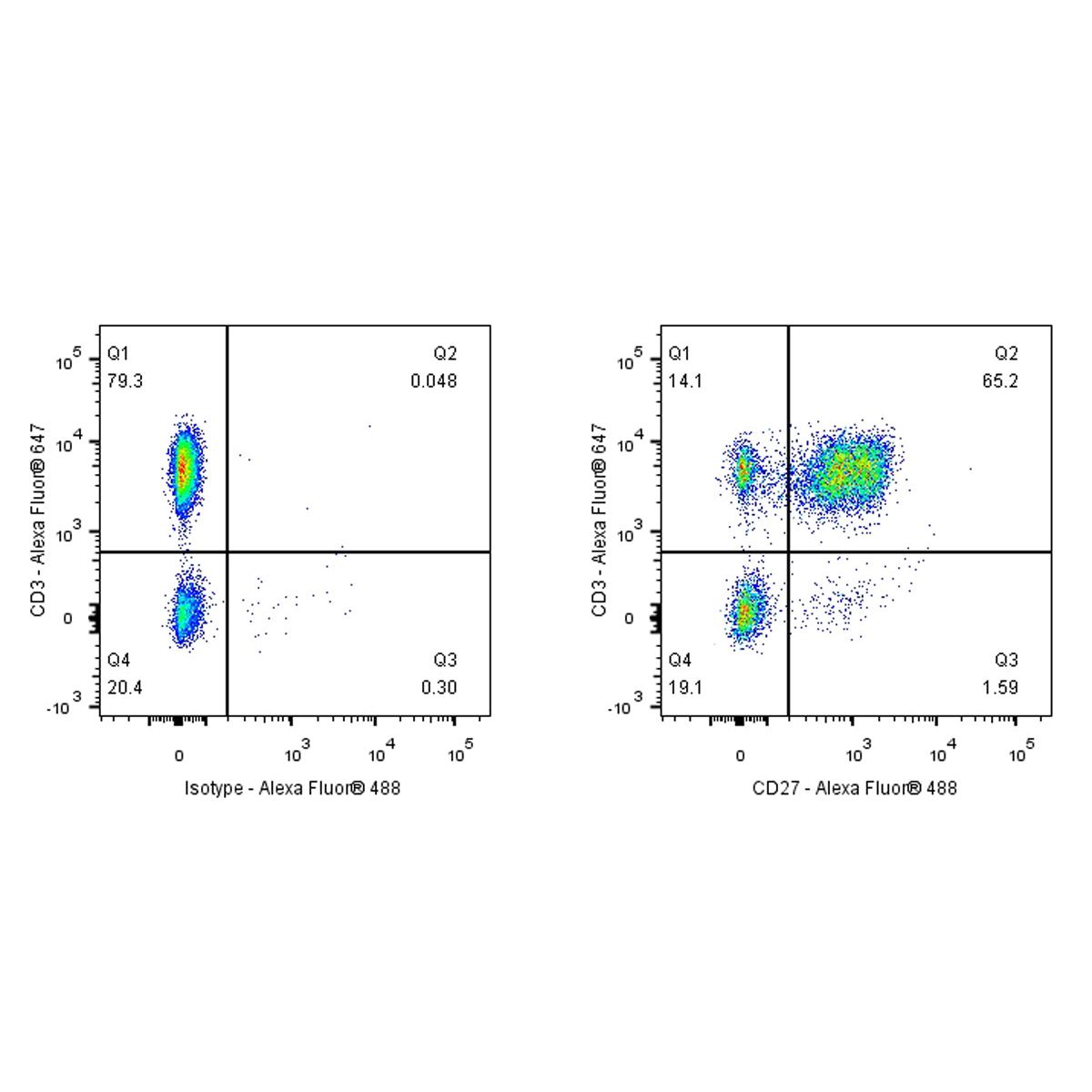
Flow cytometric analysis of human PBMC (human peripheral blood mononuclear cell) labelling CD27 antibody at 1/2000 (0.1 μg) dilution (Right) compared with a Mouse monoclonal IgG isotype control (Left). Goat Anti - Mouse IgG Alexa Fluor® 488 was used as the secondary antibody. Then cells were stained with CD3 - Alexa Fluor® 647 separately. Gated on total viable cells.
Immunohistochemistry
IHC shows positive staining in paraffin-embedded human tonsil. Anti-CD27 antibody was used at 1/2000 dilution, followed by a HRP Polymer for Mouse & Rabbit IgG (ready to use). Counterstained with hematoxylin. Heat mediated antigen retrieval with Tris/EDTA buffer pH9.0 was performed before commencing with IHC staining protocol.
IHC shows positive staining in paraffin-embedded human spleen. Anti-CD27 antibody was used at 1/2000 dilution, followed by a HRP Polymer for Mouse & Rabbit IgG (ready to use). Counterstained with hematoxylin. Heat mediated antigen retrieval with Tris/EDTA buffer pH9.0 was performed before commencing with IHC staining protocol.
IHC shows positive staining in paraffin-embedded human colon. Anti-CD27 antibody was used at 1/2000 dilution, followed by a HRP Polymer for Mouse & Rabbit IgG (ready to use). Counterstained with hematoxylin. Heat mediated antigen retrieval with Tris/EDTA buffer pH9.0 was performed before commencing with IHC staining protocol.
IHC shows positive staining in paraffin-embedded human stomach. Anti-CD27 antibody was used at 1/2000 dilution, followed by a HRP Polymer for Mouse & Rabbit IgG (ready to use). Counterstained with hematoxylin. Heat mediated antigen retrieval with Tris/EDTA buffer pH9.0 was performed before commencing with IHC staining protocol.
IHC shows positive staining in paraffin-embedded human cervical squamous cell carcinoma. Anti-CD27 antibody was used at 1/2000 dilution, followed by a HRP Polymer for Mouse & Rabbit IgG (ready to use). Counterstained with hematoxylin. Heat mediated antigen retrieval with Tris/EDTA buffer pH9.0 was performed before commencing with IHC staining protocol.
IHC shows positive staining in paraffin-embedded human endometrial carcinoma. Anti-CD27 antibody was used at 1/2000 dilution, followed by a HRP Polymer for Mouse & Rabbit IgG (ready to use). Counterstained with hematoxylin. Heat mediated antigen retrieval with Tris/EDTA buffer pH9.0 was performed before commencing with IHC staining protocol.
IHC shows positive staining in paraffin-embedded human lung cancer. Anti-CD27 antibody was used at 1/2000 dilution, followed by a HRP Polymer for Mouse & Rabbit IgG (ready to use). Counterstained with hematoxylin. Heat mediated antigen retrieval with Tris/EDTA buffer pH9.0 was performed before commencing with IHC staining protocol.
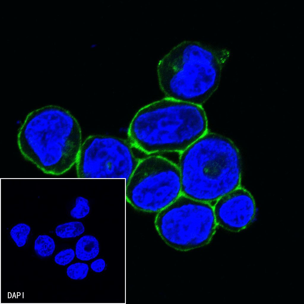
Immunocytochemistry

ICC shows positive staining in Ramos cells. Anti-CD27 antibody was used at 1/500 dilution (Green) and incubated overnight at 4°C. Goat polyclonal Antibody to mouse IgG - H&L (Alexa Fluor® 488) was used as secondary antibody at 1/1000 dilution. The cells were fixed with 4% PFA and permeabilized with 0.1% PBS-Triton X-100. Nuclei were counterstained with DAPI (Blue).
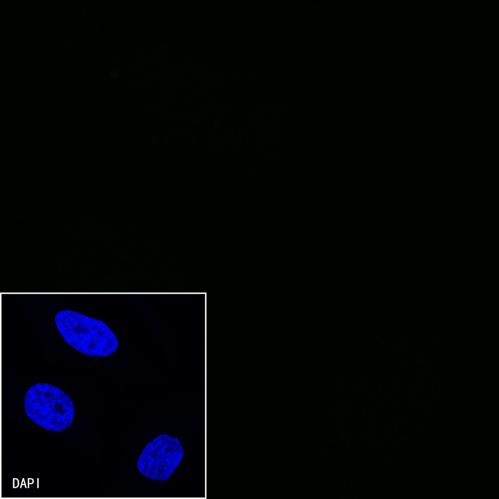
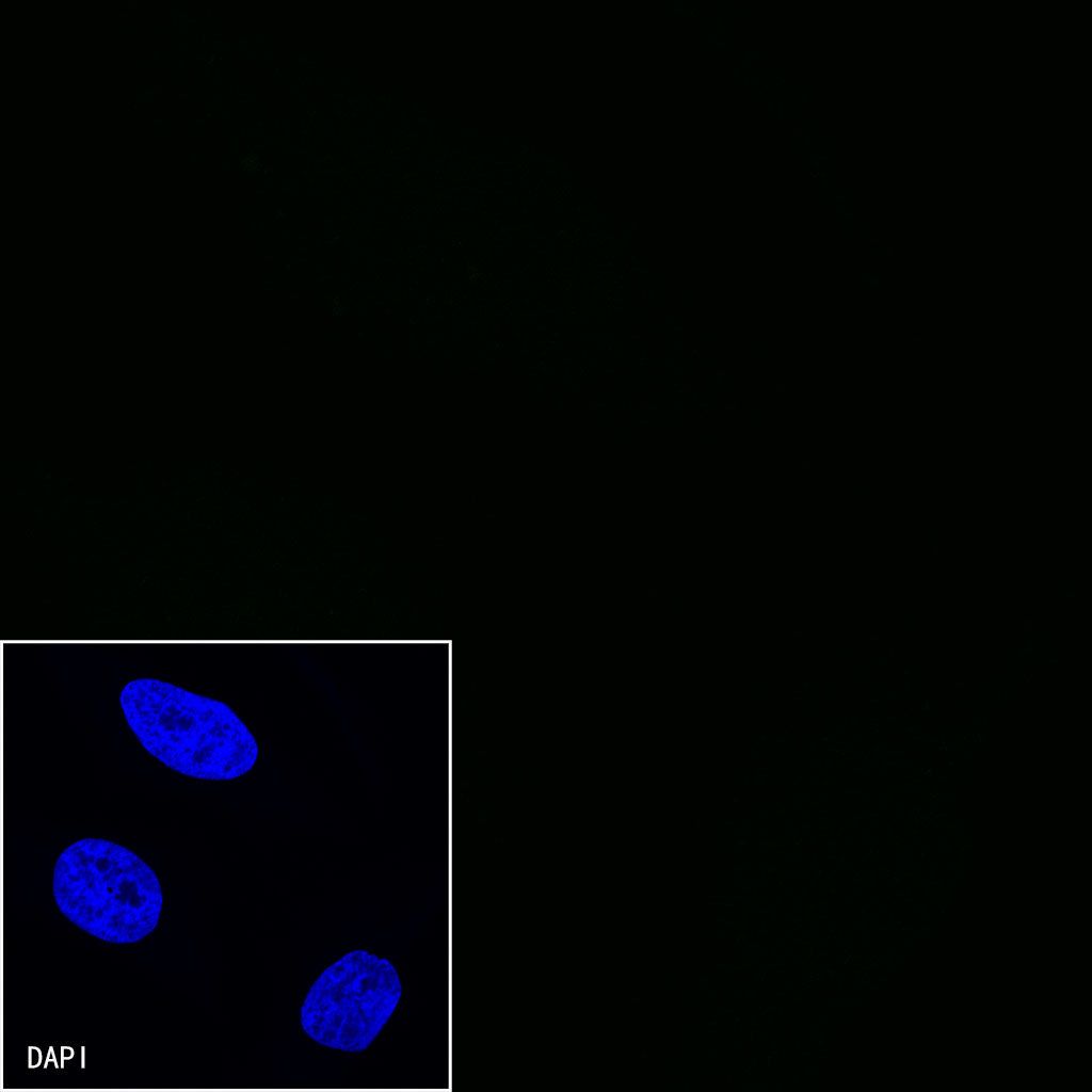
Negative control: ICC shows negative staining in HeLa cells.Anti-CD27 antibody was used at 1/500 dilution and incubated overnight at 4°C. Goat polyclonal Antibody to mouse IgG - H&L (Alexa Fluor® 488) was used as secondary antibody at 1/1000 dilution. The cells were fixed with 4% PFA and permeabilized with 0.1% PBS-Triton X-100. Nuclei were counterstained with DAPI (Blue).