Product Specification
| Host |
Rabbit |
| Antigen |
Calprotectin |
| Synonyms |
Protein S100-A8, Calgranulin-A, MRP-8, S100 calcium-binding protein A8 |
| Immunogen |
Recombinant Protein |
| Location |
Secreted |
| Accession |
P05109 |
| Clone Number |
SDT-049-44 |
| Antibody Type |
Rabbit mAb |
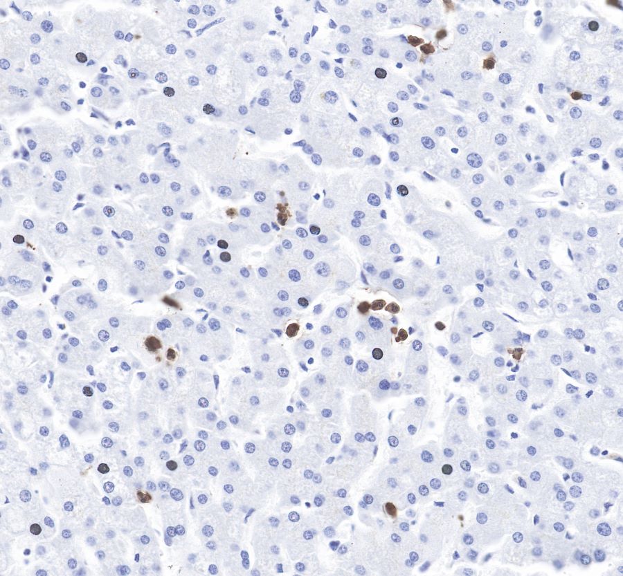
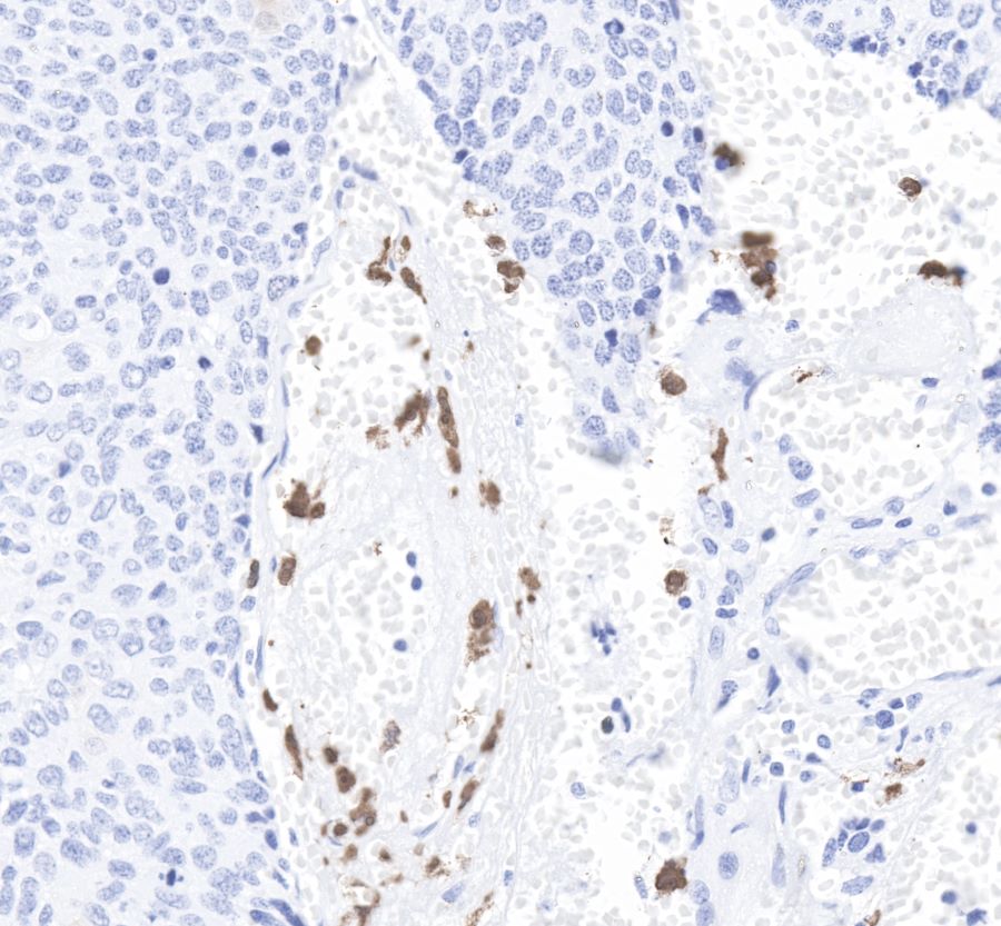
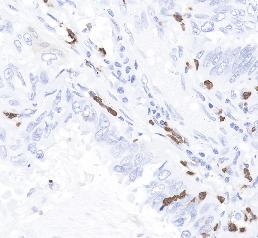
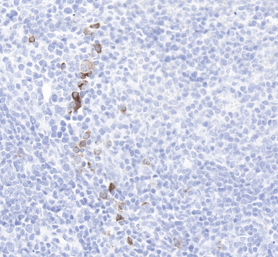
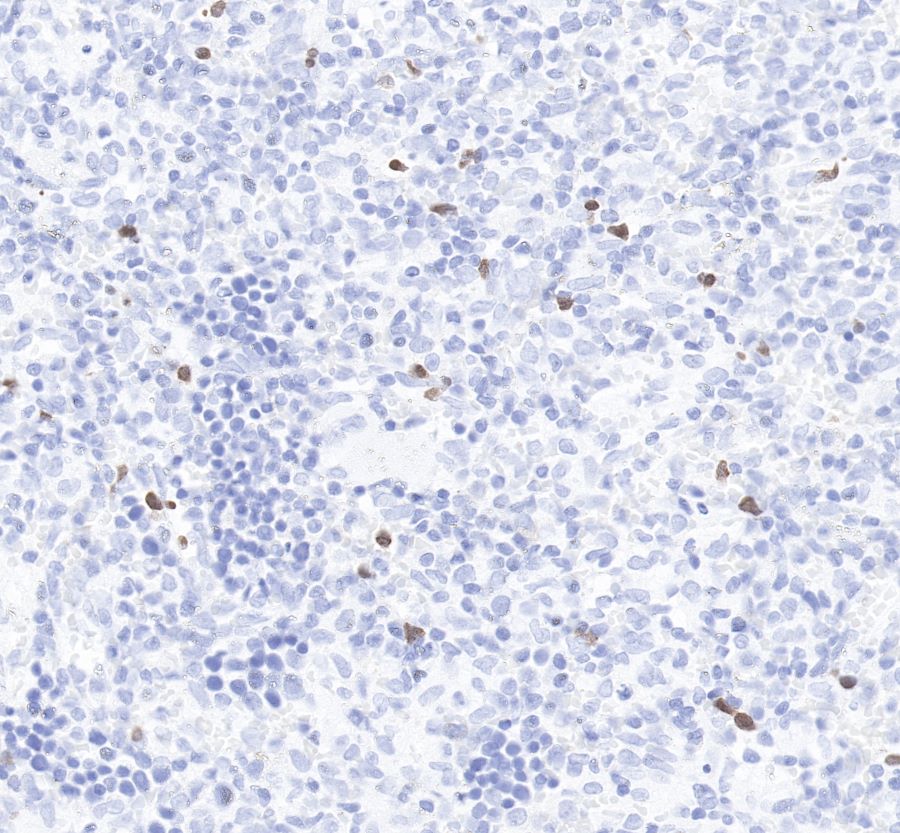
| Application |
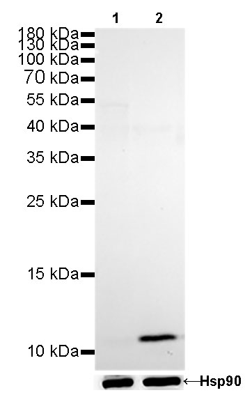
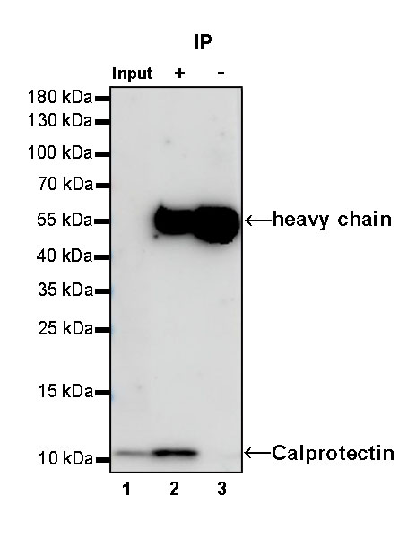
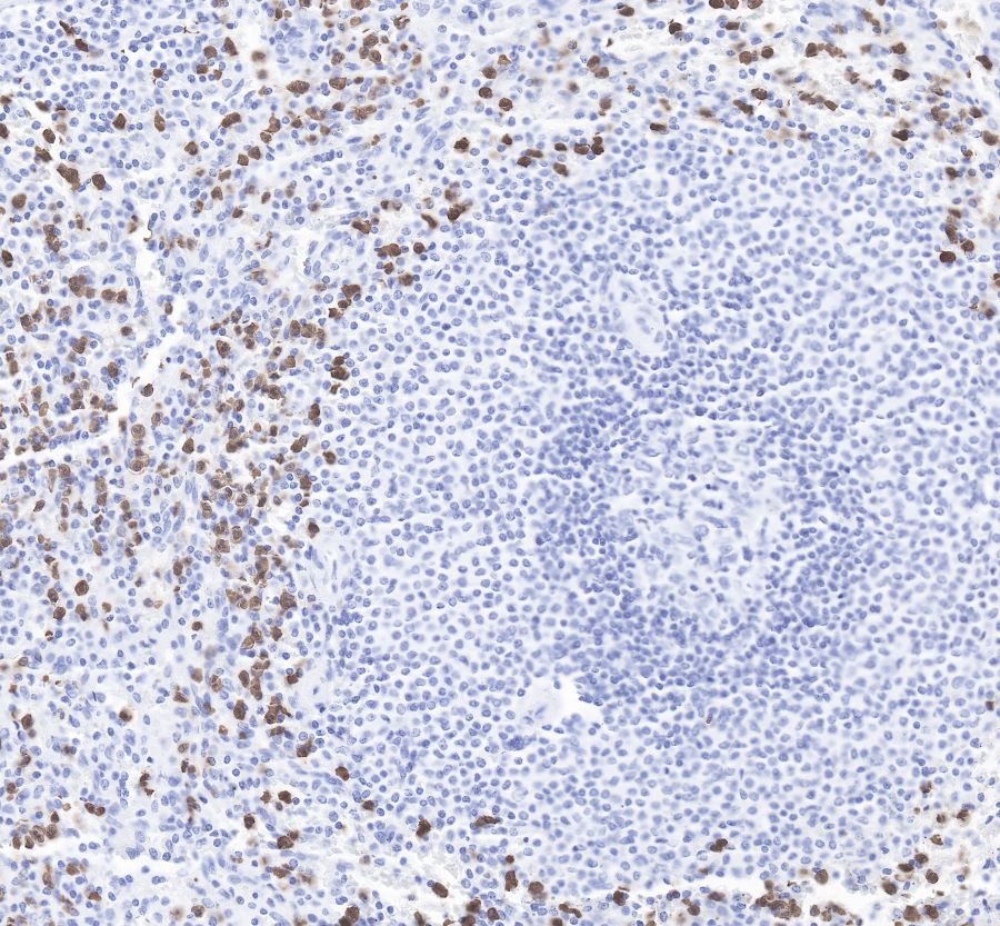
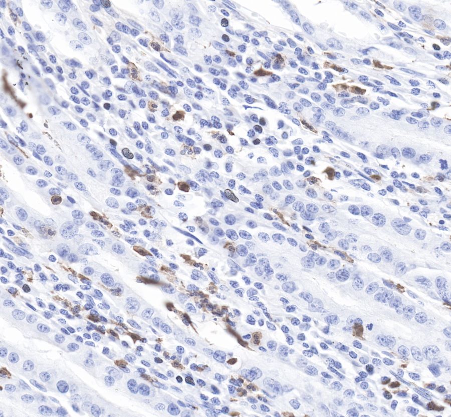
WB, IHC-P, IP |
| Reactivity |
Hu, Ms, Rt |
| Purification |
Protein A |
| Concentration |
0.1mg/ml |
| Physical Appearance |
Liquid |
| Storage Buffer |
PBS, 40% Glycerol, 0.05%BSA, 0.03% Proclin 300 |
| Stability & Storage |
12 months from date of receipt / reconstitution, -20 °C as supplied |
Dilution
| application |
dilution |
species |
| IHC-P |
1:3200 |
null |
| WB |
1:200 |
null |
| IP |
1:25 |
null |
Background
Calprotectin is a complex of the mammalian proteins S100A8 and S100A9. The proteins exist as homodimers but preferentially exist as S100A8/A9 heterodimers or heterotetramers (calprotectin) with antimicrobial, proinflammatory and prothrombotic properties. In the presence of calcium, calprotectin is capable of sequestering the transition metals iron, manganese and zinc via chelation. This metal sequestration affords the complex antimicrobial properties. Calprotectin is the only known antimicrobial manganese sequestration protein complex. Calprotectin comprises as much as 60% of the soluble protein content of the cytosol of a neutrophil, and it is secreted by an unknown mechanism during inflammation Faecal calprotectin has been used to detect intestinal inflammation (colitis or enteritis) and can serve as a biomarker for inflammatory bowel diseases. Blood based calprotectin (in serum and plasma) is used in diagnostics of multiple inflammatory diseases, including autoimmune diseases, like arthritis, and severe infections including sepsis.