WB result of c-Rel Recombinant Rabbit mAb
Primary antibody: c-Rel Recombinant mAb at 1/1000 dilution
Lane 1: RAW264.7 whole cell lysate 20 µg
Lane 2: Neuro-2a whole cell lysate 20 µg
Lane 3: mouse spleen lysate 20 µg
Lane 4: mouse thymus lysate 20 µg
Secondary antibody: Goat Anti-rabbit IgG, (H+L), HRP conjugated at 1/10000 dilution
Predicted MW: 65 kDa
Observed MW: 70 kDa
c-Rel Recombinant Rabbit mAb (S-1150-52)
c-Rel Recombinant Rabbit mAb (S-1150-52)
Price:
Regular price
$100 USD
Regular price
Sale price
$100 USD
Unit price
per
For shipping services or bulk orders, you may request a quotation.
Secure checkout with

View full details
Product Details
Product Details
Product Specification
| Host | Rabbit |
| Antigen | c-Rel |
| Synonyms | Proto-oncogene c-Rel |
| Immunogen | Synthetic Peptide |
| Location | Cytoplasm, Membrane |
| Accession | P15307 |
| Clone Number | S-1150-52 |
| Antibody Type | Recombinant mAb |
| Isotype | IgG |
| Application | WB, IHC-P, ICC, ICFCM, IP |
| Reactivity | Ms, Rt |
| Purification | Protein A |
| Concentration | 0.5 mg/ml |
| Conjugation | Unconjugated |
| Physical Appearance | Liquid |
| Storage Buffer | PBS, 40% Glycerol, 0.05% BSA, 0.03% Proclin 300 |
| Stability & Storage | 12 months from date of receipt / reconstitution, -20 °C as supplied |
Dilution
| application | dilution | species |
| WB | 1:1000 | |
| IHC-P | 1:200 | |
| ICC | 1:50 | |
| ICFCM | 1:50 | |
| IP | 1:50 |
Background
c-Rel is a member of the NF-κB family, predominantly expressed in lymphocytes and myeloid cells. As a transcription factor, it regulates the proinflammatory polarization of myeloid cells and modulates the antitumor immune response. Research has found that c-Rel serves as an important checkpoint for the immunosuppressive function of myeloid-derived suppressor cells (MDSCs). MDSCs hinder the normal function of immune cells in a tumor environment, promoting tumor immune evasion. c-Rel controls the polarization of myeloid cells in tumors, and specific inhibition of c-Rel significantly inhibits tumor growth. In c-Rel-deficient (Rel–/–) mice, melanoma and lymphoma tumor growth was significantly suppressed, with a 80% reduction in tumor size and body weight compared to controls. Inhibiting c-Rel through small molecule inhibitors or conditional knockout in MDSCs can promote the body's antitumor immune response. Combining c-Rel inhibitors with PD-1 functional blocking antibodies can further enhance the activation of the body's antitumor immune response.
Picture
Picture
Western Blot
FC
Flow cytometric analysis of 4% PFA fixed 90% methanol permeabilized Neuro-2a (Mouse neuroblastoma neuroblast) labelling c-Rel antibody at 1/50 dilution (1 μg)/ (Red) compared with a Rabbit monoclonal IgG (Black) isotype control and an unlabelled control (cells without incubation with primary antibody and secondary antibody) (Blue). Goat Anti - Rabbit IgG Alexa Fluor® 488 was used as the secondary antibody.
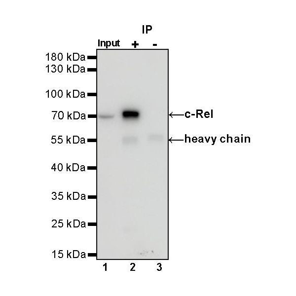
IP
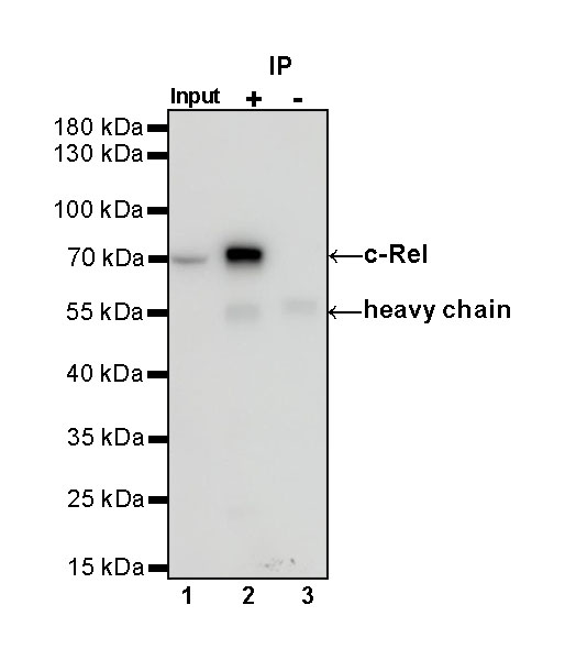
c-Rel Rabbit mAb at 1/50 dilution (1 µg) immunoprecipitating c-Rel in 0.4 mg Neuro-2a whole cell lysate.
Western blot was performed on the immunoprecipitate using c-Rel Rabbit mAb at 1/1000 dilution.
Secondary antibody (HRP) for IP was used at 1/1000 dilution.
Lane 1: Neuro-2a whole cell lysate 20 µg (Input)
Lane 2: c-Rel Rabbit mAb IP in Neuro-2a whole cell lysate
Lane 3: Rabbit monoclonal IgG IP in Neuro-2a whole cell lysate
Predicted MW: 65 kDa
Observed MW: 70 kDa
Immunohistochemistry
IHC shows positive staining in paraffin-embedded mouse spleen. Anti- c-Rel antibody was used at 1/200 dilution, followed by a HRP Polymer for Mouse & Rabbit IgG (ready to use). Counterstained with hematoxylin. Heat mediated antigen retrieval with Tris/EDTA buffer pH9.0 was performed before commencing with IHC staining protocol.
IHC shows positive staining in paraffin-embedded mouse testis. Anti- c-Rel antibody was used at 1/200 dilution, followed by a HRP Polymer for Mouse & Rabbit IgG (ready to use). Counterstained with hematoxylin. Heat mediated antigen retrieval with Tris/EDTA buffer pH9.0 was performed before commencing with IHC staining protocol.
IHC shows positive staining in paraffin-embedded rat spleen. Anti- c-Rel antibody was used at 1/200 dilution, followed by a HRP Polymer for Mouse & Rabbit IgG (ready to use). Counterstained with hematoxylin. Heat mediated antigen retrieval with Tris/EDTA buffer pH9.0 was performed before commencing with IHC staining protocol.
Immunocytochemistry
ICC shows positive staining in Neuro-2a cells. Anti- c-Rel antibody was used at 1/50 dilution (Green) and incubated overnight at 4°C. Goat polyclonal Antibody to Rabbit IgG - H&L (Alexa Fluor® 488) was used as secondary antibody at 1/1000 dilution. The cells were fixed with 100% ice-cold methanol and permeabilized with 0.1% PBS-Triton X-100. Nuclei were counterstained with DAPI (Blue). Counterstain with tubulin (Red).