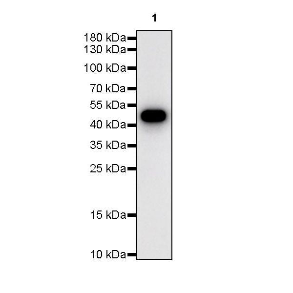
WB result of Beta II tubulin Rabbit pAb
Primary antibody: Beta II tubulin Rabbit pAb at 1/1000 dilution
Lane 1: HeLa whole cell lysate 20 µg
Lane 2: Jurkat whole cell lysate 20 µg
Secondary antibody: Goat Anti-rabbit IgG, (H+L), HRP conjugated at 1/10000 dilution
Predicted MW: 50 kDa
Observed MW: 50 kDa
Beta II tubulin Rabbit polyclonal antibody
Beta II tubulin Rabbit polyclonal antibody
Price:
Regular price
$100 USD
Regular price
Sale price
$100 USD
Unit price
per
For shipping services or bulk orders, you may request a quotation.
Secure checkout with

View full details
Product Details
Product Details
Product Specification
| Host | Rabbit |
| Synonyms | Tubulin beta-2B chain, TUBB2B |
| Immunogen | Synthetic Peptide |
| Location | Cytoplasm, Cytoskeleton |
| Accession | Q9BVA1 |
| Antibody Type | Polyclonal antibody |
| Isotype | IgG |
| Application | WB, IHC-P, ICC, ICFCM |
| Reactivity | Hu, Ms, Rt |
| Predicted Reactivity | Dr, Lob, Ar, Av, Pl, Fu, Ys, SeUr, Fs, Bv, Xe, Pg, Pz |
| Purification | Immunogen Affinity |
| Concentration | 0.5 mg/ml |
| Conjugation | Unconjugated |
| Physical Appearance | Liquid |
| Storage Buffer | PBS, 40% Glycerol, 0.05% BSA, 0.03% Proclin 300 |
| Stability & Storage | 12 months from date of receipt / reconstitution, -20 °C as supplied. |
Dilution
| application | dilution | species |
| WB | 1:1000 | |
| IHC-P | 1:500 | |
| ICFCM | 1:500 | |
| ICC | 1:500 |
Background
Beta II tubulin, also known as β tubulin protein, is a major component of microtubules, a crucial element of the cellular cytoskeleton. Microtubules are formed by the alternating arrangement of αtubulin and βtubulin proteins. Structurally, β tubulin protein mainly consists of a core β-helix domain and several auxiliary structures. The core β-helix domain serves as the primary functional region, capable of binding with αtubulin protein to form αβ-dimers, which further polymerize into microtubules. The auxiliary structures, including the variable N-terminal and C-terminal regions, contribute to the functional diversity among different β tubulin proteins. Moreover, β tubulin protein is encoded by multiple homologous gene families, such as TUBB1, TUBB2, and TUBB3. While these genes encode proteins with potential sequence variations, they share similarities in structure and function. βtubulin protein plays a pivotal role in the formation and function of microtubules. It is involved in the assembly and stabilization of microtubules, crucial for cellular structural support, intracellular transport, DNA segregation, and other vital processes.
Picture
Picture
Western Blot
WB result of Beta II tubulin Rabbit pAb
Primary antibody: Beta II tubulin Rabbit pAb at 1/1000 dilution
Lane 1: NIH/3T3 whole cell lysate 20 µg
Lane 2: mouse brain lysate 20 µg
Secondary antibody: Goat Anti-rabbit IgG, (H+L), HRP conjugated at 1/10000 dilution
Predicted MW: 50 kDa
Observed MW: 50 kDa
WB result of Beta II tubulin Rabbit pAb
Primary antibody: Beta II tubulin Rabbit pAb at 1/1000 dilution
Lane 1: C6 whole cell lysate 20 µg
Lane 2: rat brain lysate 20 µg
Secondary antibody: Goat Anti-rabbit IgG, (H+L), HRP conjugated at 1/10000 dilution
Predicted MW: 50 kDa
Observed MW: 50 kDa
WB result of Beta II tubulin Rabbit pAb
Primary antibody : Beta II tubulin Rabbit pAb at 1/1000 dilution
Lane 1 : Zebra fish lysate 20 µg
Secondary antibody: Goat Anti-Rabbit IgG, (H+L), HRP conjugated at 1/10000 dilution
Predicted MW: 50 kDa
Observed MW: 50 kDa
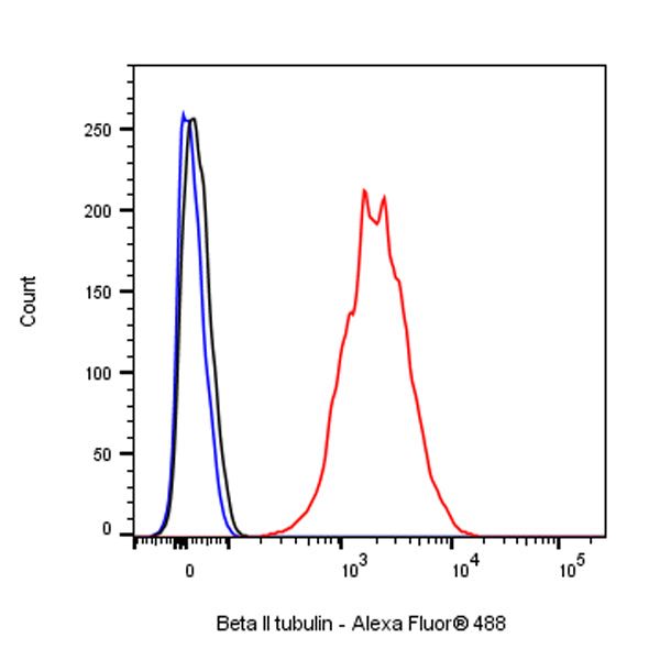
FC
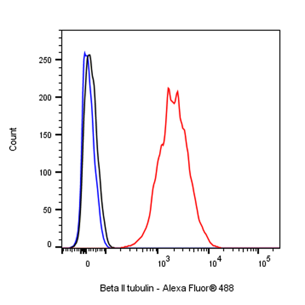
Flow cytometric analysis of 4% PFA fixed 90% methanol permeabilized HeLa (Human cervix adenocarcinoma epithelial cell) labelling Beta II tubulin antibody at 1/500 dilution (0.1 μg)/ (Red) compared with a Rabbit monoclonal IgG (Black) isotype control and an unlabelled control (cells without incubation with primary antibody and secondary antibody) (Blue). Goat Anti - Rabbit IgG Alexa Fluor® 488 was used as the secondary antibody.
Immunohistochemistry
IHC shows positive staining in paraffin-embedded human cerebral cortex. Anti-Beta II tubulin antibody was used at 1/500 dilution, followed by a HRP Polymer for Mouse & Rabbit IgG (ready to use). Counterstained with hematoxylin. Heat mediated antigen retrieval with Tris/EDTA buffer pH9.0 was performed before commencing with IHC staining protocol.
IHC shows positive staining in paraffin-embedded human kidney. Anti-Beta II tubulin antibody was used at 1/500 dilution, followed by a HRP Polymer for Mouse & Rabbit IgG (ready to use). Counterstained with hematoxylin. Heat mediated antigen retrieval with Tris/EDTA buffer pH9.0 was performed before commencing with IHC staining protocol.
IHC shows positive staining in paraffin-embedded rat cerebral cortex. Anti-Beta II tubulin antibody was used at 1/500 dilution, followed by a HRP Polymer for Mouse & Rabbit IgG (ready to use). Counterstained with hematoxylin. Heat mediated antigen retrieval with Tris/EDTA buffer pH9.0 was performed before commencing with IHC staining protocol.
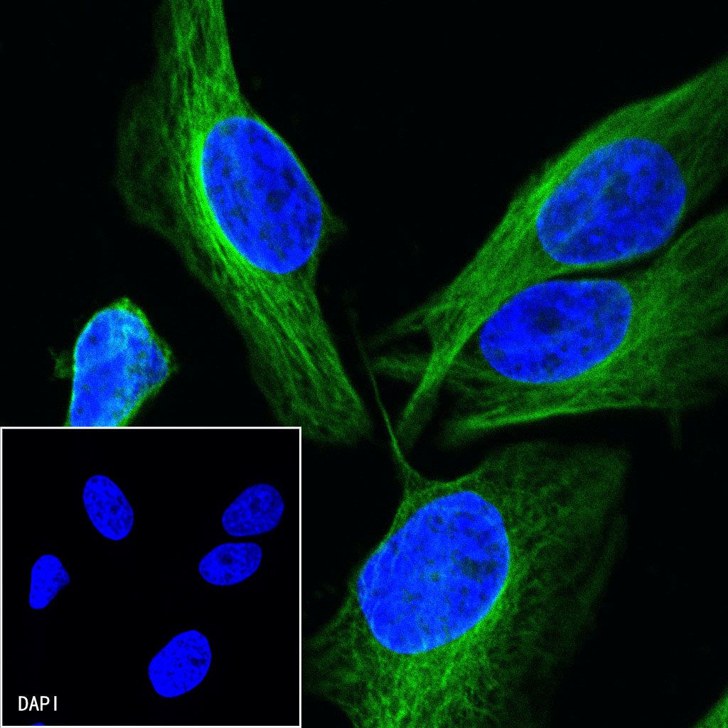
Immunocytochemistry

ICC shows positive staining in HeLa cells. Anti-Beta II tubulin antibody was used at 1/500 dilution (Green) and incubated overnight at 4°C. Goat polyclonal Antibody to Rabbit IgG - H&L (Alexa Fluor® 488) was used as secondary antibody at 1/1000 dilution. The cells were fixed with 4% PFA and permeabilized with 0.1% PBS-Triton X-100. Nuclei were counterstained with DAPI (Blue).

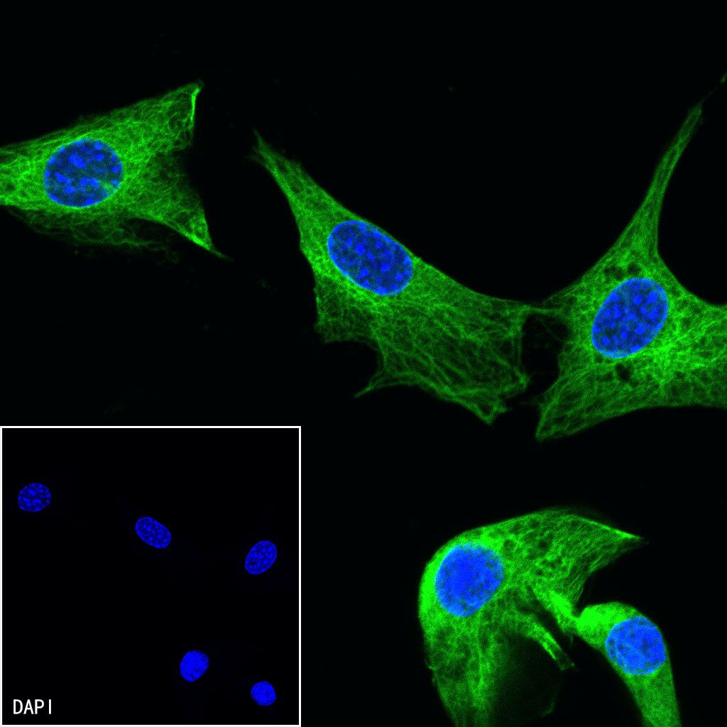
ICC shows positive staining in NIH/3T3 cells. Anti-Beta II tubulin antibody was used at 1/500 dilution (Green) and incubated overnight at 4°C. Goat polyclonal Antibody to Rabbit IgG - H&L (Alexa Fluor® 488) was used as secondary antibody at 1/1000 dilution. The cells were fixed with 4% PFA and permeabilized with 0.1% PBS-Triton X-100. Nuclei were counterstained with DAPI (Blue).