Product Specification
| Host |
Rabbit |
| Antigen |
Aurora-B |
| Synonyms |
Aurora 1, AIM-1, ARK-2, STK-1, Aurora-related kinase 2 |
| Immunogen |
Synthetic Peptide |
| Accession |
Q96GD4 |
| Clone Number |
SDT-079-32 |
| Antibody Type |
Rabbit mAb |
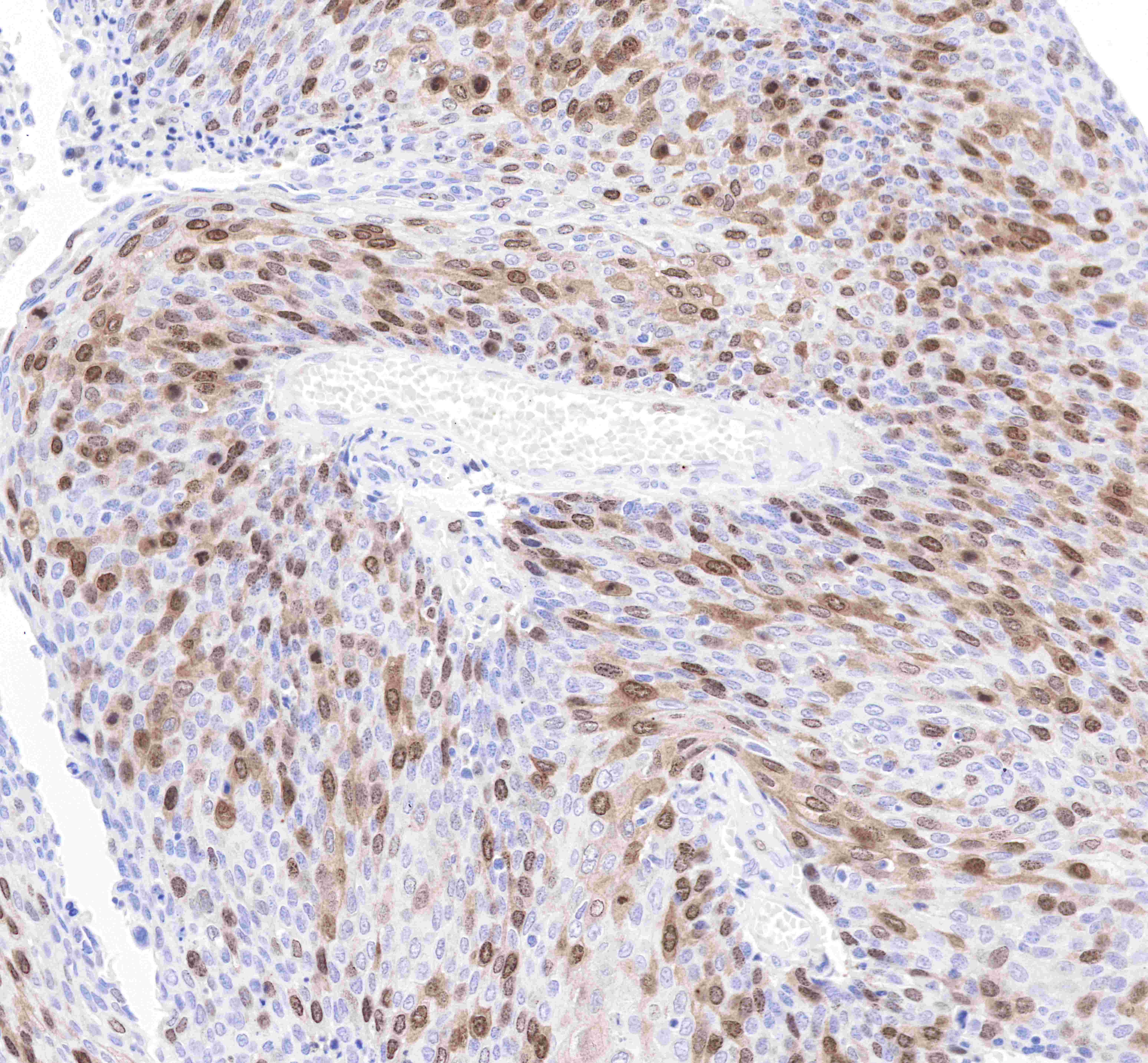
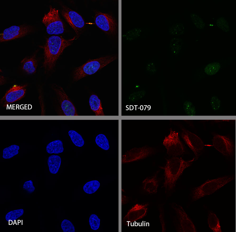
| Application |
WB, IHC-P, ICC, ICFCM, IP |
| Reactivity |
Hu |
| Purification |
Protein A |
| Concentration |
0.5mg/ml |
| Conjugation |
Unconjugated |
| Physical Appearance |
Liquid |
| Storage Buffer |
PBS, 40% Glycerol, 0.05%BSA, 0.03% Proclin 300 |
| Stability & Storage |
12 months from date of receipt / reconstitution, -20 °C as supplied |
Dilution
| application |
dilution |
species |
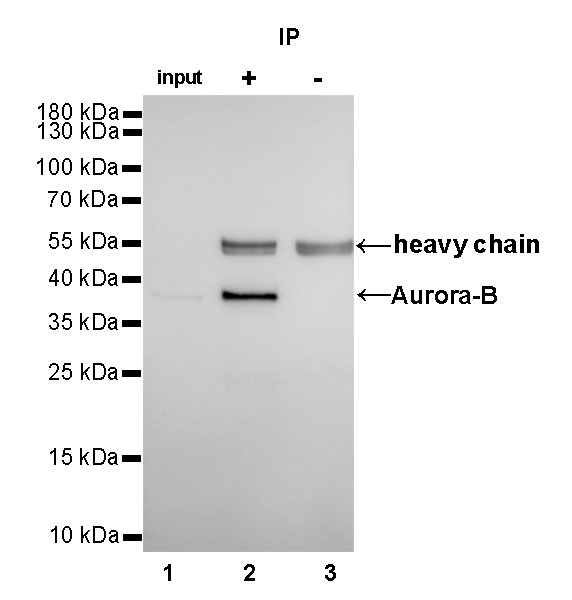
| IP |
1:25 |
|
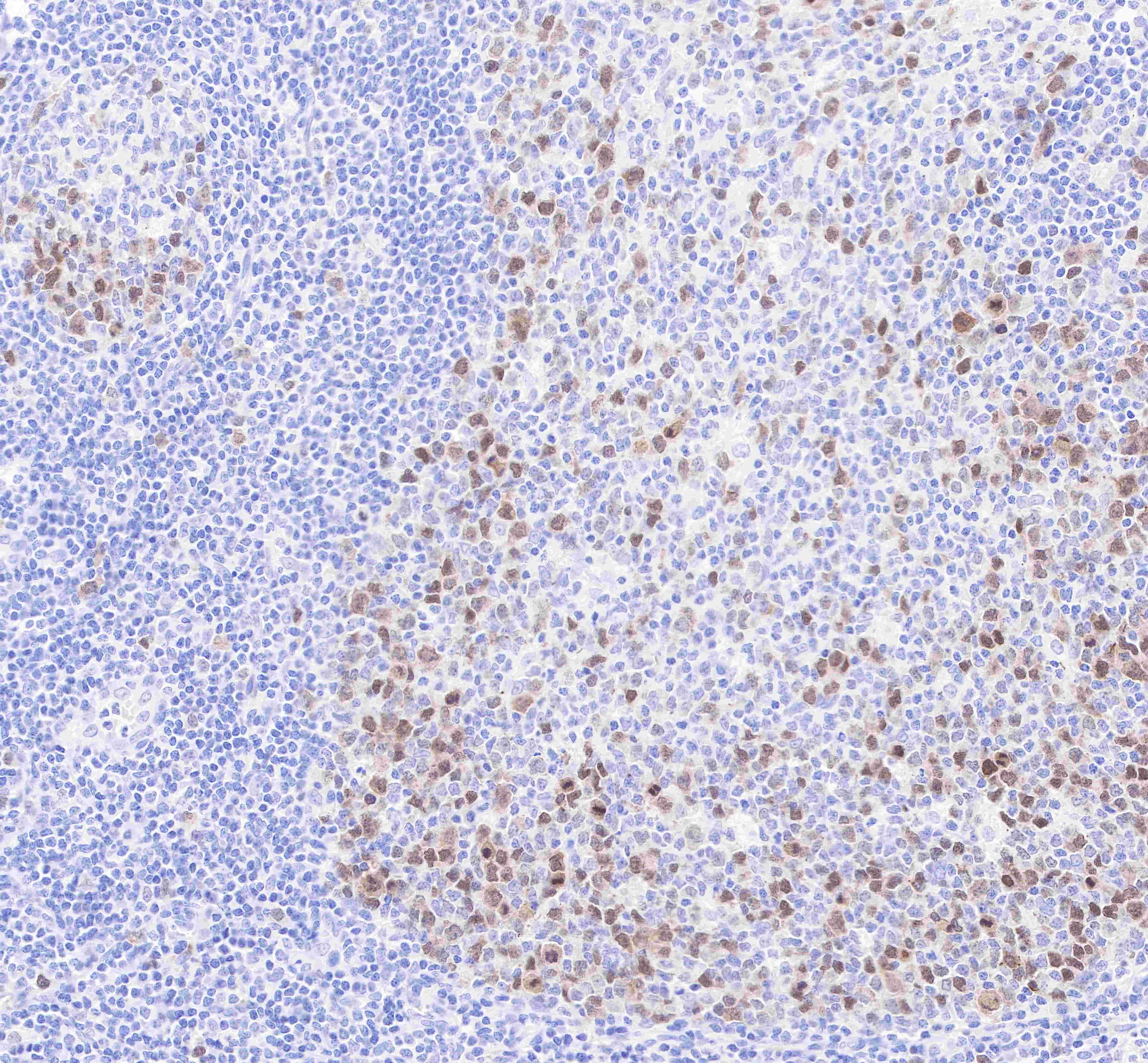
| IHC-P |
1:500-2000 |
|
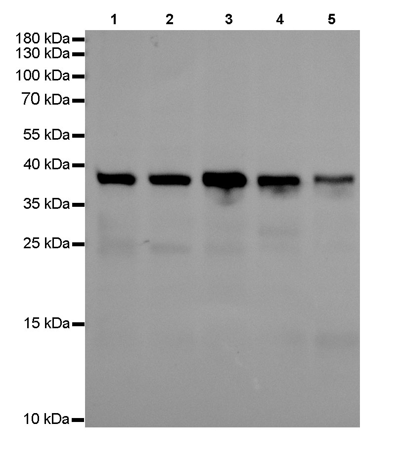
| WB |
1:1000 |
|
| ICC |
1:500 |
|
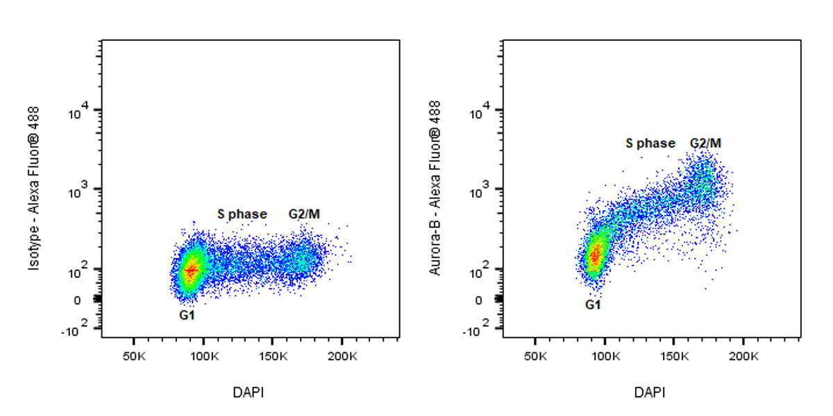
| ICFCM |
1:5000 |
|
Background
Aurora kinase B is a protein that functions in the attachment of the mitotic spindle to the centromere. The expression and activity of Aurora B are regulated according to the cell cycle. Expression of Aurora B reaches a maximum at the G2-M transition, whereas Aurora B protein is most active during mitosis. The Aurora kinases associate with microtubules during chromosome movement and segregation. Aurora kinase B localizes to microtubules near kinetochores, specifically to the specialized microtubules called K-fibers, and Aurora kinase A localizes to centrosomes. In cancerous cells, over-expression of these enzymes causes unequal distribution of genetic information, creating aneuploid cells, a hallmark of cancer.