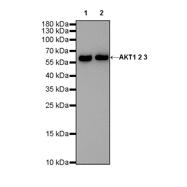
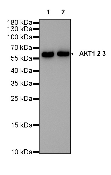
WB result of Akt (pan) Rabbit mAb
Primary antibody: Akt (pan) Rabbit mAb at 1/1000 dilution
Lane 1: Jurkat whole cell lysate 20 µg
Lane 2: MCF7 whole cell lysate 20 µg
Secondary antibody: Goat Anti-Rabbit IgG, (H+L), HRP conjugated at 1/10000 dilution
Predicted MW: 60kDa
Observed MW: 60kDa
Akt (pan) Recombinant Rabbit mAb (SDT-R169)
Akt (pan) Recombinant Rabbit mAb (SDT-R169)
Price:
Regular price
$100 USD
Regular price
Sale price
$100 USD
Unit price
per
For shipping services or bulk orders, you may request a quotation.
Secure checkout with

View full details
Product Details
Product Details
Product Specification
| Host | Rabbit |
| Antigen | AKT1 + AKT2 + AKT3 |
| Synonyms | N/A |
| Immunogen | N/A |
| Location | Cytoplasm, Nucleus, Cell membrane |
| Accession | P31749、 Q9Y243、P31751 |
| Clone Number | SDT-R169 |
| Antibody Type | Recombinant mAb |
| Application | WB, IHC-P, IP |
| Reactivity | Hu, Ms, Rt |
| Purification | Protein A |
| Concentration | 0.5 mg/ml |
| Tag | N/A |
| Physical Appearance | Liquid |
| Storage Buffer | PBS, 40% Glycerol, 0.05% BSA, 0.03% Proclin 300 |
| Stability & Storage | 12 months from date of receipt / reconstitution, -20 °C as supplied |
Dilution
| application | dilution | species |
| WB | 1:1000 | null |
| IHC-P | 1:500-1:2000 | null |
| IP | 1:50 | null |
Background
Akt is a family of three serine-threonine kinases, Akt1, Akt2, and Akt3 [PMID: 28115590]. Akt is a serine/threonine kinase and it participates in the key role of the PI3K signaling pathway. The Akt can be activated by a wide range of growth signals and the biochemical mechanisms leading to Akt activation are well defined [PMID: 31173856].
Picture
Picture
Western Blot

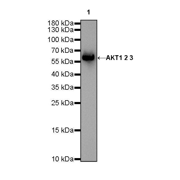
WB result of Akt (pan) Rabbit mAb
Primary antibody: Akt (pan) Rabbit mAb at 1/1000 dilution
Lane 1: NIH/3T3 whole cell lysate 20 µg
Secondary antibody: Goat Anti-Rabbit IgG, (H+L), HRP conjugated at 1/10000 dilution
Predicted MW: 60kDa
Observed MW: 60kDa
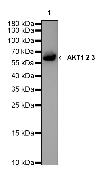
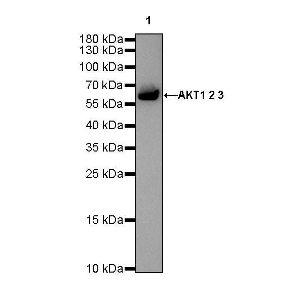
WB result of Akt (pan) Rabbit mAb
Primary antibody: Akt (pan) Rabbit mAb at 1/1000 dilution
Lane 1: C6 whole cell lysate 20 µg
Secondary antibody: Goat Anti-Rabbit IgG, (H+L), HRP conjugated at 1/10000 dilution
Predicted MW: 60kDa
Observed MW: 60kDa
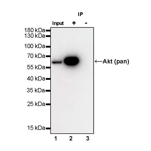
IP
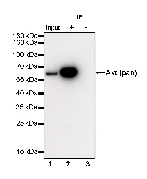
Akt (pan) Rabbit mAb at 1/50 dilution (1 µg) immunoprecipitating Akt (pan) in 0.4 mg MCF-7 whole cell lysate.
Western blot was performed on the immunoprecipitate using Akt (pan) Rabbit mAb at 1/1000 dilution.
Secondary antibody (HRP) for IP was used at 1/400 dilution.
Lane 1: MCF-7 whole cell lysate 20 µg (Input)
Lane 2: Akt (pan) Rabbit mAb IP in MCF-7 whole cell lysate
Lane 3: Rabbit monoclonal IgG IP in MCF-7 whole cell lysate
Predicted MW: 60 kDa
Observed MW: 60 kDa
Immunohistochemistry

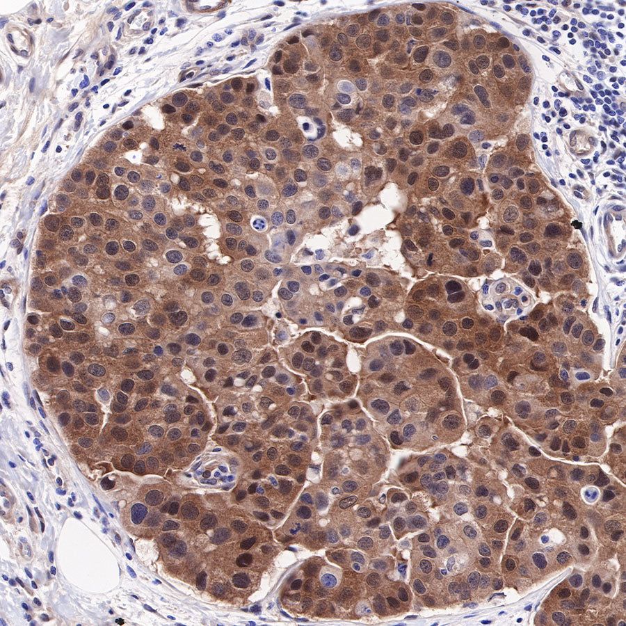
IHC shows positive staining in paraffin-embedded human breast cancer. Anti-Akt (pan) antibody was used at 1/2000 dilution, followed by a HRP Polymer for Mouse & Rabbit IgG (ready to use). Counterstained with hematoxylin. Heat mediated antigen retrieval with Tris/EDTA buffer pH9.0 was performed before commencing with IHC staining protocol.
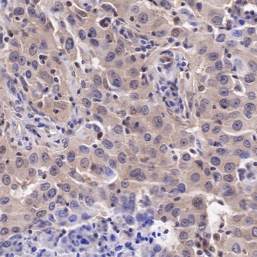
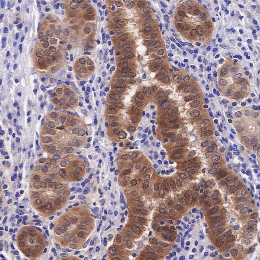
IHC shows positive staining in paraffin-embedded human lung adenocarcinoma. Anti-Akt (pan) antibody was used at 1/500 dilution, followed by a HRP Polymer for Mouse & Rabbit IgG (ready to use). Counterstained with hematoxylin. Heat mediated antigen retrieval with Tris/EDTA buffer pH9.0 was performed before commencing with IHC staining protocol.
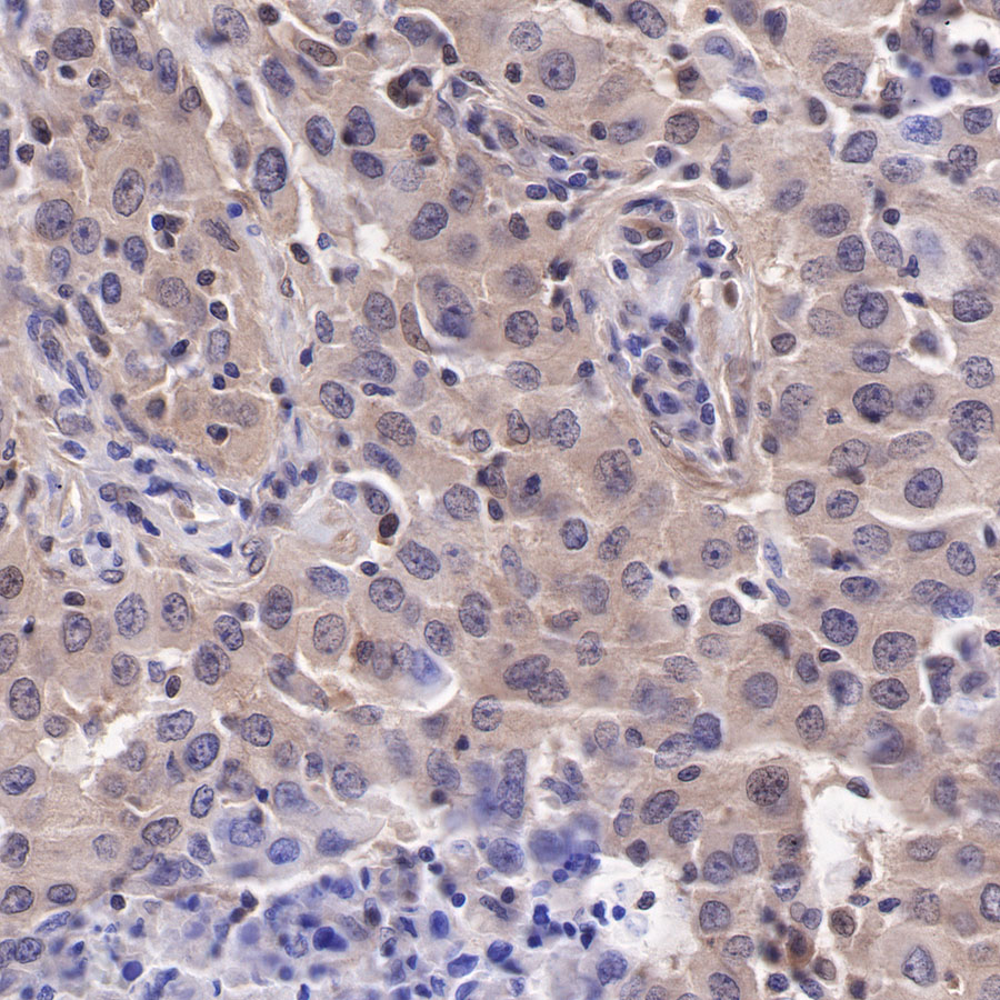
IHC shows positive staining in paraffin-embedded human thyroid cancer. Anti-Akt (pan) antibody was used at 1/2000 dilution, followed by a HRP Polymer for Mouse & Rabbit IgG (ready to use). Counterstained with hematoxylin. Heat mediated antigen retrieval with Tris/EDTA buffer pH9.0 was performed before commencing with IHC staining protocol.