Storage Stability Test: A549 cells at a concentration of 1×10^5/mL were seeded into a 96-well white-bottom transparent culture plate, 100 µL per well. After 24 hours, the plates were equilibrated to room temperature, and UA-Glo cell viability assay reagent stored at room temperature for varying durations was added at 50 µL per well, followed by mixing. Fluorescence readings were taken after 10 minutes. The same batch of product stored at −20°C for 0 hours served as the control, and the relative fluorescence value percentage was calculated.
UA-Glo® Luminescent Cell Viability Assay
UA-Glo® Luminescent Cell Viability Assay
Price:
Regular price
$45 USD
Regular price
Sale price
$45 USD
Unit price
per
For shipping services or bulk orders, you may request a quotation.
Secure checkout with

View full details
Product Details
Product Details
Product Specification
| Synonyms | 细胞活力检测试剂盒 |
| Stability & Storage | Store at room temperature (22–25°C) for 48 hours, at 4°C for 1 week, at -20°C for 3 months, or at -80°C or below for 12 months; luciferase retains >90% activity. |
Background
The UA-Glo homogeneous ready-to-use cell viability assay kit is designed for quantitatively measuring ATP levels in live cells, thereby indirectly determining the number of viable cells. This reagent features high signal-to-noise ratio, excellent reproducibility, and outstanding stability. Its ready-to-use formulation eliminates the need for separate lysis and detection steps, reducing errors caused by frequent pipetting. The stable glow-type signal makes this product particularly suitable for high-throughput tumor cell proliferation assays and compound screening.
Components
The product is a mixture of luciferase, luciferin, and buffer solution, packaged in 10 ml or 100 ml brown bottles.
Product Specification |
Number of detectable 96-well plate wells |
Number of detectable 384-well plate wells |
1 x 10 ml |
200 |
1,000 |
1 x 100 ml |
2,000 |
10,000 |
10 x 100 ml |
20,000 |
100,000 |
Protocol
1. Cell Preparation 1) Seed the cells to be tested at an appropriate density in a 96-well or 384-well cell culture plate. A white plate is recommended. 2) Prepare the test compounds. Add the test compounds at appropriate concentrations to the wells of the cell plate. Keep the organic solvent concentration in the culture medium below 1-2%. Establish a control group without compounds. Continue incubation for an appropriate duration based on project requirements. 2. Cell Viability Assay 1) Take out the UA-Glo cell viability assay reagent and equilibrate at room temperature for 20 minutes. Gently shake to mix. 2) Take out the cell culture plate to be tested and equilibrate at room temperature for 20 minutes. 3) Add 50 µl of the assay reagent to 100 µl of cells in a 96-well plate, or 10 µl of reagent to 20 µl of cells in a 384-well plate. Gently shake for 2 minutes, then incubate in the dark for an additional 10 minutes for lysis. 4) Read the luminescence signal on a luminescence plate reader.
Guidelines
1. Temperature: Unless otherwise specified, the luciferase assay is performed at room temperature (22-25°C). Temperature significantly affects enzymatic reactions.
2. Reagent Volume: Unless rigorously validated, it is not recommended to arbitrarily alter the reaction reagent volumes.
3. Plate Reading Time: After adding reagents, shake for 2 minutes, incubate in the dark for 10 minutes, then read the plate. It is advisable to complete the reading within two hours after reagent addition.
4. Inter-Plate Controls: If inter-plate data comparison is required, it is recommended to include two wells of positive controls (without compounds) on all reaction plates as inter-plate controls. Normalize the plate readings using these controls before downstream data analysis.
5. Reaction Plate: High fluorescence readings may cause significant interference (e.g., signal bleed-through) to adjacent wells. If necessary, use opaque white or black plates for experiments or validation.
6. Application: This product is designed for live-cell assays. If used in cell-free systems with recombinant proteins, additional ATP must be supplied.
7. Reagent Mixing: Avoid mixing reagents from different batches, partially used but stored reagents, or reagents with significantly different storage conditions.
8. Storage: Aliquot and store reaction reagents as instructed to ensure stability.
Picture
Picture
Bioactivity
Sensitivity test: HEK293 cells were seeded in a 96-well clear-bottom white plate at two-fold serial dilutions, 100 µL per well. After 24 hours, the plates were equilibrated to room temperature, and UA-Glo cell viability assay reagent, also equilibrated to room temperature, was added at 50 µL per well and mixed thoroughly. Fluorescence readings were taken after 10 minutes. A strong linear relationship was observed between the fluorescence signal and the number of cells (r² > 0.999), and as few as 100 cells per well could be detected.
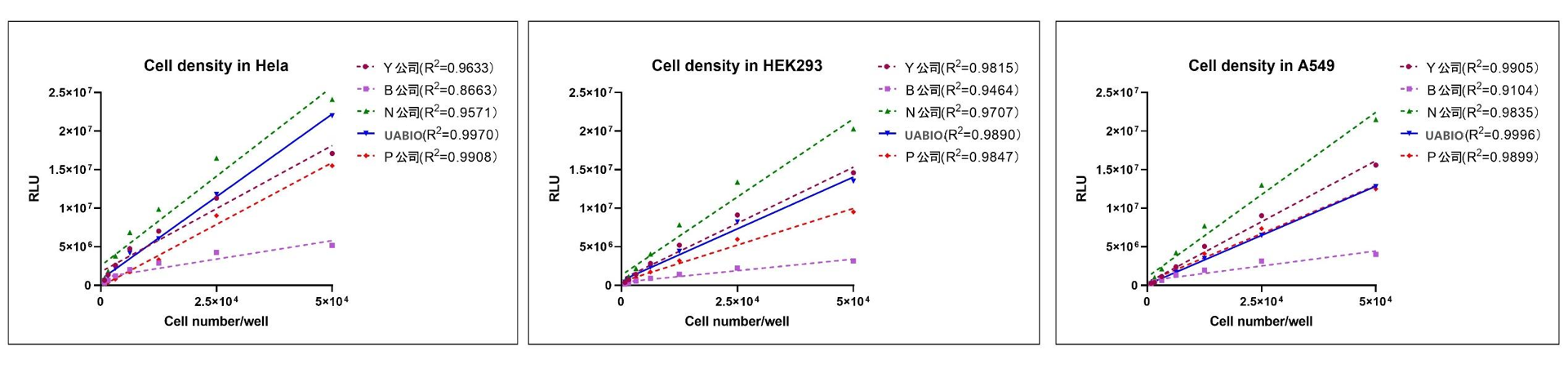
Linear comparison with similar competitive products: Hela cells, HEK293 cells, and A549 cells were counted using a hemocytometer, and their concentrations were adjusted to 5 × 10^5 cells/mL. Cells subjected to two-fold serial dilutions were each seeded into 96-well clear-bottom white plates at 100 µL per well. UA-Glo cell viability assay reagent, a domestic comparable product, and a foreign comparable product (Company P), all equilibrated to room temperature, were added at 50 µL per well and gently mixed. Fluorescence readings were taken after 10 minutes. The results showed that, for all three cell lines, the UA-Glo cell viability assay reagent (Neubo) exhibited the best linearity.
Assessment of the activity of the compound Staurosporine in different tumor cell lines: A549, HeLa, MCF-7, and HCT116 cells were seeded at 1–5 × 10^4 cells/mL into 384-well white-bottom clear-bottom plates, 20 µL per well. After 24 hours, Staurosporine at various concentrations was added, and the cultures were incubated at 37°C for an additional 72 hours. The cells were then brought to room temperature, followed by the addition of UA-Glo Cell Viability Assay reagent equilibrated to room temperature, 10 µL per well, and mixed thoroughly. Fluorescence readings were taken after 10 minutes. The IC50 values of Staurosporine in A549, HeLa, MCF-7, and HCT116 cells were 2.9 nM, 5.4 nM, 16.5 nM, and 33 nM, respectively.
*: In the Z' experiment, cells of each type were seeded at a specified concentration into a 384-well white-bottom transparent plate, 20 µL per well. In experiments 1, 2, and 3, 24 hours after cell plating, 1 µM staurosporine (with a final DMSO concentration of 0.5%) was added to the left half of the plate, while the right half received 0.5% DMSO. In experiments 4 and 5, 24 hours after cell plating, 1 µM staurosporine (with a final DMSO concentration of 0.5%) was added to the right half of the plate, and the left half received 0.5% DMSO. Seventy-two hours after adding staurosporine and DMSO, the cells were brought to room temperature, and UA-Glo cell viability reagent, equilibrated to room temperature, was added at 10 µL per well and mixed thoroughly. Fluorescence readings were taken after 10 minutes.
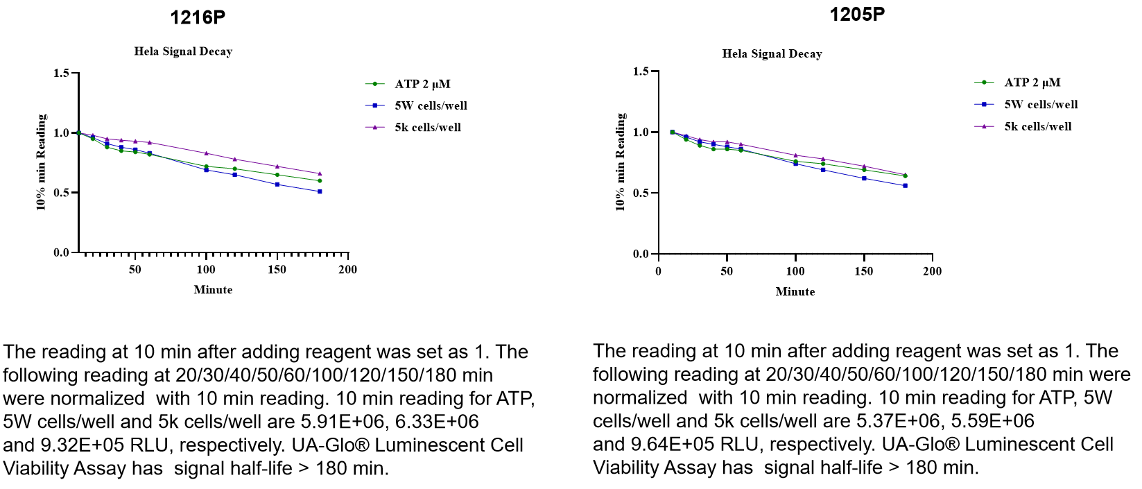
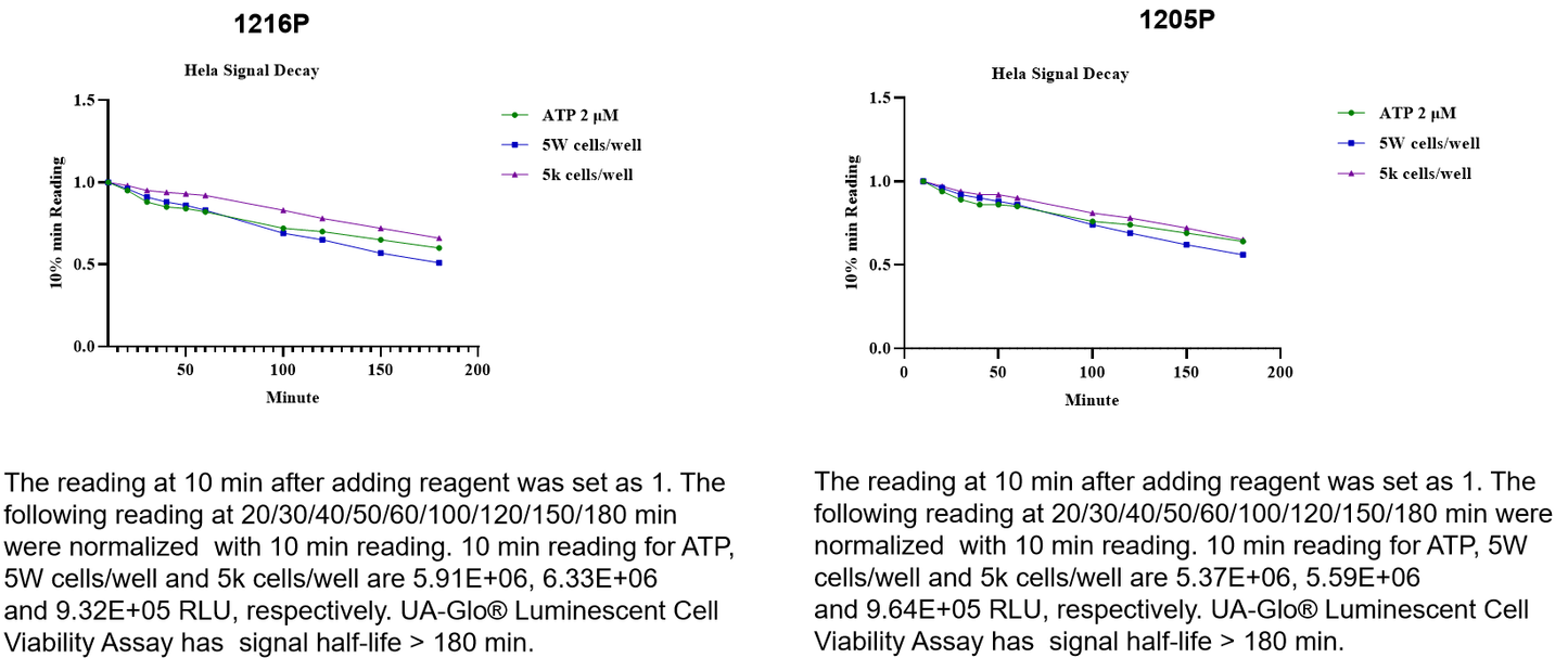
Stability test under 22℃ conditions