Product Specification
| Host |
Rabbit |
| Antigen |
TCL1 |
| Synonyms |
Oncogene TCL-1, Protein p14 TCL1 |
| Immunogen |
Synthetic Peptide |
| Location |
Cytoplasm, Nucleus |
| Accession |
P56279 |
| Clone Number |
SDT-052-12 |
| Antibody Type |
Rabbit mAb |
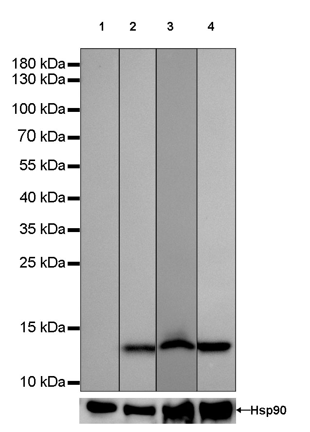
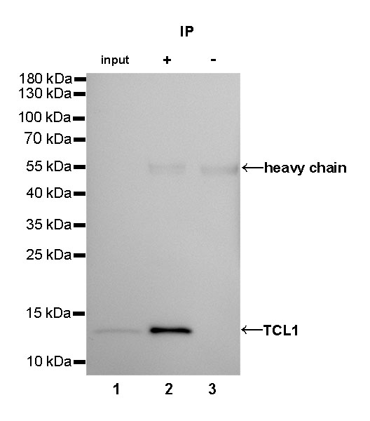
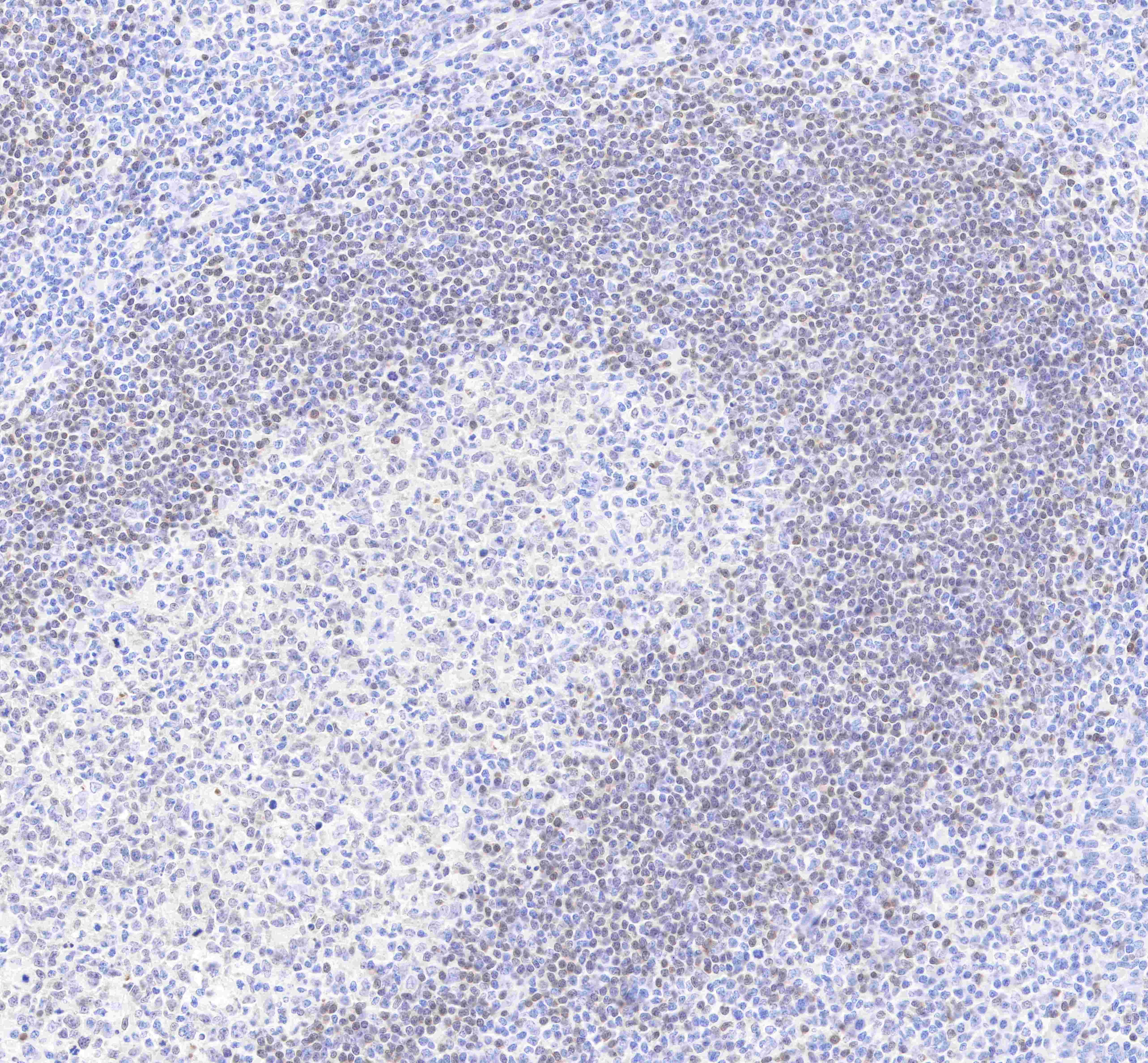
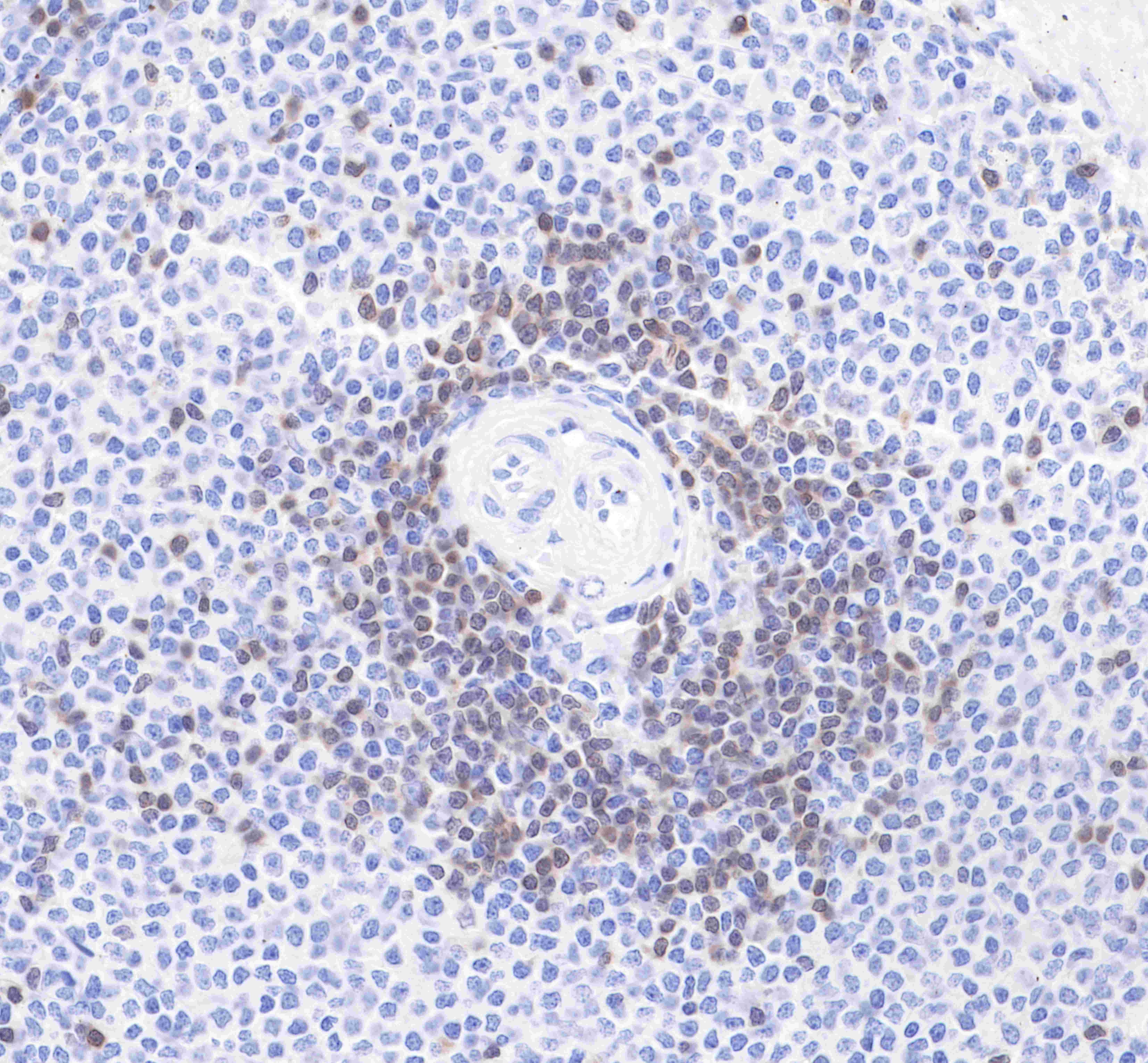
| Application |
WB, IHC-P, IP |
| Reactivity |
Hu |
| Purification |
Protein A |
| Concentration |
0.5mg/ml |
| Conjugation |
Unconjugated |
| Physical Appearance |
Liquid |
| Storage Buffer |
PBS, 40% Glycerol, 0.05%BSA, 0.03% Proclin 300 |
| Stability & Storage |
12 months from date of receipt / reconstitution, -20 °C as supplied |
Dilution
| application |
dilution |
species |
| IHC-P |
1:2000 |
|
| WB |
1:1000 |
|
| IP |
1:25 |
|
Background
T-cell leukemia/lymphoma protein 1A is a protein that in humans is encoded by the TCL1A gene. The TCL1 locus on human chromosome 14q32.1 is activated in T-cell leukemias by translocations and inversions that juxtapose it to regulatory elements of T-cell receptor genes. TCL1A has been shown to interact with AKT1 and AKT2. Protein expression in immature T and B lymphoid cells and overexpression of TCL1 is considered critical in the oncogenetic transformation.