WB result of Pan Trk Rabbit mAb
Primary antibody: Pan Trk Rabbit mAb at 1/1000 dilution
Lane 1: mouse kidney lysate 20 µg
Lane 2: mouse brain lysate 20 µg
Negative control: mouse kidney lysate
Secondary antibody: Goat Anti-Rabbit IgG, (H+L), HRP conjugated at 1/10000 dilution
Predicted MW: 87 kDa
Observed MW: 120 kDa
S-RMab® Pan Trk Recombinant Rabbit mAb (SDT-512-5)
S-RMab® Pan Trk Recombinant Rabbit mAb (SDT-512-5)
Price:
Regular price
$100 USD
Regular price
Sale price
$100 USD
Unit price
per
For shipping services or bulk orders, you may request a quotation.
Secure checkout with

View full details
Product Details
Product Details
Product Specification
| Host | Rabbit |
| Antigen | Pan Trk |
| Synonyms | Trk A + Trk B + Trk C |
| Immunogen | Synthetic Peptide |
| Location | Cytoplasm, Cell membrane |
| Accession | Q16620 |
| Clone Number | SDT-512-5 |
| Antibody Type | Recombinant mAb |
| Application | WB, IHC-P, ICC, IP |
| Reactivity | Hu, Ms, Rt |
| Predicted Reactivity | Av |
| Purification | Protein A |
| Concentration | 0.5 mg/ml |
| Conjugation | Unconjugated |
| Physical Appearance | Liquid |
| Storage Buffer | PBS, 40% Glycerol, 0.05%BSA, 0.03% Proclin 300 |
| Stability & Storage | 12 months from date of receipt / reconstitution, -20 °C as supplied |
Dilution
| application | dilution | species |
| WB | 1:1000 | |
| IP | 1:50 | |
| IHC | 1:500 | |
| ICC | 1:50 |
Background
Neurotrophic tyrosine kinase receptor (NTRK) is a family of 3 proto-oncogenes including NTRK1, NTRK2, and NTRK3 which encode Trk A, Trk B, and Trk C proteins, respectively [PMID: 28719467]. Pan-TRK antibodies have been used to detect gene fusions in diverse types of tumors [PMID: 37017998].
Picture
Picture
Western Blot
WB result of Pan Trk Rabbit mAb
Primary antibody: Pan Trk Rabbit mAb at 1/1000 dilution
Lane 1: rat kidney lysate 20 µg
Lane 2: rat brain lysate 20 µg
Negative control: rat kidney lysate
Secondary antibody: Goat Anti-Rabbit IgG, (H+L), HRP conjugated at 1/10000 dilution
Predicted MW: 87 kDa
Observed MW: 120~140 kDa
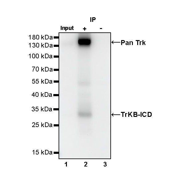
IP
Pan Trk Rabbit mAb at 1/50 dilution (1 µg) immunoprecipitating Pan Trk in 0.4 mg mouse brain lysate.
Western blot was performed on the immunoprecipitate using Pan Trk Rabbit mAb at 1/1000 dilution.
Secondary antibody (HRP) for IP was used at 1/400 dilution.
Lane 1: mouse brain lysate 20 µg (Input)
Lane 2: Pan Trk Rabbit mAb IP in mouse brain lysate
Lane 3: Rabbit monoclonal IgG IP in mouse brain lysate
Predicted MW: 87 kDa
Observed MW: 120 kDa
Immunohistochemistry
IHC shows positive staining in paraffin-embedded human cerebral cortex. Anti-Pan Trk antibody was used at 1/500 dilution, followed by a HRP Polymer for Mouse & Rabbit IgG (ready to use). Counterstained with hematoxylin. Heat mediated antigen retrieval with Tris/EDTA buffer pH9.0 was performed before commencing with IHC staining protocol.
Negative control: IHC shows negative staining in paraffin-embedded human liver. Anti- Pan Trk antibody was used at 1/500 dilution, followed by a HRP Polymer for Mouse & Rabbit IgG (ready to use). Counterstained with hematoxylin. Heat mediated antigen retrieval with Tris/EDTA buffer pH9.0 was performed before commencing with IHC staining protocol.
IHC shows positive staining in paraffin-embedded human diffuse astrocytoma. Anti-Pan Trk antibody was used at 1/500 dilution, followed by a HRP Polymer for Mouse & Rabbit IgG (ready to use). Counterstained with hematoxylin. Heat mediated antigen retrieval with Tris/EDTA buffer pH9.0 was performed before commencing with IHC staining protocol.
IHC shows positive staining in paraffin-embedded mouse cerebral cortex. Anti-Pan Trk antibody was used at 1/500 dilution, followed by a HRP Polymer for Mouse & Rabbit IgG (ready to use). Counterstained with hematoxylin. Heat mediated antigen retrieval with Tris/EDTA buffer pH9.0 was performed before commencing with IHC staining protocol.
Negative control: IHC shows negative staining in paraffin-embedded mouse liver. Anti- Pan Trk antibody was used at 1/500 dilution, followed by a HRP Polymer for Mouse & Rabbit IgG (ready to use). Counterstained with hematoxylin. Heat mediated antigen retrieval with Tris/EDTA buffer pH9.0 was performed before commencing with IHC staining protocol.
IHC shows positive staining in paraffin-embedded rat cerebral cortex. Anti-Pan Trk antibody was used at 1/500 dilution, followed by a HRP Polymer for Mouse & Rabbit IgG (ready to use). Counterstained with hematoxylin. Heat mediated antigen retrieval with Tris/EDTA buffer pH9.0 was performed before commencing with IHC staining protocol.
Negative control: IHC shows negative staining in paraffin-embedded rat liver. Anti- Pan Trk antibody was used at 1/500 dilution, followed by a HRP Polymer for Mouse & Rabbit IgG (ready to use). Counterstained with hematoxylin. Heat mediated antigen retrieval with Tris/EDTA buffer pH9.0 was performed before commencing with IHC staining protocol.
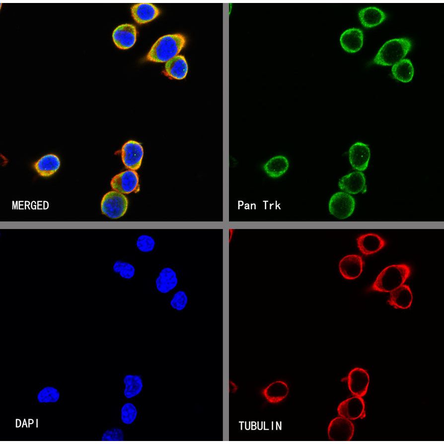
Immunocytochemistry
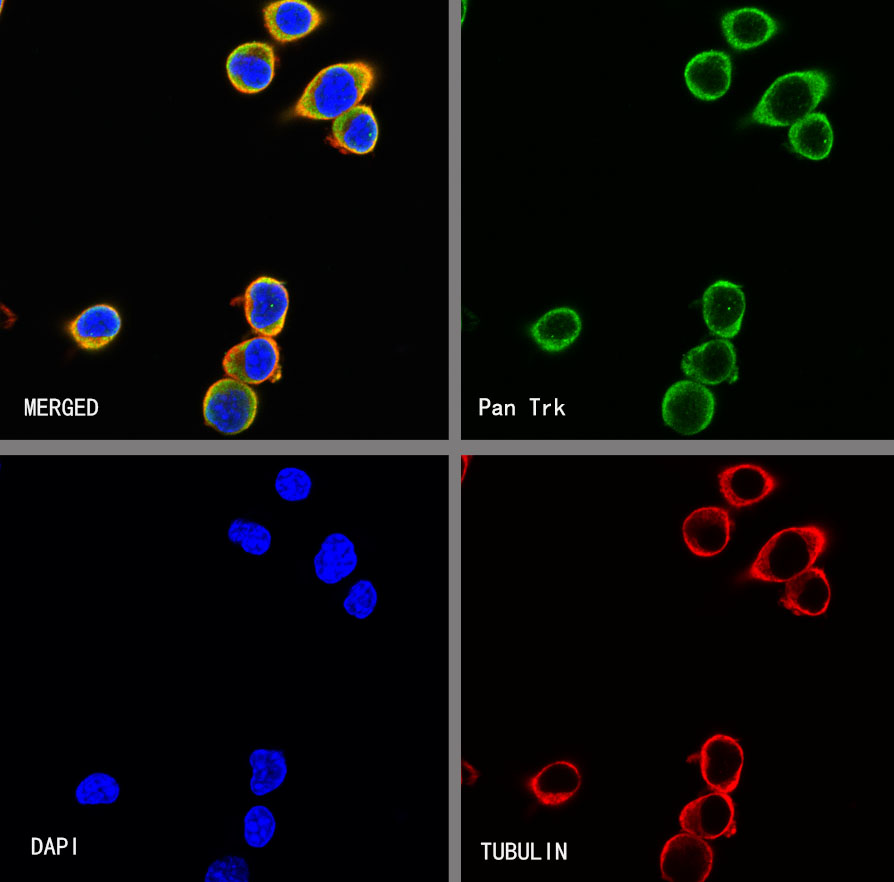
ICC shows positive staining in Neuro-2a cells. Anti- Pan Trk antibody was used at 1/50 dilution (Green) and incubated overnight at 4°C. Goat polyclonal Antibody to Rabbit IgG - H&L (Alexa Fluor® 488) was used as secondary antibody at 1/1000 dilution. The cells were fixed with 100% ice-cold methanol and permeabilized with 0.1% PBS-Triton X-100. Nuclei were counterstained with DAPI (Blue). Counterstain with tubulin (Red).