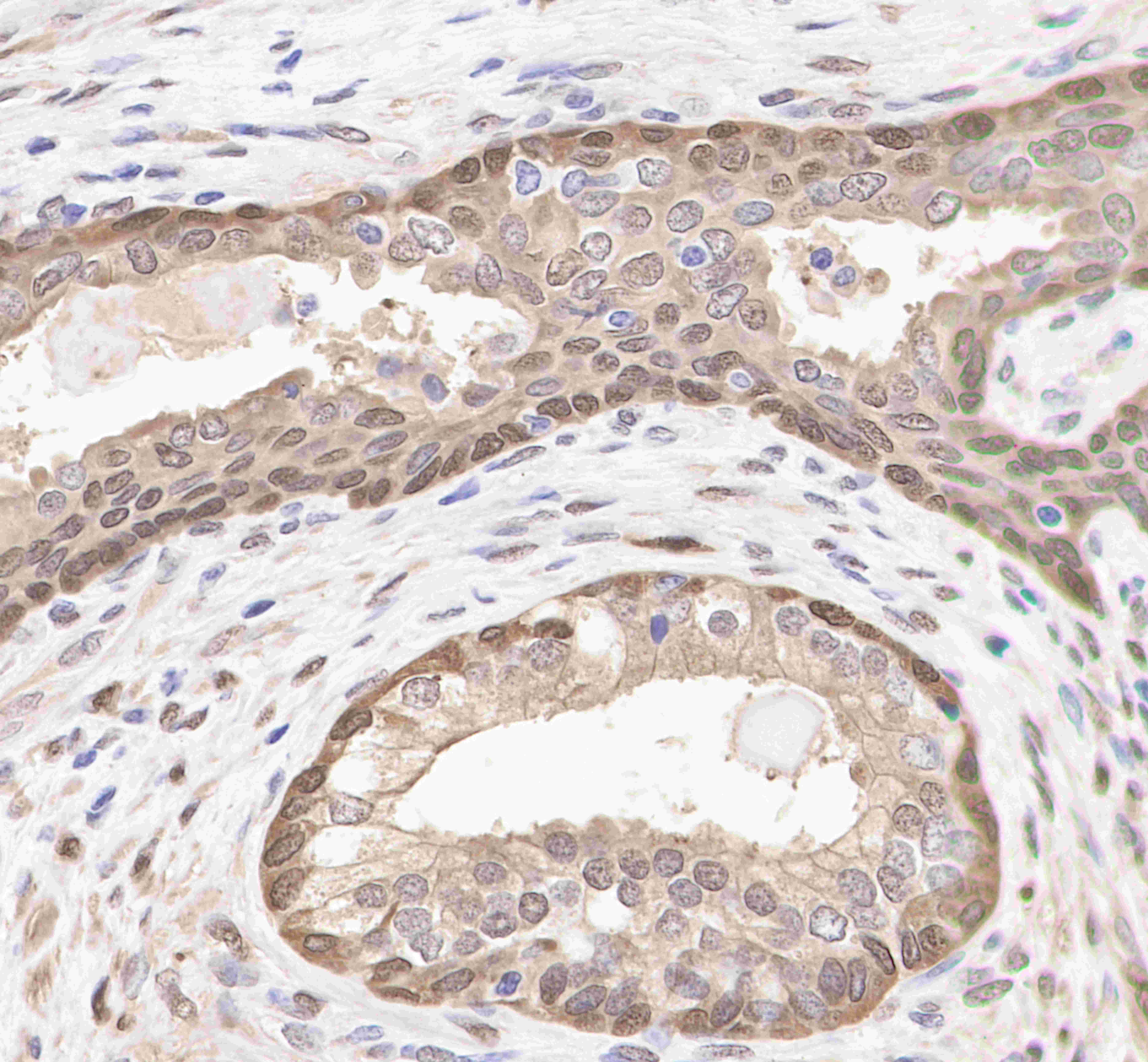
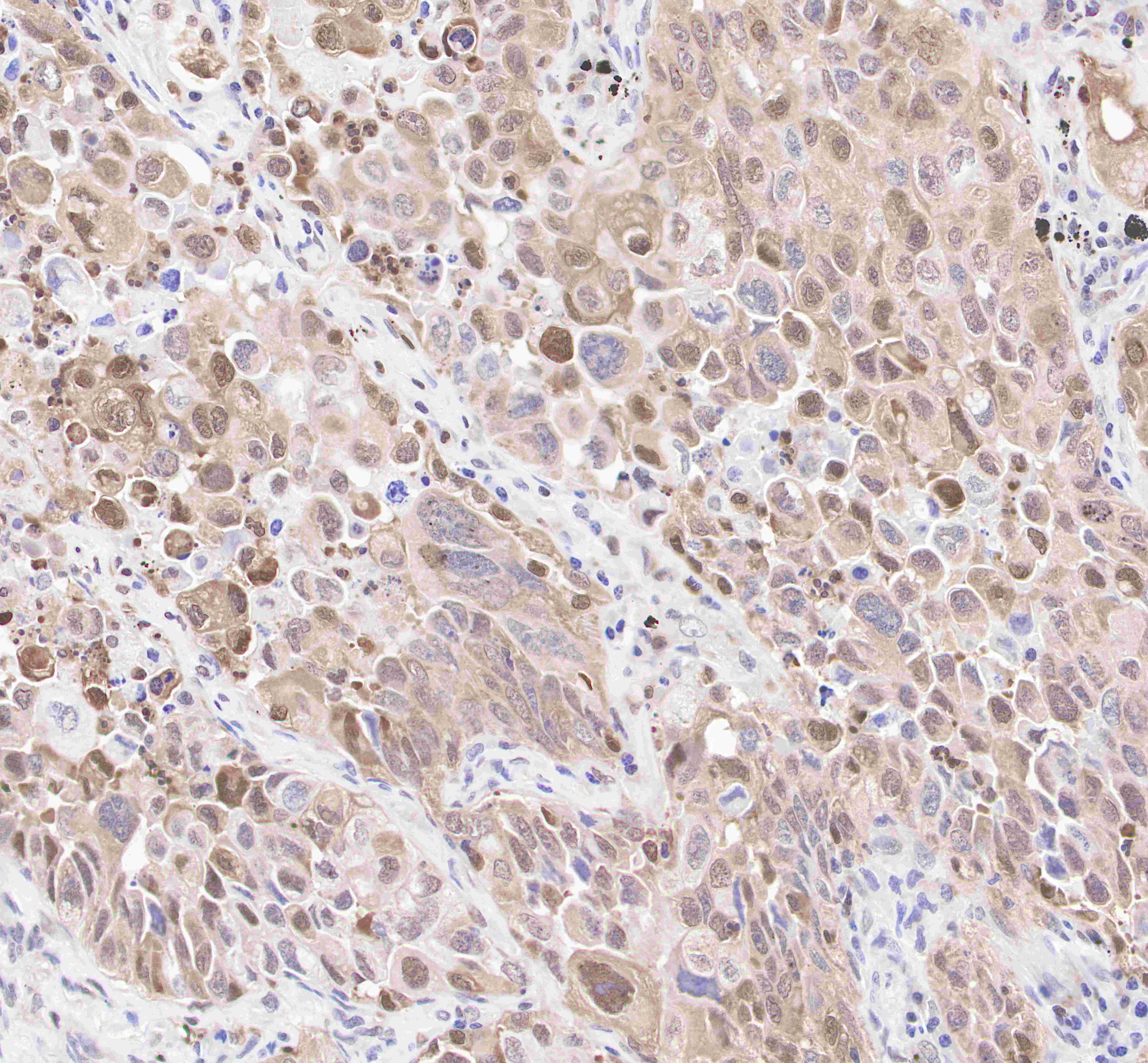
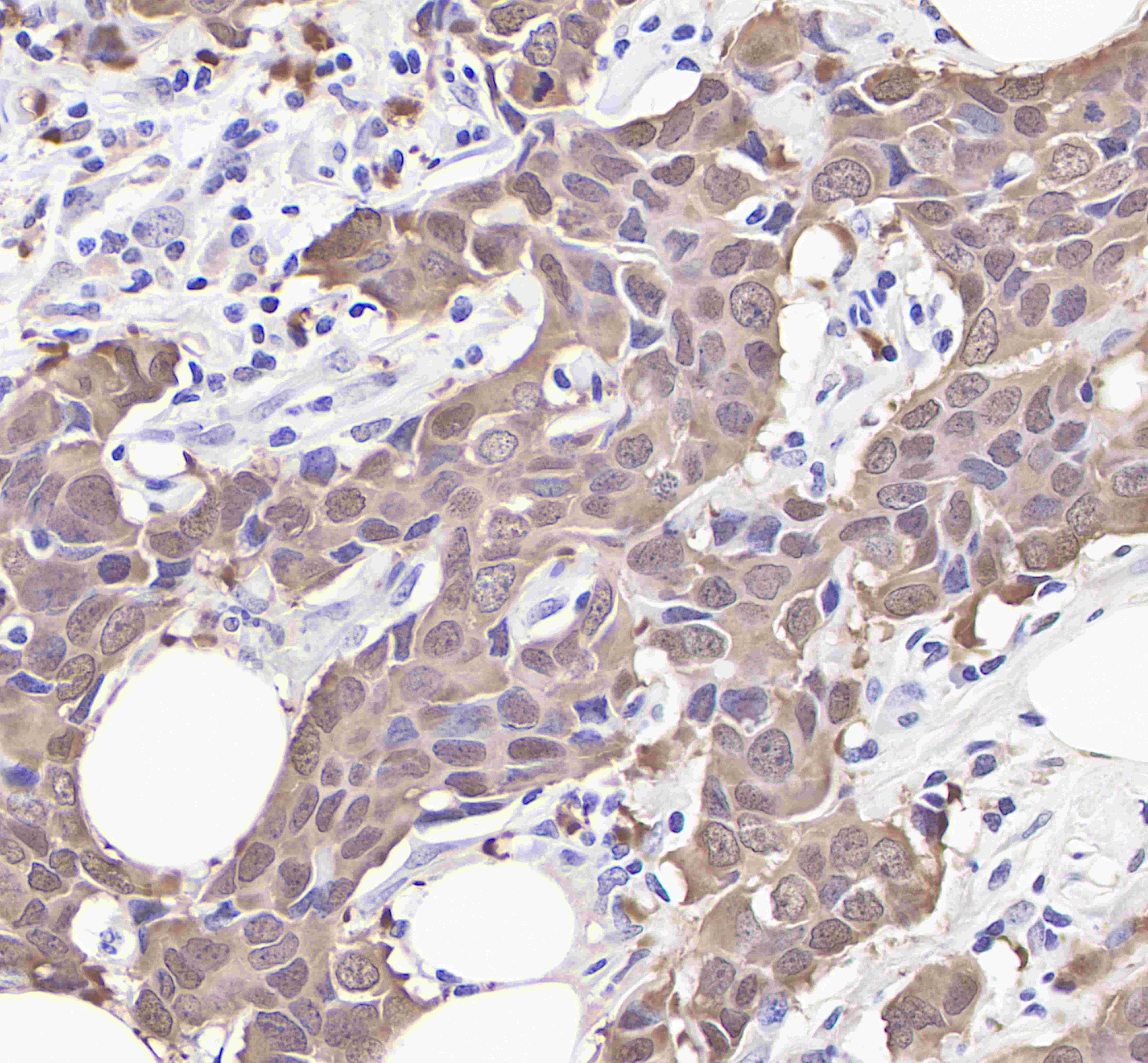
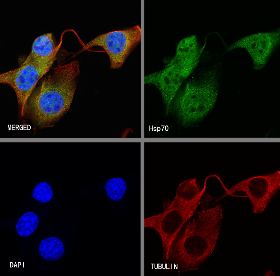
Product Specification
| Host |
Rabbit |
| Antigen |
Hsp70 |
| Synonyms |
Heat shock 70 kDa protein |
| Immunogen |
Recombinant Protein |
| Location |
Cytoplasm, Nucleus |
| Accession |
P0DMV8,P0DMV9 |
| Clone Number |
SDT-R016 |
| Antibody Type |
Rabbit mAb |
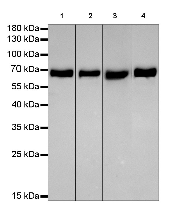
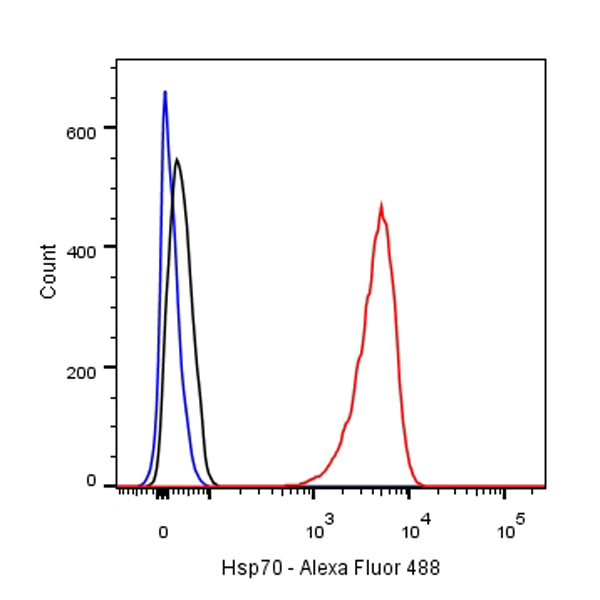
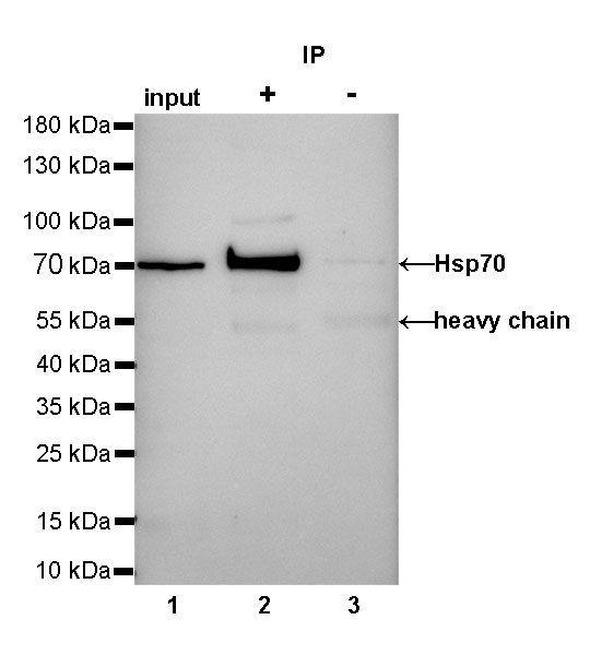
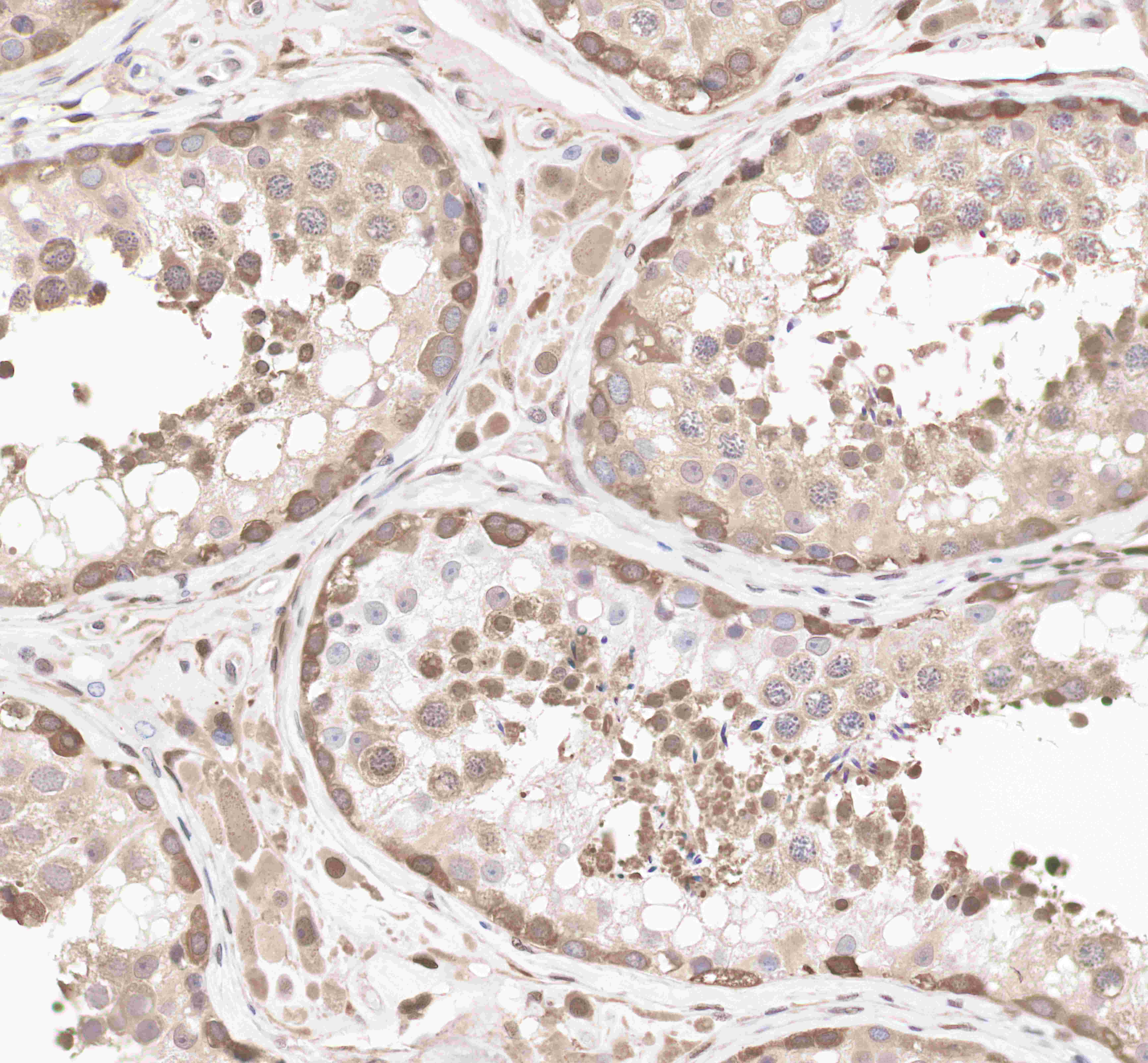
| Application |
WB, IHC-P, ICC, ICFCM, IP |
| Reactivity |
Hu |
| Purification |
Protein A |
| Concentration |
0.5mg/ml |
| Conjugation |
Unconjugated |
| Physical Appearance |
Liquid |
| Storage Buffer |
PBS, 40% Glycerol, 0.05%BSA, 0.03% Proclin 300 |
| Stability & Storage |
12 months from date of receipt / reconstitution, -20 °C as supplied. |
Dilution
| application |
dilution |
species |
| ICFCM |
1:500 |
null |
| IHC-P |
1:1000 |
null |
| IP |
1:25 |
null |
| WB |
1:2000-1:50000 |
null |
| ICC |
1:100-1:500 |
null |
Background
The 70 kilodalton heat shock proteins (Hsp70s or DnaK) are a family of conserved ubiquitously expressed heat shock proteins. Proteins with similar structure exist in virtually all living organisms. Intracellularly localized Hsp70s are an important part of the cell's machinery for protein folding, performing chaperoning functions, and helping to protect cells from the adverse effects of physiological stresses. Additionally, membrane-bound Hsp70s have been identified as a potential target for cancer therapies and their extracellularly localized counterparts have been identified as having both membrane-bound and membrane-free structures.