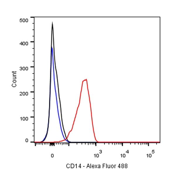
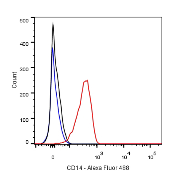
Flow cytometric analysis of THP-1 cells labelled with CD14 antibody at 1/50 dilution (1 μg)/ (red) compared with a Rabbit monoclonal IgG (Black) isotype control and an unlabelled control (cells without incubation with primary antibody and secondary antibody) (Blue).
Goat Anti-Rabbit IgG Alexa Fluor® 488 at 1/1000 dilution was used as the secondary antibody
S-RMab® CD14 Recombinant Rabbit mAb(SDT-060-50)
S-RMab® CD14 Recombinant Rabbit mAb(SDT-060-50)
Price:
Regular price
$100 USD
Regular price
Sale price
$100 USD
Unit price
per
For shipping services or bulk orders, you may request a quotation.
Secure checkout with

View full details
Product Details
Product Details
Product Specification
| Host | Rabbit |
| Antigen | CD14 |
| Synonyms | Myeloid cell-specific leucine-rich glycoprotein |
| Immunogen | Synthetic Peptide |
| Location | Membrane |
| Accession | P08571 |
| Clone Number | SDT-060-50 |
| Antibody Type | Rabbit mAb |
| Application | WB, IHC-P, ICC, FC, IP |
| Reactivity | Hu |
| Purification | Protein A |
| Concentration | 0.5 mg/ml |
| Physical Appearance | Liquid |
| Storage Buffer | PBS, 40% Glycerol, 0.05%BSA, 0.03% Proclin 300 |
| Stability & Storage | 12 months from date of receipt / reconstitution, -20 °C as supplied |
Dilution
| application | dilution | species |
| IHC-P | 1:2000 | |
| WB | 1:1000 | |
| IP | 1:25 | |
| FACS | 1:500 | |
| ICC | 1:500 |
Background
CD14 (cluster of differentiation 14) is a human protein made mostly by macrophages as part of the innate immune system. It helps to detect bacteria in the body by binding lipopolysaccharide (LPS), a pathogen-associated molecular pattern (PAMP). CD14 exists in two forms, one anchored to the membrane by a glycosylphosphatidylinositol (GPI) tail (mCD14), the other a soluble form (sCD14). Soluble CD14 either appears after shedding of mCD14 (48 kDa) or is directly secreted from intracellular vesicles (56 kDa).
Picture
Picture
Validation Data
Western Blot
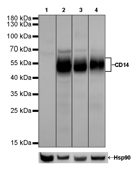
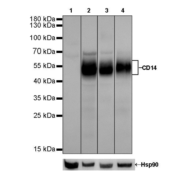
WB result of CD14 Rabbit mAb
Primary antibody: CD14 Rabbit mAb at 1/1000 dilution
Lane 1: Neuro-2a whole cell lysate 20 µg
Lane 2: THP-1 whole cell lysate 20 µg
Lane 3: A549 whole cell lysate 20 µg
Lane 4: HL-60 whole cell lysate 20 µg
Negative control: Neuro-2a whole cell lysate
Secondary antibody: Goat Anti-Rabbit IgG, (H+L), HRP conjugated at 1/10000 dilution
Predicted MW: 40 kDa
Observed MW: 47~57 kDa
Exposure time: Lane 1、lane 2 and lane 3: 120s; Lane 4: 30s
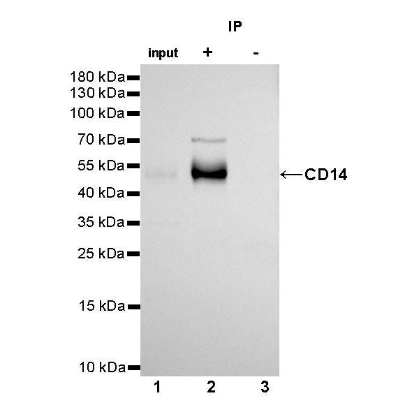
IP
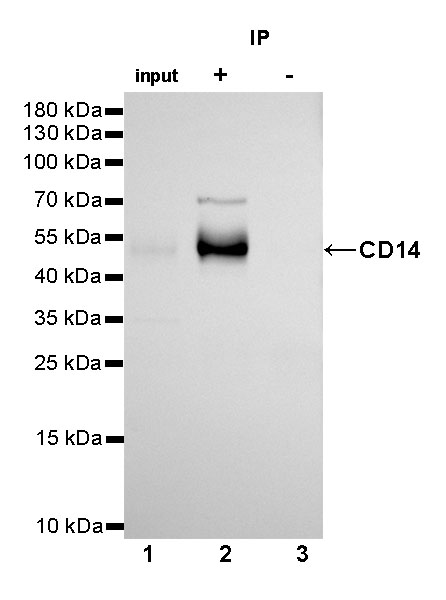
CD14 Rabbit mAb at 1/25 dilution (2µg) immunoprecipitating CD14 in 0.4mg A549 whole cell lysate.
Western blot was performed on the immunoprecipitate using CD14 Rabbit mAb at 1/1000 dilution.
Secondary antibody (HRP) for IP was used at 1/400 dilution.
Lane 1 : A549 whole cell lysate 10µg (input)
Lane 2 : CD14 Rabbit mAb IP in A549 whole cell lysate
Lane 3 : Rabbit monoclonal IgG IP in A549 whole cell lysate
Predicted MW: 40 kDa
Observed MW: 47~57 kDa
Exposure time: 180s
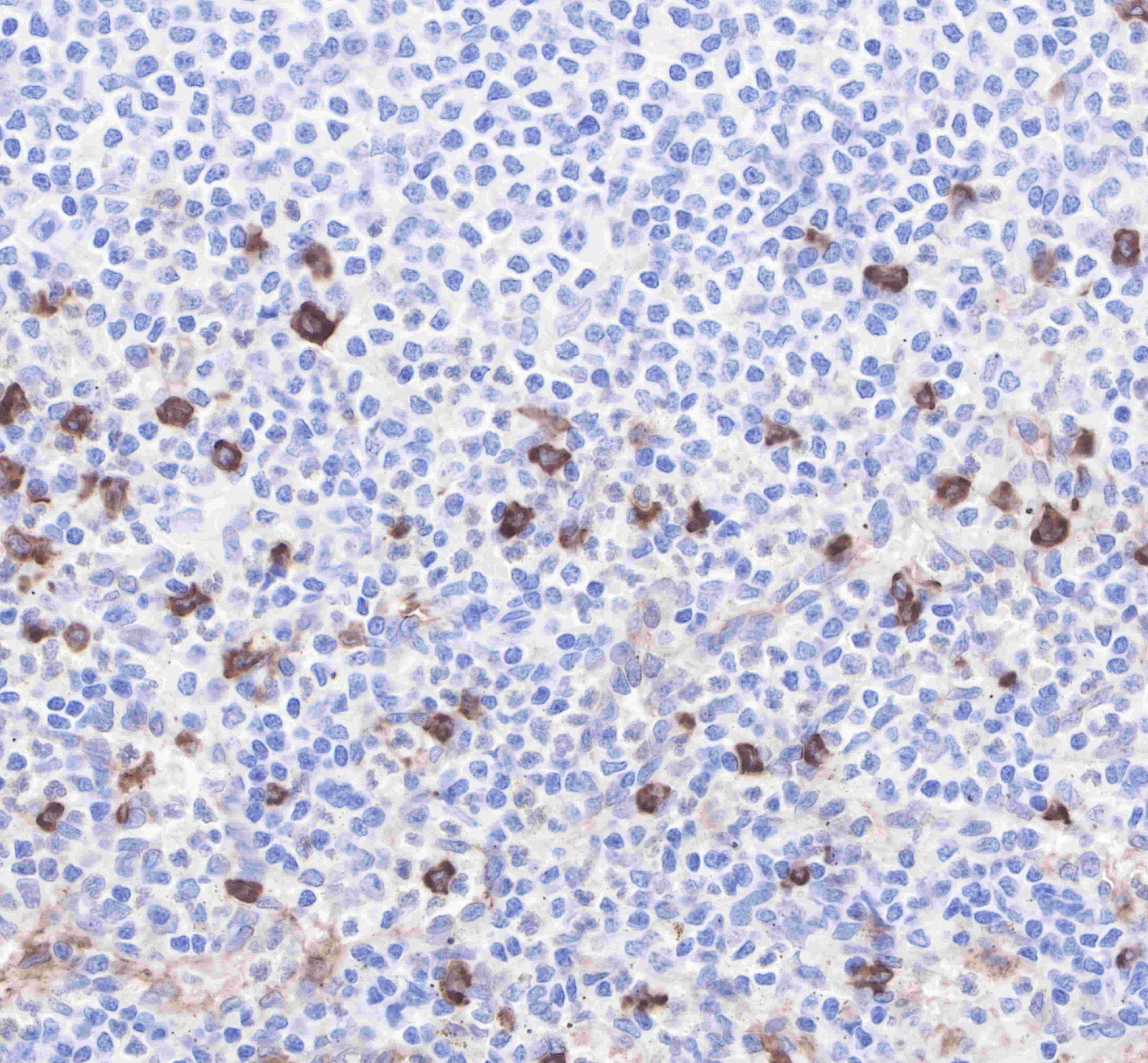
Immunohistochemistry
IHC shows positive staining in paraffin-embedded human tonsil. Anti-CD14 antibody was used at 1/2000 dilution, followed by a Goat Anti-Rabbit IgG H&L (HRP) ready to use.
Counterstained with hematoxylin.
Heat mediated antigen retrieval with Tris/EDTA buffer pH9.0 was performed before commencing with IHC staining protocol.
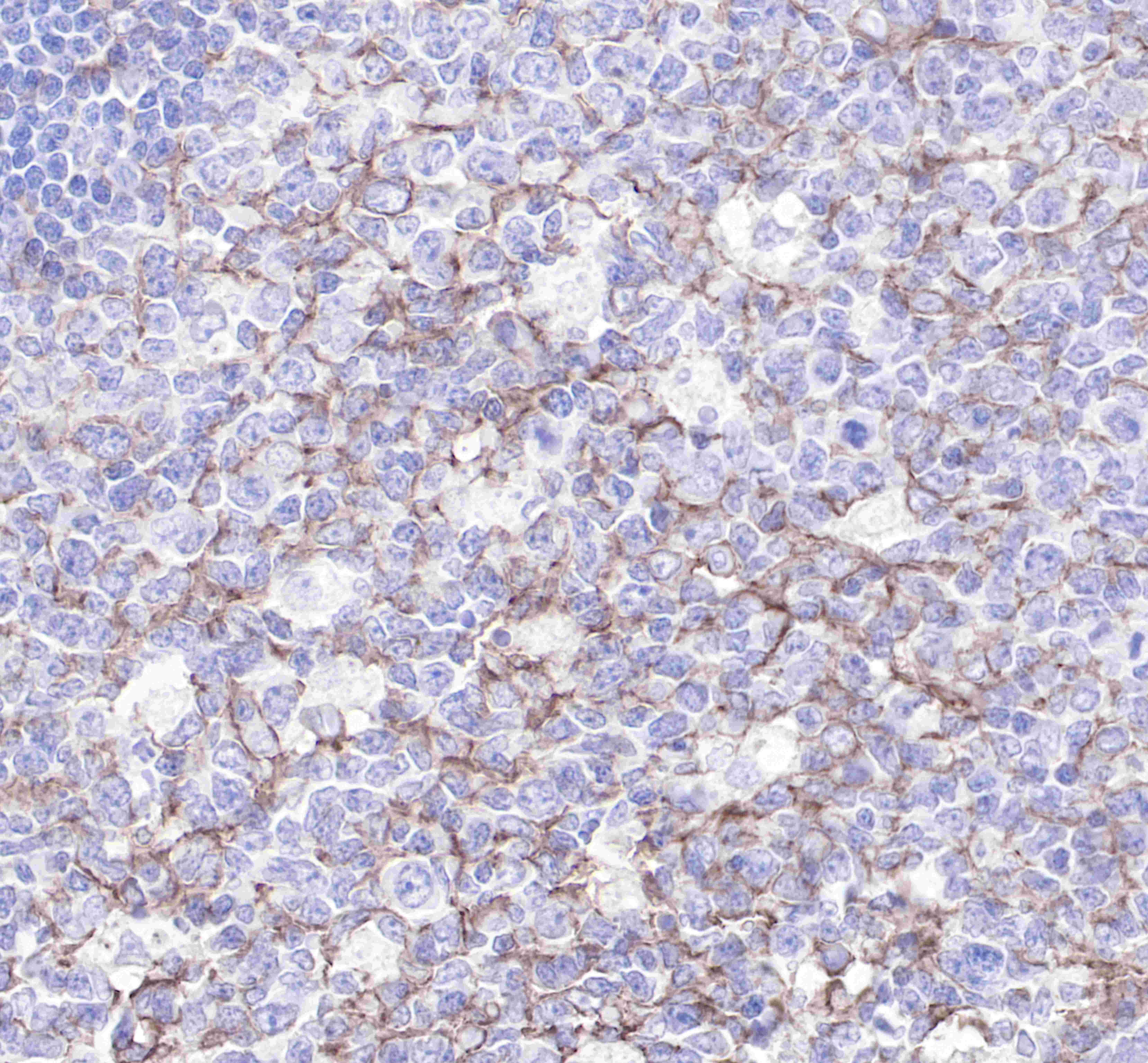
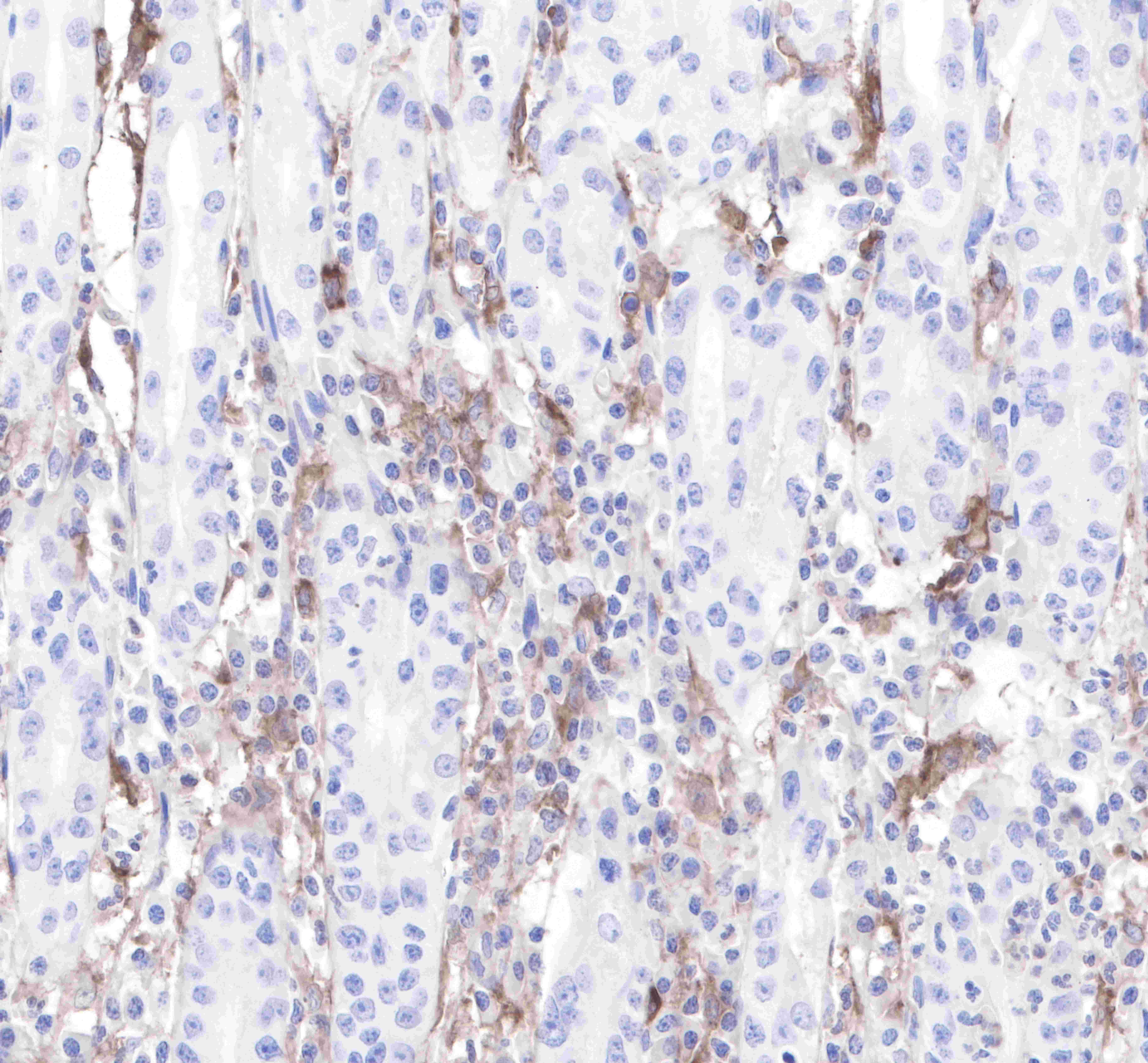
IHC shows positive staining in paraffin-embedded human spleen. Anti-CD14 antibody was used at 1/2000 dilution, followed by a Goat Anti-Rabbit IgG H&L (HRP) ready to use.
Counterstained with hematoxylin.
Heat mediated antigen retrieval with Tris/EDTA buffer pH9.0 was performed before commencing with IHC staining protocol.
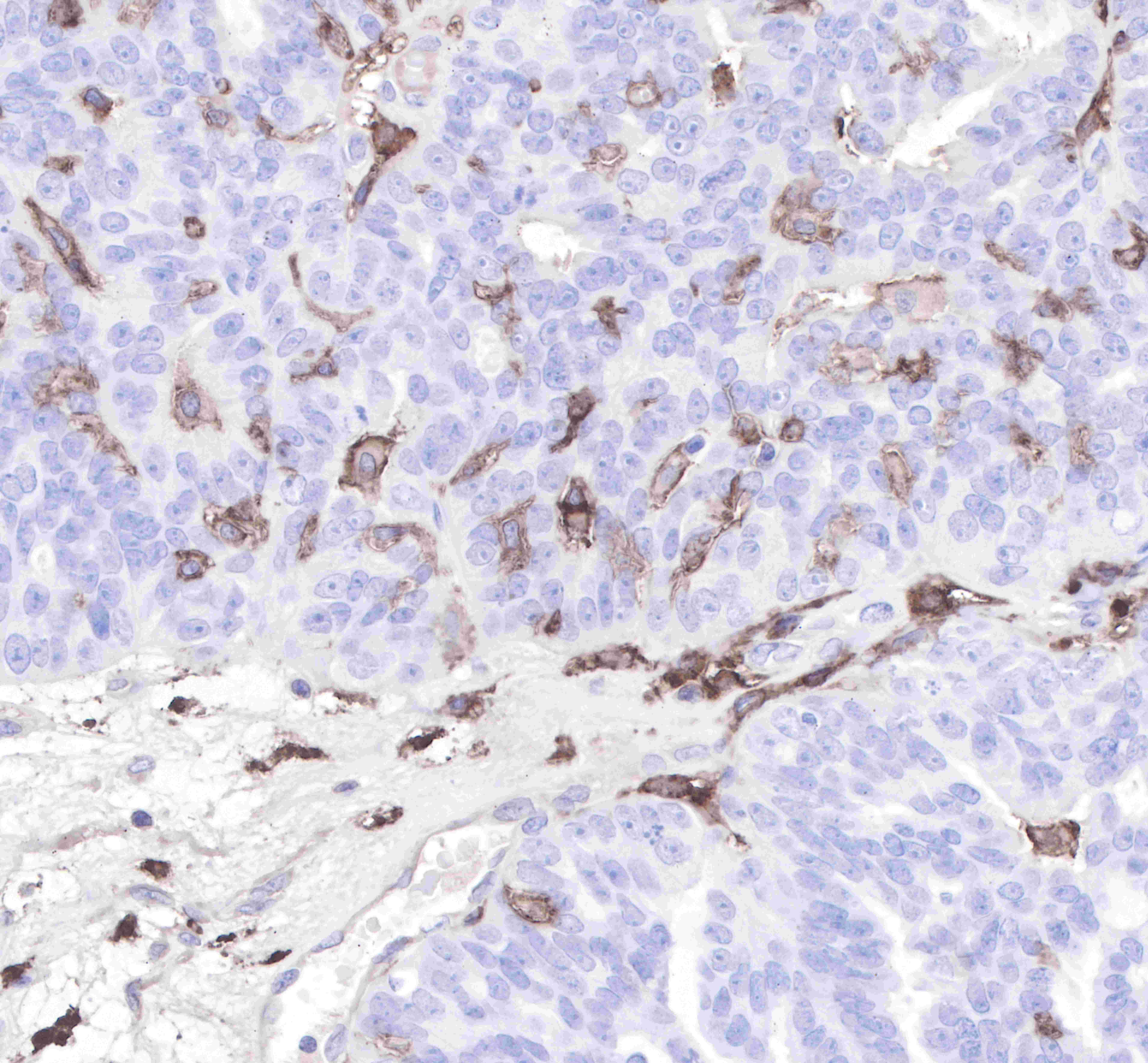
IHC shows positive staining in paraffin-embedded human stomach. Anti-CD14 antibody was used at 1/2000 dilution, followed by a Goat Anti-Rabbit IgG H&L (HRP) ready to use.
Counterstained with hematoxylin.
Heat mediated antigen retrieval with Tris/EDTA buffer pH9.0 was performed before commencing with IHC staining protocol.
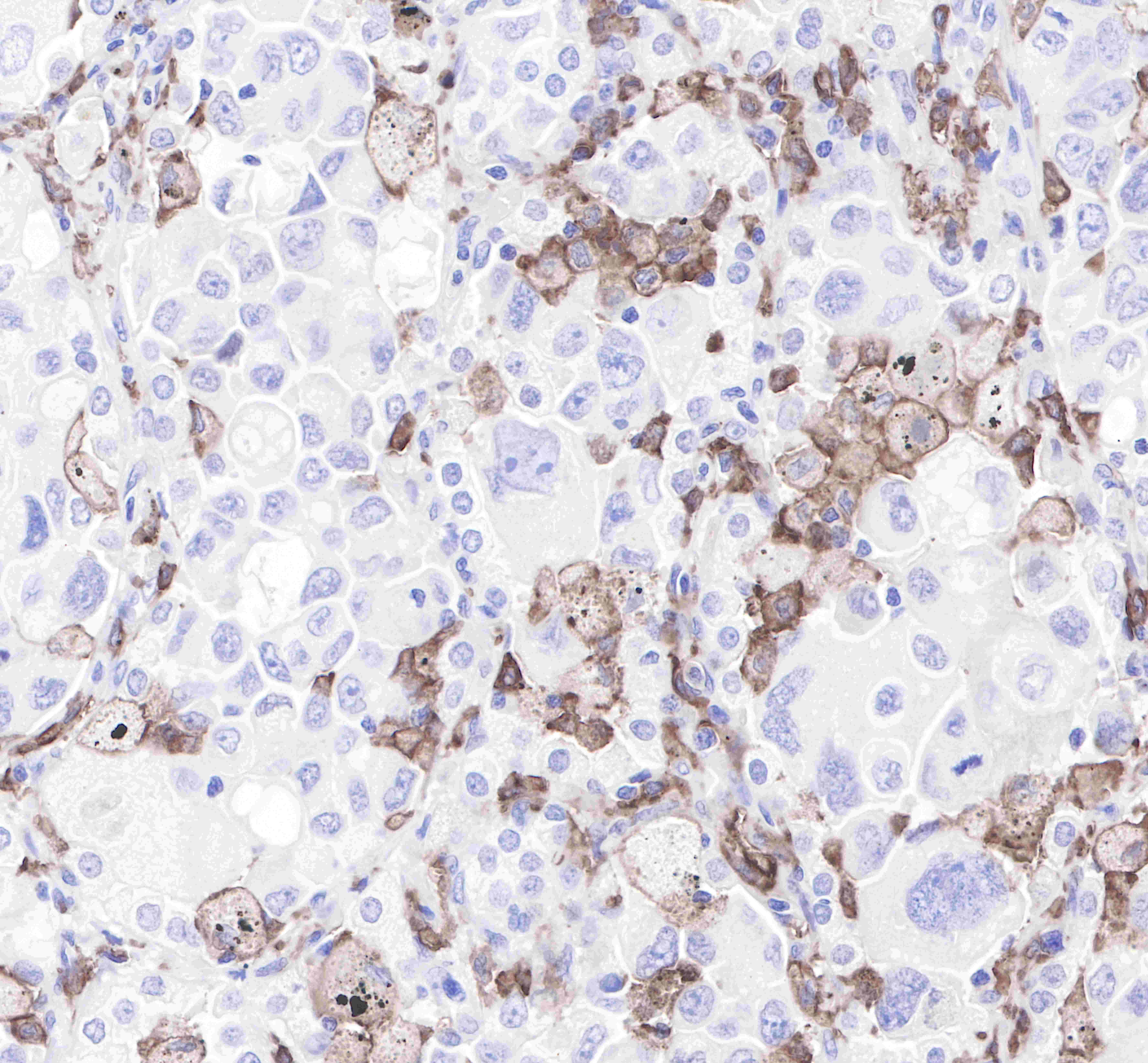
IHC shows positive staining in paraffin-embedded human ovarian cancer. Anti-CD14 antibody was used at 1/2000 dilution, followed by a Goat Anti-Rabbit IgG H&L (HRP) ready to use.
Counterstained with hematoxylin.
Heat mediated antigen retrieval with Tris/EDTA buffer pH9.0 was performed before commencing with IHC staining protocol.
IHC shows positive staining in paraffin-embedded human lung adenocarcinoma.
Anti-CD14 antibody was used at 1/2000 dilution, followed by a Goat Anti-Rabbit IgG H&L (HRP) ready to use.
Counterstained with hematoxylin.
Heat mediated antigen retrieval with Tris/EDTA buffer pH9.0 was performed before commencing with IHC staining protocol.
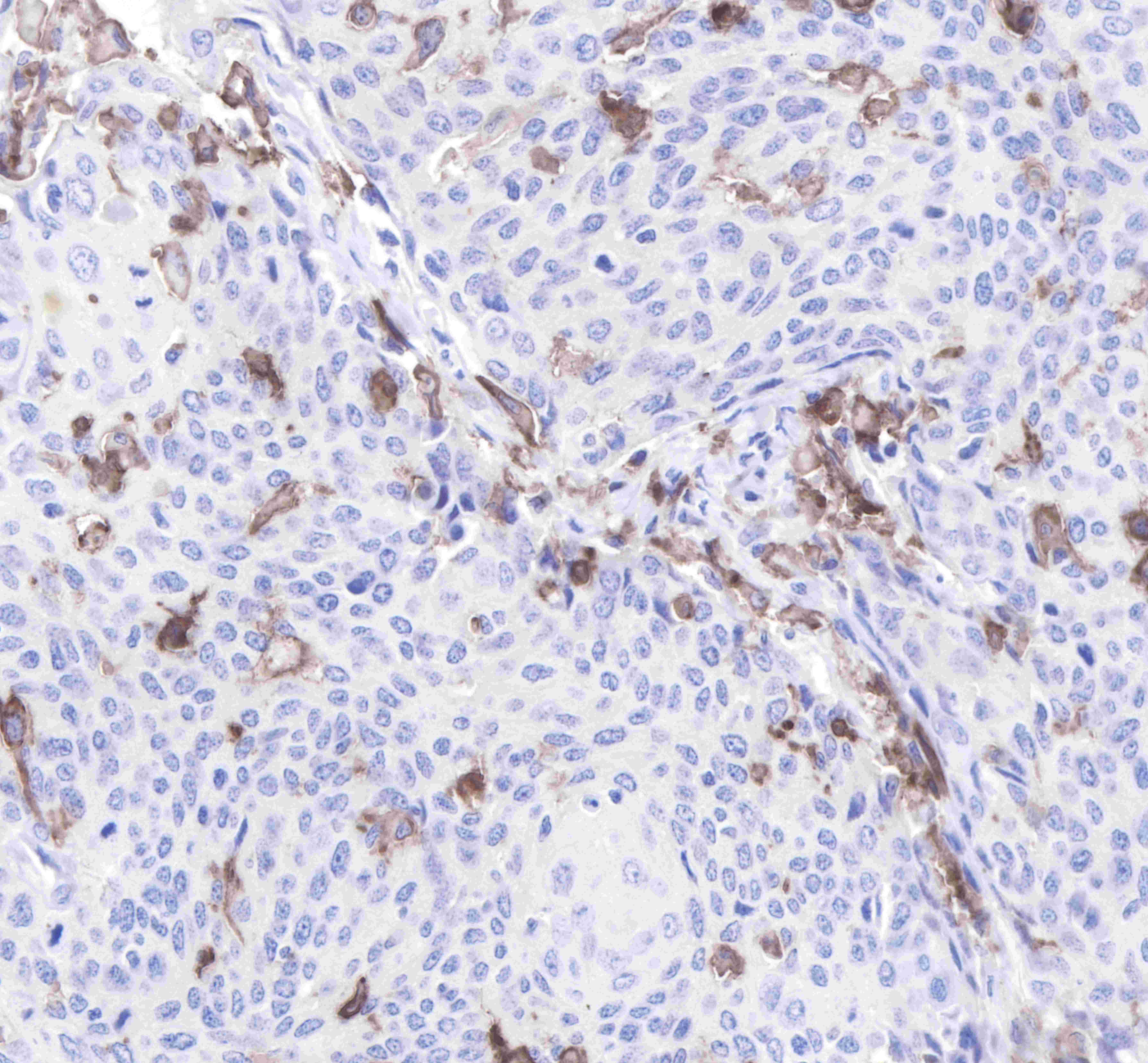
IHC shows positive staining in paraffin-embedded human cervix cancer.
Anti-CD14 antibody was used at 1/2000 dilution, followed by a Goat Anti-Rabbit IgG H&L (HRP) ready to use.
Counterstained with hematoxylin.
Heat mediated antigen retrieval with Tris/EDTA buffer pH9.0 was performed before commencing with IHC staining protocol.
Immunocytochemistry
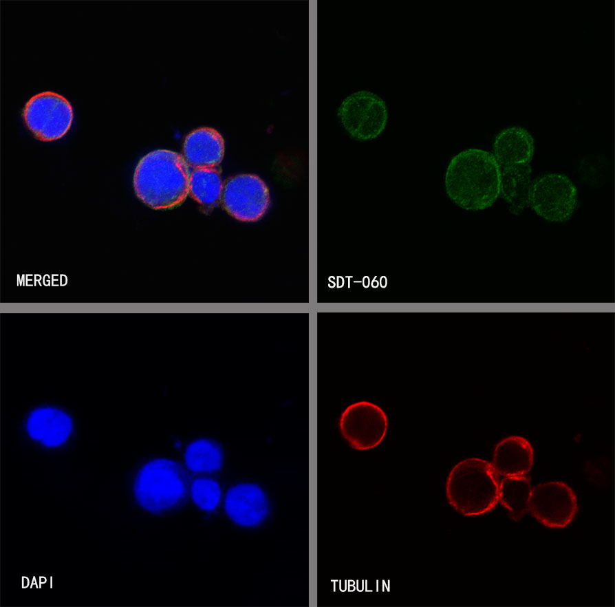
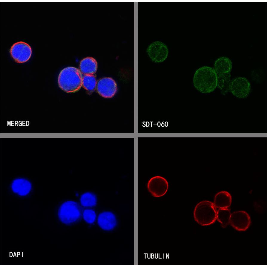
ICC shows positive staining in THP-1 cells. Anti-CD14 antibody was used at 1/500 dilution (Green) and incubated overnight at 4°C. Goat polyclonal Antibody to Rabbit IgG - H&L (Alexa Fluor® 488) was used as secondary antibody at 1/1000 dilution. The cells were fixed with 100% ice-cold methanol and permeabilized with 0.1% PBS-Triton X-100. Nuclei were counterstained with DAPI (Blue).Counterstain with tubulin (red).