WB result of LAMP1 Rabbit mAb Primary antibody: LAMP1 Rabbit mAb at 1/1000 dilution Lane 1: HeLa whole cell lysate 20 µg Secondary antibody: Goat Anti-Rabbit IgG, (H+L), HRP conjugated at 1/10000 dilution Predicted MW: 45 kDa Observed MW: 90~120 kDa
LAMP1 Recombinant Rabbit mAb (SDT-405-39)
LAMP1 Recombinant Rabbit mAb (SDT-405-39)
Price:
Regular price
$100 USD
Regular price
Sale price
$100 USD
Unit price
per
For shipping services or bulk orders, you may request a quotation.
Secure checkout with

View full details
Product Details
Product Details
Product Specification
| Host | Rabbit |
| Antigen | LAMP1 |
| Synonyms | Lysosome-associated membrane glycoprotein 1, CD107 antigen-like family member A, CD107a |
| Immunogen | Recombinant Protein |
| Accession | P11279 |
| Clone Number | SDT-405-39 |
| Antibody Type | Recombinant mAb |
| Application | WB, IHC-P, ICC, FC, IP |
| Reactivity | Hu |
| Purification | Protein A |
| Concentration | 0.5 mg/ml |
| Conjugation | Unconjugated |
| Physical Appearance | Liquid |
| Storage Buffer | PBS, 40% Glycerol, 0.05% BSA, 0.03% Proclin 300 |
| Stability & Storage | 12 months from date of receipt / reconstitution, -20 °C as supplied. |
Dilution
| application | dilution | species |
| WB | 1:1000 | null |
| ICC | 1:100 | null |
| IP | 1:50 | null |
| IHC-P | 1:500 | null |
| FC | 1:500 | null |
Background
Lysosomal-associated membrane protein 1 (LAMP-1) also known as lysosome-associated membrane glycoprotein 1 and CD107a (Cluster of Differentiation 107a), is a protein that in humans is encoded by the LAMP1 gene. The LAMP-1 glycoprotein is a type I transmembrane protein [PMID: 16973206] which is expressed at high or medium levels in at least 76 different normal tissue cell types. It resides primarily across lysosomal membranes [PMID: 2584229], and functions to provide selectins with carbohydrate ligands. LAMP-1 has also been shown to be a marker of degranulation on lymphocytes such as CD8+ and NK cells, and may also play a role in tumor cell differentiation and metastasis.
Picture
Picture
Western Blot
FC
Flow cytometric analysis of HeLa (Human cervix adenocarcinoma epithelial cell) labelling LAMP1 antibody at 1/500 dilution(0.1 μg) / (red) compared with a Rabbit monoclonal IgG (Black) isotype control and an unlabelled control (cells without incubation with primary antibody and secondary antibody) (Blue). Goat Anti-Rabbit IgG Alexa Fluor® 488 was used as the secondary antibody.
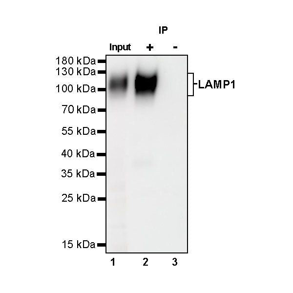
IP
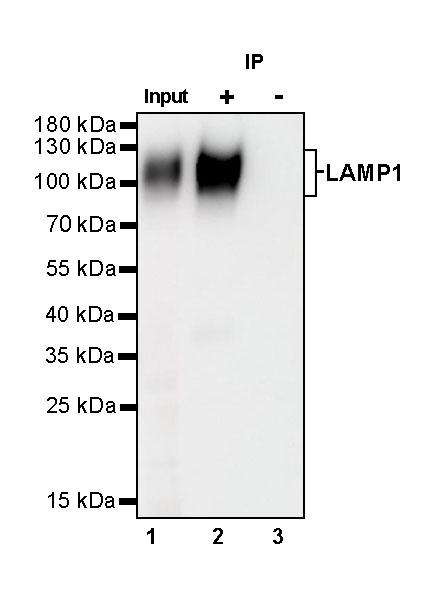
LAMP1 Rabbit mAb at 1/50 dilution (1 µg) immunoprecipitating LAMP1 in 0.4 mg HeLa whole cell lysate.
Western blot was performed on the immunoprecipitate using LAMP1 Rabbit mAb at 1/1000 dilution.
Secondary antibody (HRP) for IP was used at 1/400 dilution.
Lane 1: HeLa whole cell lysate 20 µg (Input)
Lane 2: LAMP1 Rabbit mAb IP in HeLa whole cell lysate
Lane 3: Rabbit monoclonal IgG IP in HeLa whole cell lysate
Predicted MW: 45 kDa
Observed MW: 90~120 kDa
Immunohistochemistry
IHC shows positive staining in paraffin-embedded human tonsil. Anti-LAMP1 antibody was used at 1/500 dilution, followed by a HRP Polymer for Mouse & Rabbit IgG (ready to use). Counterstained with hematoxylin. Heat mediated antigen retrieval with Tris/EDTA buffer pH9.0 was performed before commencing with IHC staining protocol.
IHC shows positive staining in paraffin-embedded human kidney. Anti-LAMP1 antibody was used at 1/500 dilution, followed by a HRP Polymer for Mouse & Rabbit IgG (ready to use). Counterstained with hematoxylin. Heat mediated antigen retrieval with Tris/EDTA buffer pH9.0 was performed before commencing with IHC staining protocol.
IHC shows positive staining in paraffin-embedded human breast cancer. Anti-LAMP1 antibody was used at 1/500 dilution, followed by a HRP Polymer for Mouse & Rabbit IgG (ready to use). Counterstained with hematoxylin. Heat mediated antigen retrieval with Tris/EDTA buffer pH9.0 was performed before commencing with IHC staining protocol.
Immunocytochemistry
ICC shows positive staining in HeLa cells. Anti-LAMP1 antibody was used at 1/100 dilution (Green) and incubated overnight at 4°C. Goat polyclonal Antibody to Rabbit IgG - H&L (Alexa Fluor® 488) was used as secondary antibody at 1/1000 dilution. The cells were fixed with 100% ice-cold methanol and permeabilized with 0.1% PBS-Triton X-100. Nuclei were counterstained with DAPI (Blue).