Product Specification
| Host |
Rabbit |
| Antigen |
Keratin 8 |
| Synonyms |
Cytokeratin-8,Type-II keratin Kb8, KRT8,CYK8,CK-8,K8 |
| Immunogen |
Synthetic Peptide |
| Accession |
P05787 |
| Clone Number |
SDT-016-46 |
| Antibody Type |
Rabbit mAb |
| Isotype |
IgG2b |
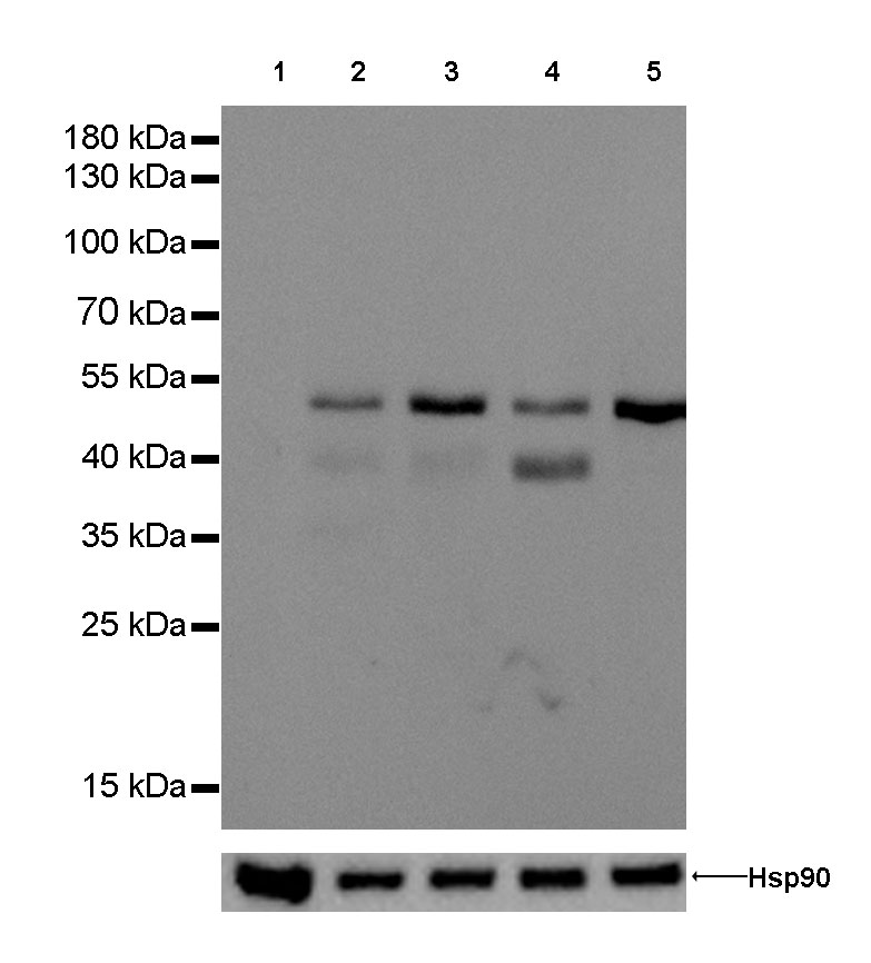
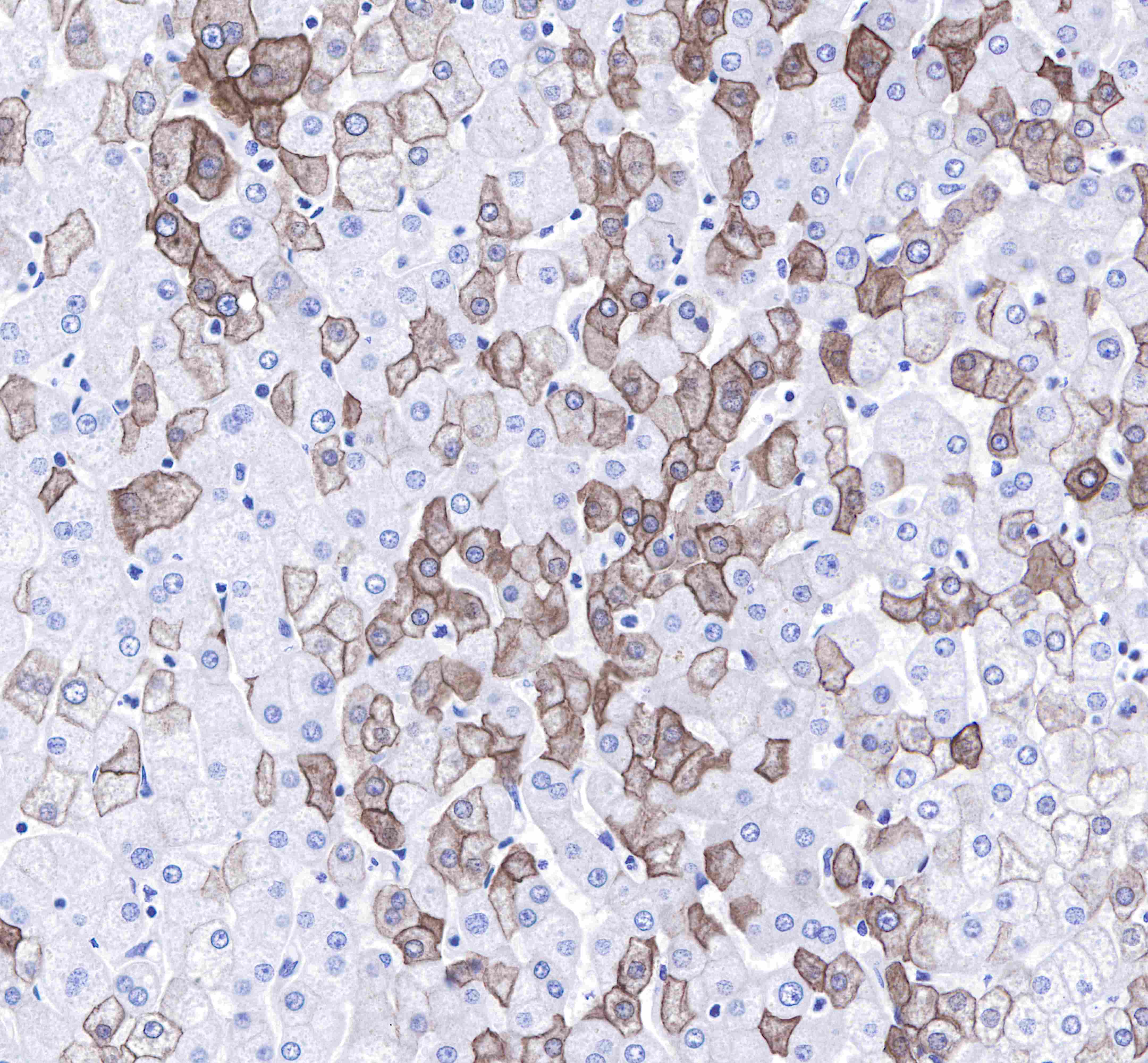
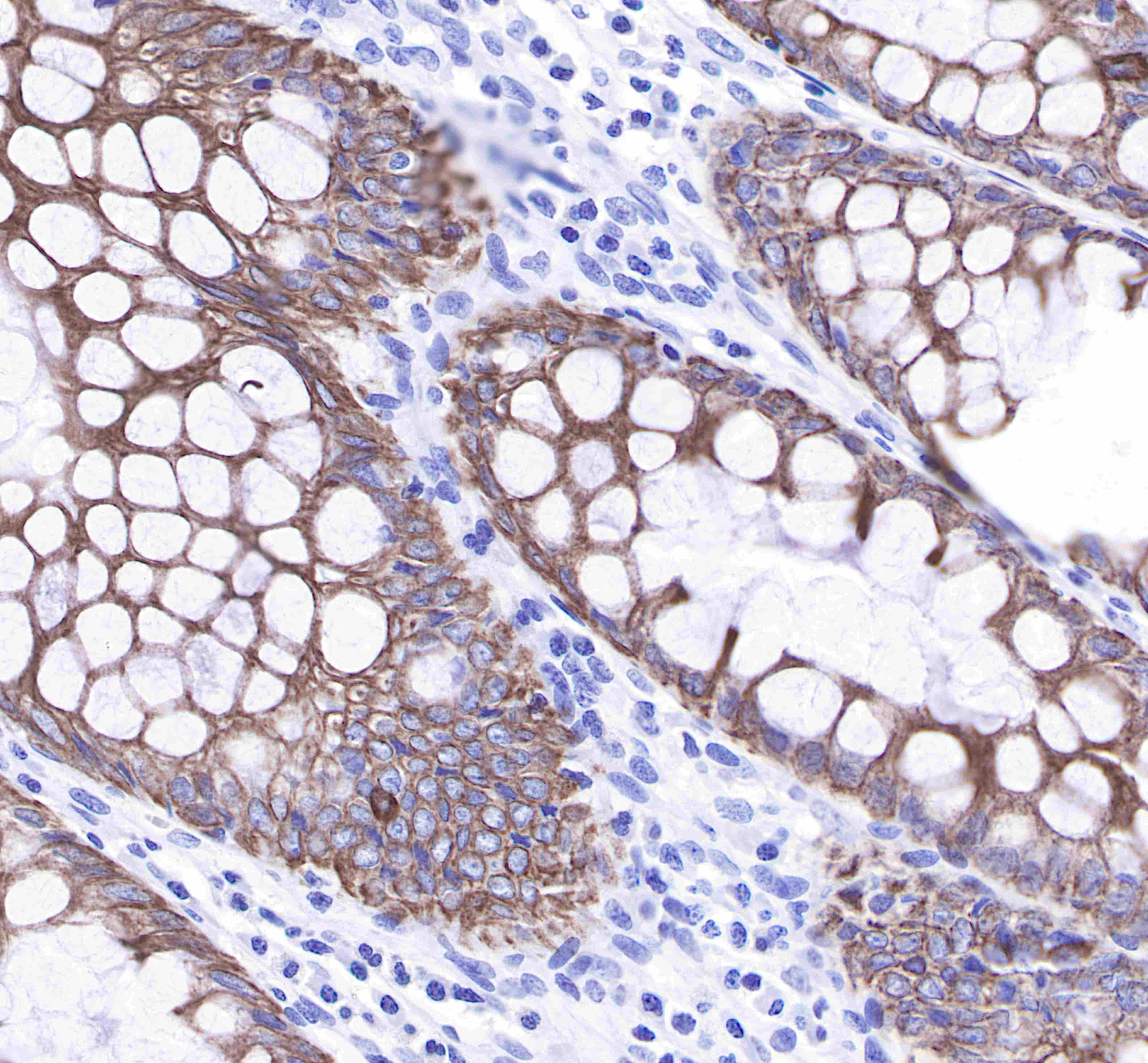
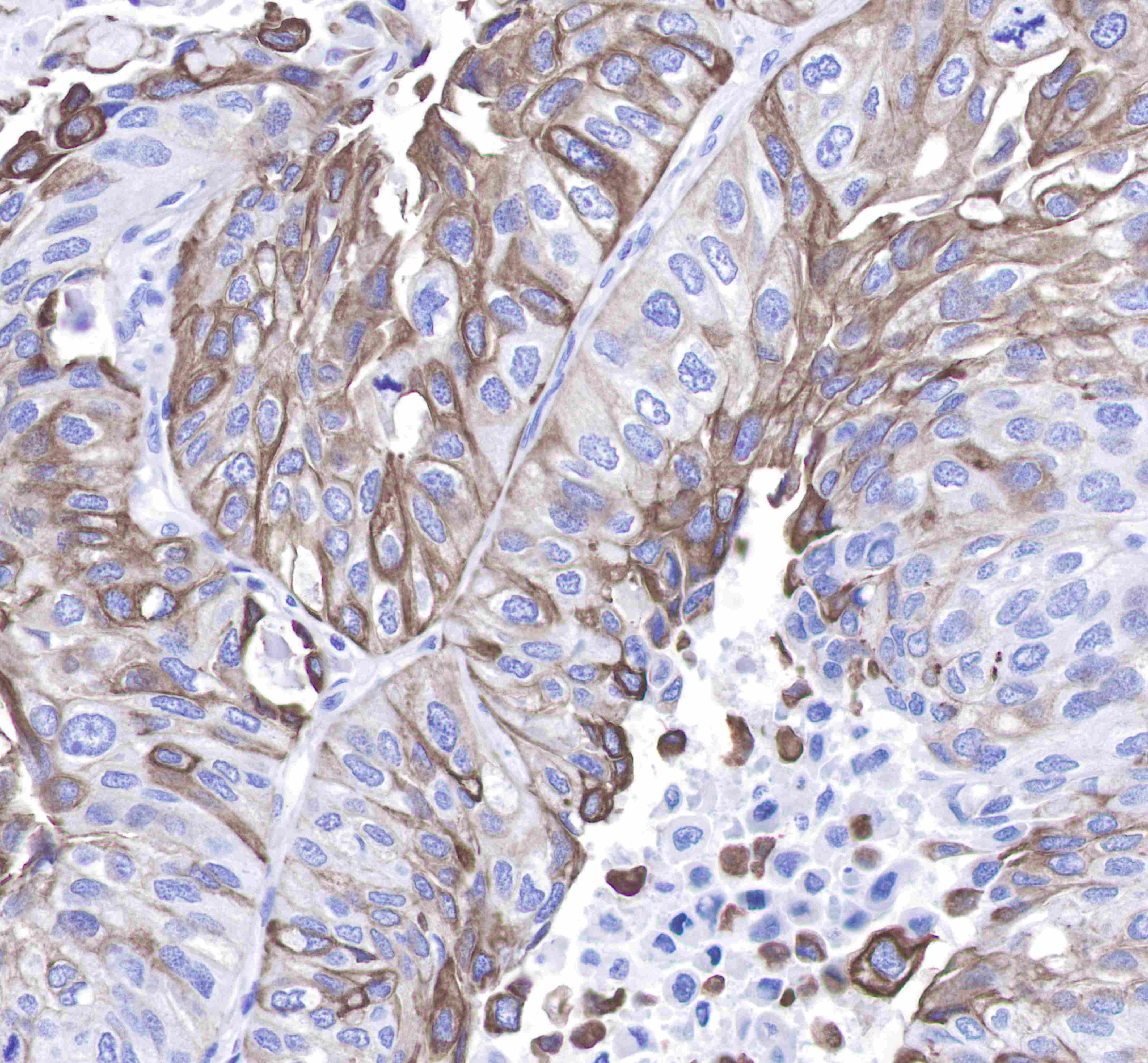
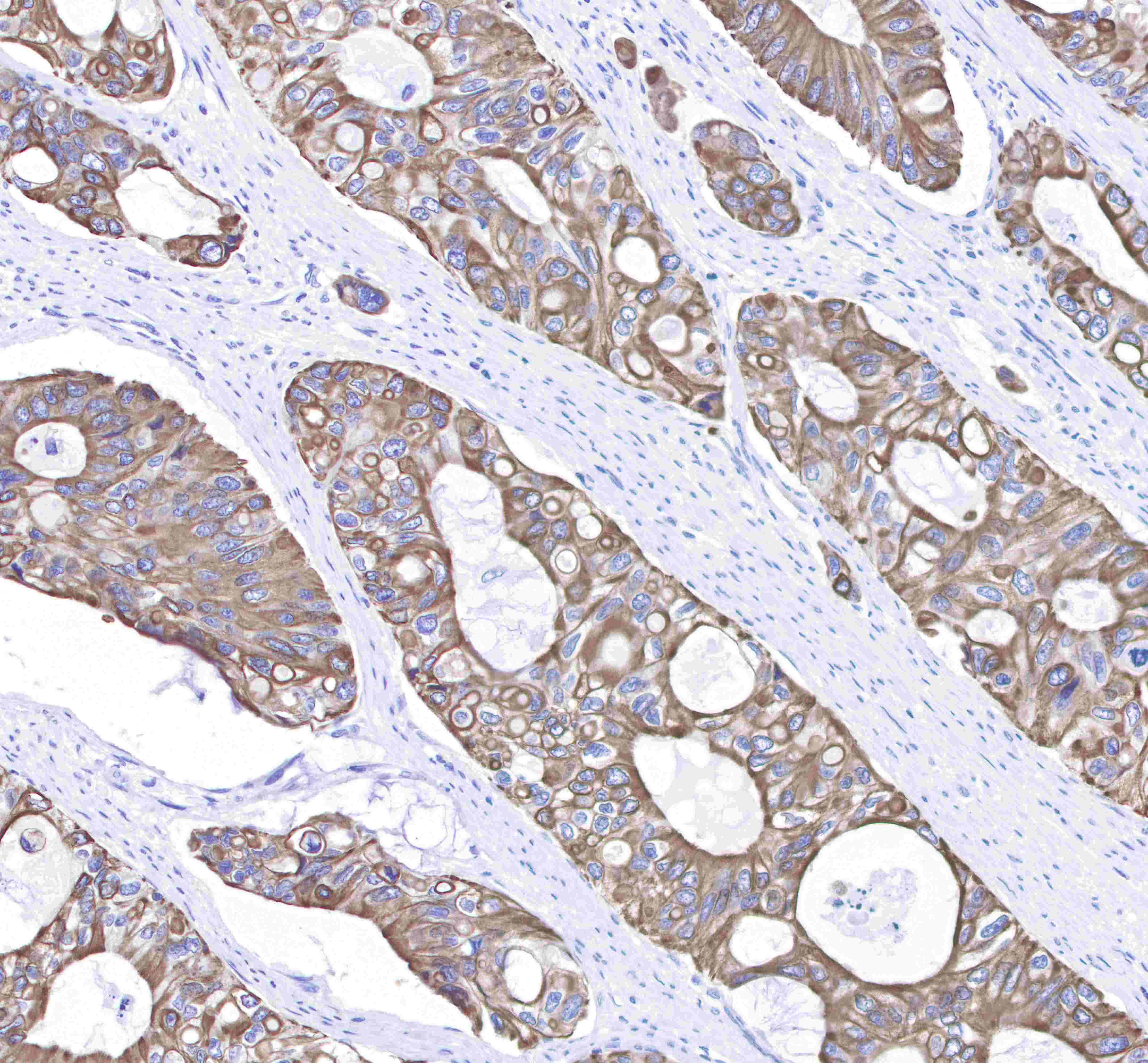
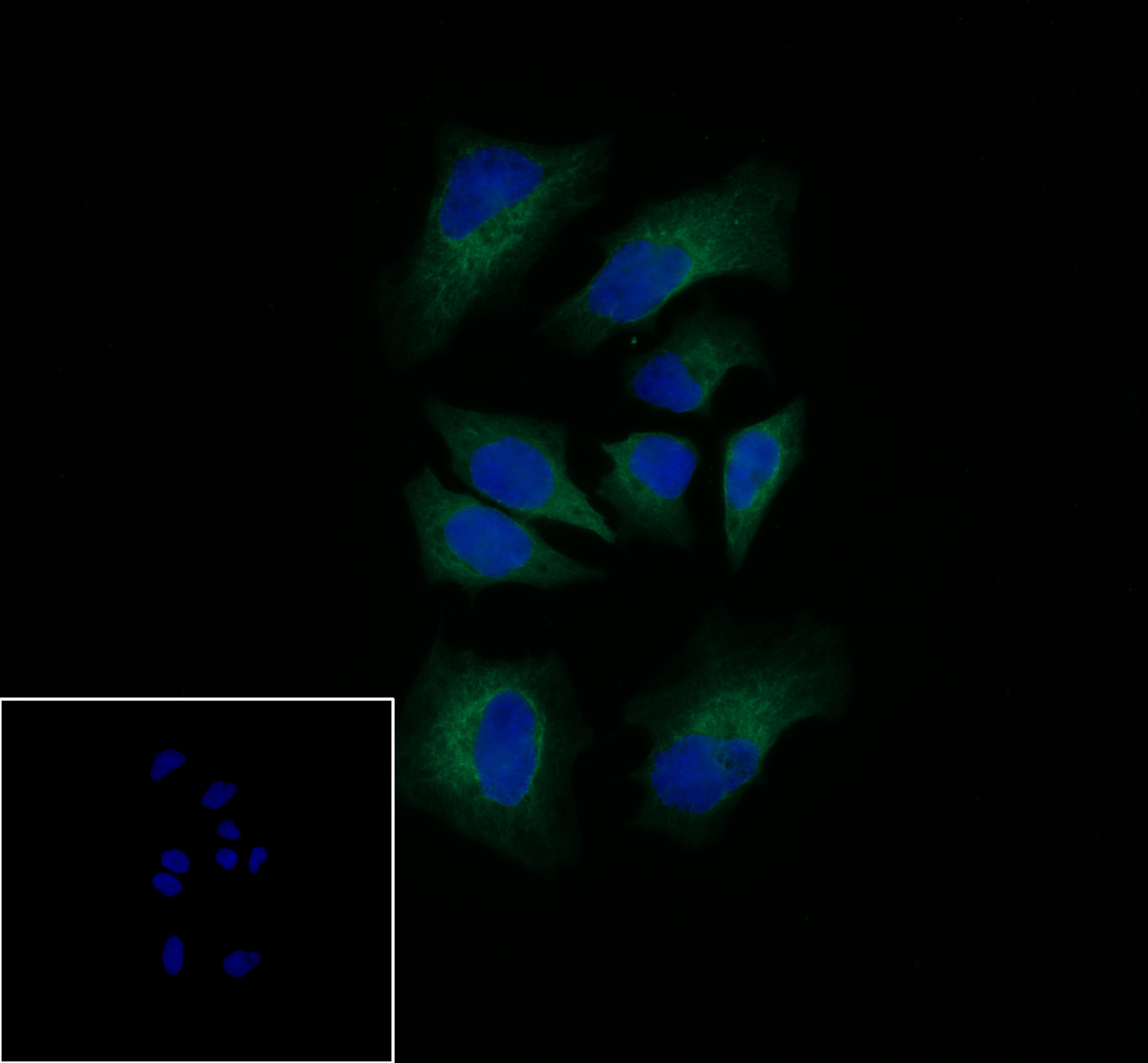
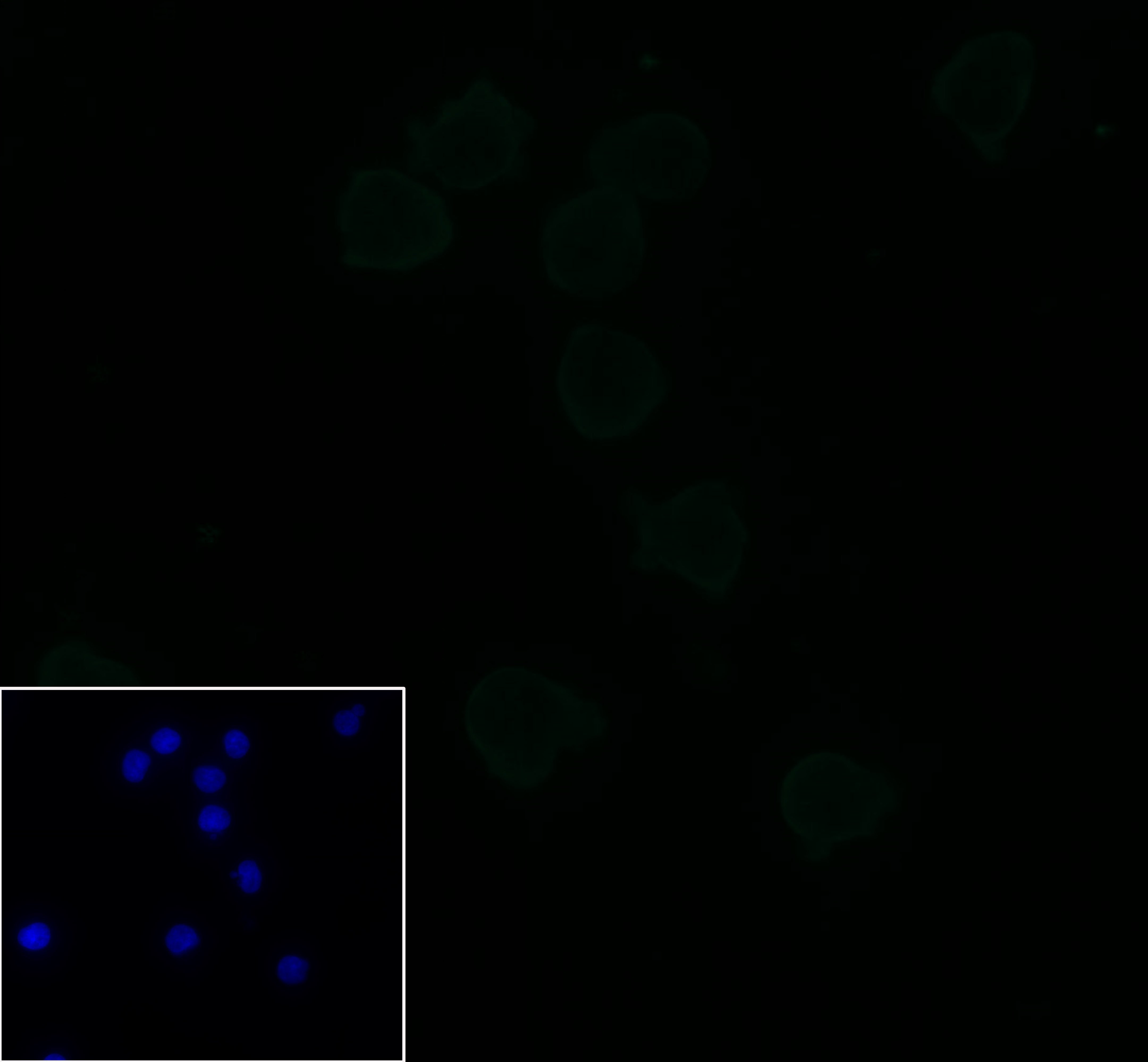
| Application |
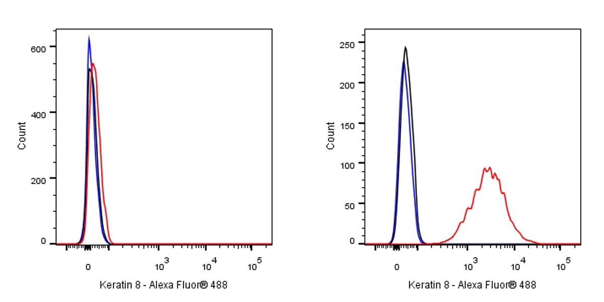
WB, IHC-P, ICC, FCM |
| Reactivity |
Hu |
| Predicted Reactivity |
Cz |
| Purification |
Protein A |
| Research Area |
Signal Transduction |
| Concentration |
0.5mg/ml |
| Molecular Weight |
53kDa |
| Physical Appearance |
Liquid |
| Storage Buffer |
PBS, 40% Glycerol, 0.05%BSA, 0.03% Proclin 300 |
| Stability & Storage |
12 months from date of receipt / reconstitution, -20 °C as supplied |
Dilution
| application |
dilution |
species |
| WB |
1:1000 |
|
| ICC |
1:250 |
|
| IHC-P |
1:250-1:1000 |
|
| ICFCM |
1:500 |
|
Background
Keratin, type II cytoskeletal 8 also known as cytokeratin-8 (CK-8) or keratin-8 (K8) is a keratin protein that is encoded in human by the KRT8 gene. In normal tissue, it reacts mainly with secretory epithelia, but not with squamous epithelium, such as that found in the skin, cervix, and esophagus. However, it also reacts with a range of malignant cells, including those derived from secretory epithelia, but also some squamous carcinomata, such as spindle cell carcinoma. It also reacts with neuroendocrine tumors.Keratin 8 is often used together with keratin 18 and keratin 19 to differentiate cells of epithelial origin from hematopoietic cells in tests that enumerate circulating tumor cells in blood.