Product Specification
| Host |
Rabbit |
| Antigen |
Keratin 5 |
| Synonyms |
58 kDa cytokeratin, Cytokeratin-5, KRT5, Type-II keratin Kb5 |
| Immunogen |
Synthetic Peptide |
| Location |
Intracellular |
| Accession |
P09693 |
| Clone Number |
SDT-014-67 |
| Antibody Type |
Rabbit mAb |
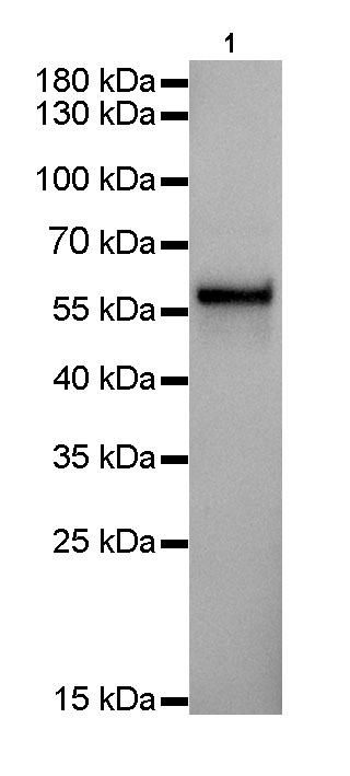
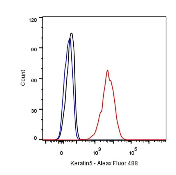
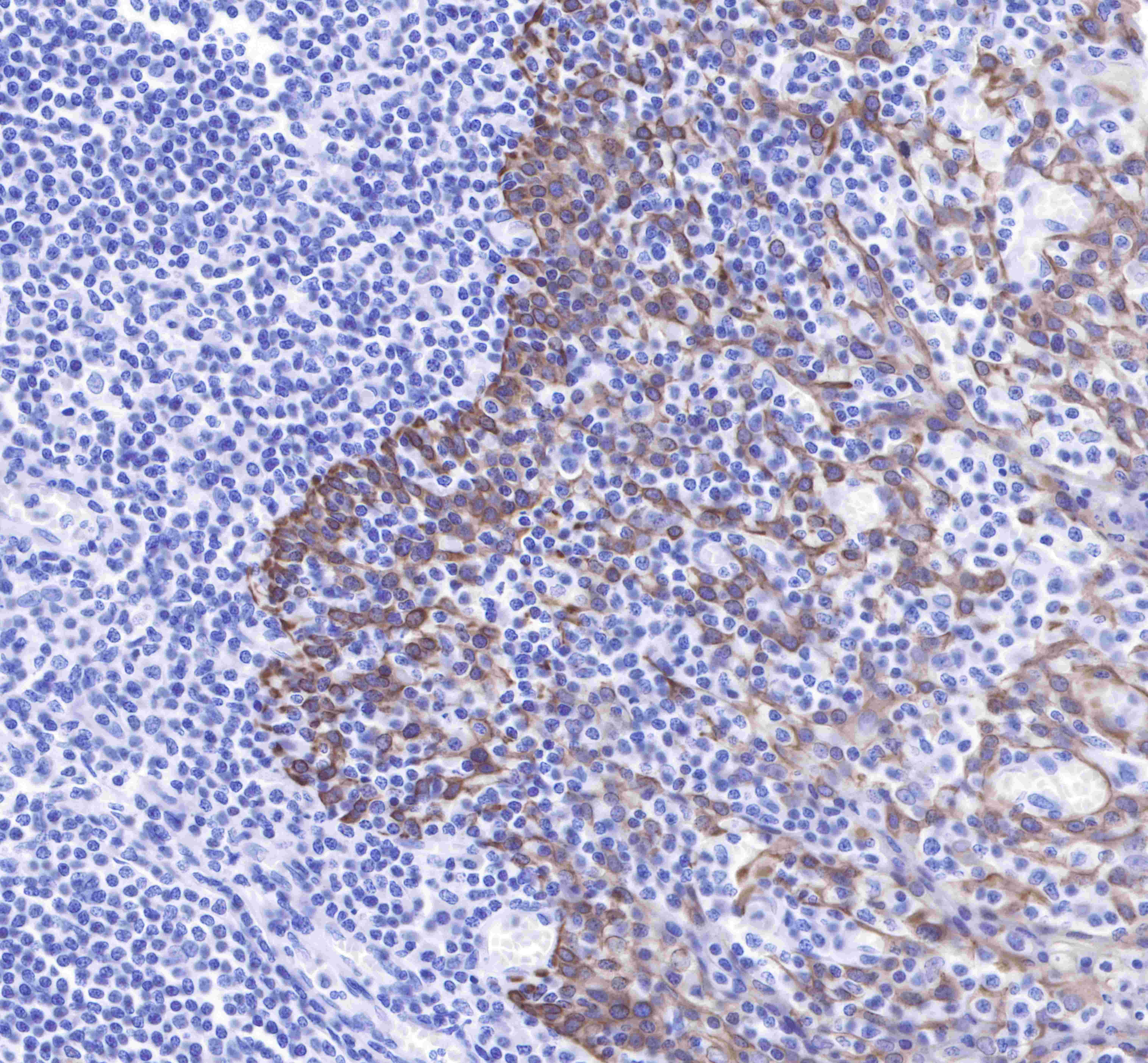
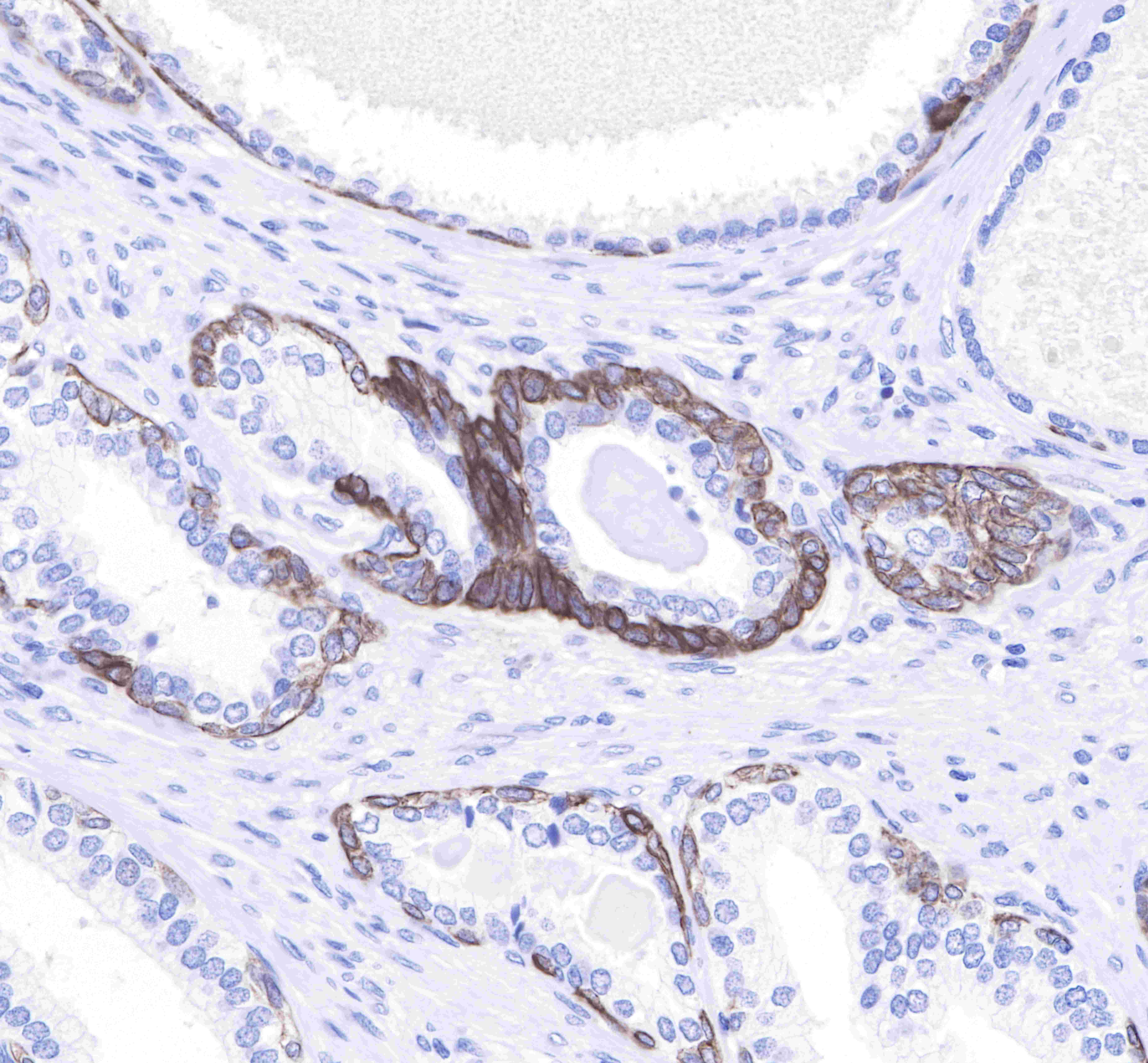
| Application |
WB, IHC-P, ICC, ICFCM |
| Reactivity |
Hu |
| Purification |
Protein A |
| Concentration |
0.5mg/ml |
| Conjugation |
Unconjugated |
| Physical Appearance |
Liquid |
| Storage Buffer |
PBS, 40% Glycerol, 0.05%BSA, 0.03% Proclin 300 |
| Stability & Storage |
12 months from date of receipt / reconstitution, -20 °C as supplied |
Dilution
| application |
dilution |
species |
| IHC-P |
1:1000-1:4000 |
|
| ICFCM |
1:500 |
|
| WB |
1:1000 |
|
| ICC |
1:1000 |
|
Background
Keratin 5, also known as KRT5, K5, or CK5, is a protein that is encoded in humans by the KRT5 gene. It dimerizes with keratin 14 and forms the intermediate filaments (IF) that make up the cytoskeleton of basal epithelial cells. Keratin 5 serves as a biomarker for several different types of cancer, including breast and lung cancers. It is often tested in conjunction with keratin 6, using CK5/6 antibodies, which target both keratin forms.