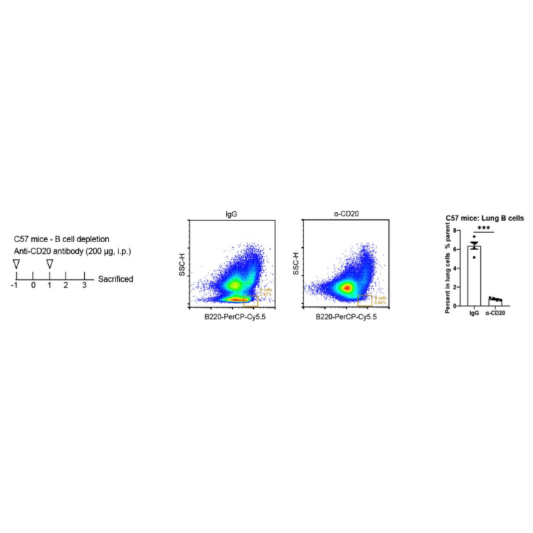
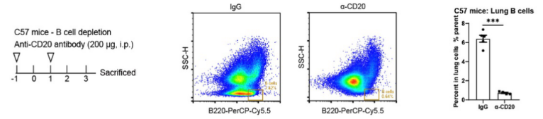
B cell depletion efficacy of S0B0547 in C57 mice
Invivo Recombinant anti-mouse CD20 antibody
Invivo Recombinant anti-mouse CD20 antibody
Price:
Regular price
$135 USD
Regular price
Sale price
$135 USD
Unit price
per
For shipping services or bulk orders, you may request a quotation.
Secure checkout with

View full details
Product Details
Product Details
Product Specification
| Host | Mouse |
| Clone Number | 18B12 |
| Antibody Type | Recombinant mAb |
| Isotype | IgG2a |
| Isotype Control | Invivo mouse IgG2a isotype control |
| Application | FCM, Functional Assay |
| Reactivity | Ms |
| Purification | Protein A |
| Concentration | 5 mg/ml |
| Purity | >95% by SDS-PAGE |
| Endotoxin | <1EU/mg |
| Physical Appearance | Liquid |
| Storage Buffer | PBS pH7.4, containing no preservative |
| Stability & Storage |
2 to 8 °C for 2 weeks under sterile conditions; -20 °C for 3 months under sterile conditions; -80 °C for 24 months under sterile conditions.
Please avoid repeated freeze-thaw cycles.
|
Dilution
| application | dilution | species |
| FCM | 1:500 |
Background
The cluster of differentiation 20 (CD20, aka Ly-44, or B-lymphocyte antigen) is a ~35 KDa cell surface protein mainly expressed by resting and activated B lymphocytes, but not plasma cells [1]. CD20 is also expressed by most malignant B cells, which makes it an ideal target for antibody-based immunotherapies against lymphoid malignancies [1-4]. For example, Rituximab, Obinutuzumab, and Ofatumumab are FDA-approved monoclonal anti-human CD20 antibodies for the treatment of B-cell non-Hodgkin’s lymphoma and B-cell chronic lymphocytic leukemia [1-4]. These antibodies mediate the target cell destruction through different mechanisms including direct signaling of apoptosis, complement activation (CDC), and cell-mediated cytotoxicity (ADCC).
The anti-mCD20 18B12 mAb is commonly used for in vivo depletion of the CD20+ B cell population to study the role of B cells in various immune responses, including auto-immune or tumoral contexts [1, 7].
Anti-mCD20-mIgG2a is provided in an Invivo grade, a high-quality standard specifically adapted to in vivo studies.
Picture
Picture
Validation Data
FC
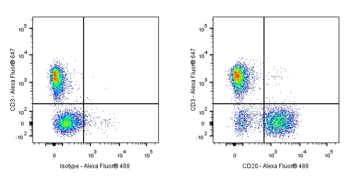
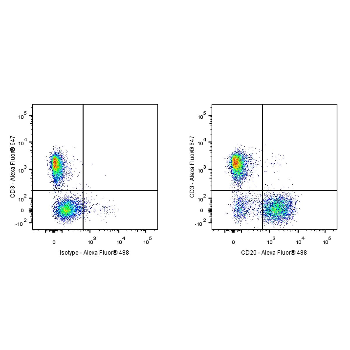
Flow cytometric analysis of mouse primary splenocytes labeling CD20 with purified antibody at 1/500 dilution (1 μg) (Right) compared with a Mouse monoclonal IgG isotype control (Left). Goat Anti-Mouse IgG Alexa Fluor® 488 was used as the secondary antibody.
Cells were surface stained with CD3-Alexa Fluor® 647, then stained with rabbit IgG (Left) / anti-CD20 (Right) separately. CD3 and CD20 are mutually exclusive expressed in mouse primary splenocytes. Gated on total viable cells.