WB result of HLA-DPB1 Rabbit mAb
Primary antibody: HLA-DPB1 Rabbit mAb at 1/1000 dilution
Lane 1: Jurkat whole cell lysate 20 µg
Lane 2: Daudi whole cell lysate 20 µg
Lane 3: Raji whole cell lysate 20 µg
Lane 4: Ramos whole cell lysate 20 µg
Lane 5: HuT 78 whole cell lysate 20 µg
Negative control: Jurkat whole cell lysate
Secondary antibody: Goat Anti-Rabbit IgG, (H+L), HRP conjugated at 1/10000 dilution
Predicted MW: 29 kDa
Observed MW: 29 kDa
HLA-DPB1 Recombinant Rabbit mAb (S-R263)
HLA-DPB1 Recombinant Rabbit mAb (S-R263)
Price:
Regular price
$130.00 SGD
Regular price
Sale price
$130.00 SGD
Unit price
per
For shipping services or bulk orders, you may request a quotation.
Secure checkout with

View full details
Product Details
Product Details
Product Specification
| Host | Rabbit |
| Antigen | HLA-DPB1 |
| Synonyms | HLA class II histocompatibility antigen DP beta 1 chain, HLA class II histocompatibility antigen DP(W4) beta chain, MHC class II antigen DPB1, HLA-DP1B, HLA DPB1 |
| Location | Cell membrane, Endoplasmic reticulum membrane |
| Accession | P04440 |
| Clone Number | S-R263 |
| Antibody Type | Recombinant mAb |
| Application | WB, IHC-P, ICC, ICFCM, IP |
| Reactivity | Hu |
| Purification | Protein A |
| Concentration | 0.5 mg/ml |
| Conjugation | Unconjugated |
| Physical Appearance | Liquid |
| Storage Buffer | PBS, 40% Glycerol, 0.05%BSA, 0.03% Proclin 300 |
| Stability & Storage | 12 months from date of receipt / reconstitution, -20 °C as supplied |
Dilution
| application | dilution | species |
| WB | 1:1000 | null |
| IP | 1:50 | null |
| IHC | 1:500-1:2000 | null |
| ICC | 1:500 | null |
| ICFCM | 1:500 | null |
Background
HLA class II histocompatibility antigen, DP(W2) beta chain is a protein that in humans is encoded by the HLA-DPB1 gene. HLA-DPB belongs to the HLA class II beta chain paralogues. This class II molecule is a heterodimer consisting of an alpha (DPA) and a beta chain (DPB), both anchored in the membrane. It plays a central role in the immune system by presenting peptides derived from extracellular proteins.
Picture
Picture
Western Blot
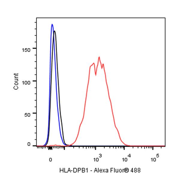
FC
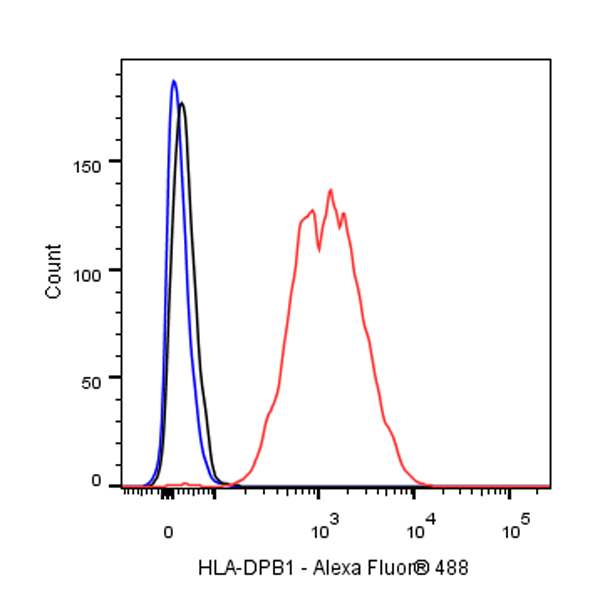
Flow cytometric analysis of Raji (Human Burkitt's lymphoma B lymphocyte) cells labelling HLA-DPB1 antibody at 1/500 (0.1 μg) dilution / (red) compared with a Rabbit monoclonal IgG (Black) isotype control and an unlabelled control (cells without incubation with primary antibody and secondary antibody) (Blue). Goat Anti –Rabbit IgG Alexa Fluor® 488 was used as the secondary antibody.
IP
HLA-DPB1 Rabbit mAb at 1/50 dilution (1 µg) immunoprecipitating HLA-DPB1 in 0.2 mg Raji whole cell lysate.
Western blot was performed on the immunoprecipitate using HLA-DPB1 Rabbit mAb at 1/1000 dilution.
Secondary antibody (HRP) for IP was used at 1/400 dilution.
Lane 1: Raji whole cell lysate 20 µg (Input)
Lane 2: HLA-DPB1 Rabbit mAb IP in Raji whole cell lysate
Lane 3: Rabbit monoclonal IgG IP in Raji whole cell lysate
Predicted MW: 29 kDa
Observed MW: 29 kDa
Immunohistochemistry
IHC shows positive staining in paraffin-embedded human spleen. Anti-HLA-DPB1 antibody was used at 1/500 dilution, followed by a HRP Polymer for Mouse & Rabbit IgG (ready to use). Counterstained with hematoxylin. Heat mediated antigen retrieval with Tris/EDTA buffer pH9.0 was performed before commencing with IHC staining protocol.
IHC shows positive staining in paraffin-embedded human tonsil. Anti-HLA-DPB1 antibody was used at 1/500 dilution, followed by a HRP Polymer for Mouse & Rabbit IgG (ready to use). Counterstained with hematoxylin. Heat mediated antigen retrieval with Tris/EDTA buffer pH9.0 was performed before commencing with IHC staining protocol.
IHC shows positive staining in paraffin-embedded human colon. Anti-HLA-DPB1 antibody was used at 1/500 dilution, followed by a HRP Polymer for Mouse & Rabbit IgG (ready to use). Counterstained with hematoxylin. Heat mediated antigen retrieval with Tris/EDTA buffer pH9.0 was performed before commencing with IHC staining protocol.
Negative control: IHC shows negative staining in paraffin-embedded human cerebral cortex. Anti-HLA-DPB1 antibody was used at 1/500 dilution, followed by a HRP Polymer for Mouse & Rabbit IgG (ready to use). Counterstained with hematoxylin. Heat mediated antigen retrieval with Tris/EDTA buffer pH9.0 was performed before commencing with IHC staining protocol.
IHC shows positive staining in paraffin-embedded human diffuse large B-cell lymphoma. Anti-HLA-DPB1 antibody was used at 1/500 dilution, followed by a HRP Polymer for Mouse & Rabbit IgG (ready to use). Counterstained with hematoxylin. Heat mediated antigen retrieval with Tris/EDTA buffer pH9.0 was performed before commencing with IHC staining protocol.
IHC shows positive staining in paraffin-embedded human cervical squamous cell carcinoma. Anti-HLA-DPB1 antibody was used at 1/500 dilution, followed by a HRP Polymer for Mouse & Rabbit IgG (ready to use). Counterstained with hematoxylin. Heat mediated antigen retrieval with Tris/EDTA buffer pH9.0 was performed before commencing with IHC staining protocol.
IHC shows positive staining in paraffin-embedded human lung cancer. Anti-HLA-DPB1 antibody was used at 1/2000 dilution, followed by a HRP Polymer for Mouse & Rabbit IgG (ready to use). Counterstained with hematoxylin. Heat mediated antigen retrieval with Tris/EDTA buffer pH9.0 was performed before commencing with IHC staining protocol.
Immunocytochemistry
ICC shows positive staining in Raji cells. Anti-HLA-DPB1 antibody was used at 1/500 dilution (Green) and incubated overnight at 4°C. Goat polyclonal Antibody to Rabbit IgG - H&L (Alexa Fluor® 488) was used as secondary antibody at 1/1000 dilution. The cells were fixed with 100% ice-cold methanol and permeabilized with 0.1% PBS-Triton X-100. Nuclei were counterstained with DAPI (Blue).
Negative control: ICC shows negative staining in Jurkat cells. Anti- HLA-DPB1 antibody was used at 1/500 dilution and incubated overnight at 4°C. Goat polyclonal Antibody to Rabbit IgG - H&L (Alexa Fluor® 488) was used as secondary antibody at 1/1000 dilution. The cells were fixed with 100% ice-cold methanol and permeabilized with 0.1% PBS-Triton X-100. Nuclei were counterstained with DAPI (Blue).