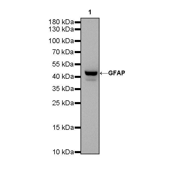
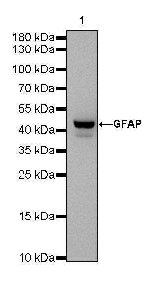
WB result of GFAP Rabbit mAb
Primary antibody: GFAP Rabbit mAb at 1/1000 dilution
Lane 1: mouse brain lysate 20 µg
Secondary antibody: Goat Anti-Rabbit IgG, (H+L), HRP conjugated at 1/10000 dilution
Predicted MW: 50kDa
Observed MW: 42kDa
GFAP Recombinant Rabbit mAb (SDT-306-10)
GFAP Recombinant Rabbit mAb (SDT-306-10)
Price:
Regular price
$45 USD
Regular price
Sale price
$45 USD
Unit price
per
For shipping services or bulk orders, you may request a quotation.
Secure checkout with

View full details
Product Details
Product Details
Product Specification
| Host | Rabbit |
| Antigen | GFAP |
| Synonyms | Glial fibrillary acidic protein |
| Immunogen | Synthetic Peptide |
| Location | Cytoplasm |
| Accession | P14136 |
| Clone Number | SDT-306-10 |
| Antibody Type | Recombinant mAb |
| Application | WB, IHC-P, IP, IF |
| Reactivity | Hu, Ms, Rt |
| Purification | Protein A |
| Concentration | 0.25 mg/ml |
| Conjugation | Unconjugated |
| Physical Appearance | Liquid |
| Storage Buffer | PBS, 40% Glycerol, 0.05% BSA, 0.03% Proclin 300 |
| Stability & Storage | 12 months from date of receipt / reconstitution, -20 °C as supplied |
Dilution
| application | dilution | species |
| WB | 1:500 | |
| IHC-P | 1:1000 | |
| IP | 1:25 | |
| IF | 1:5000 |
Background
Glial fibrillary acidic protein (GFAP) is the hallmark intermediate filament (IF; also known as nanofilament) protein in astrocytes, a main type of glial cells in the central nervous system (CNS). Astrocytes have a range of control and homeostatic functions in health and disease. Astrocytes assume a reactive phenotype in acute CNS trauma, ischemia, and in neurodegenerative diseases. This coincides with an upregulation and rearrangement of the IFs, which form a highly complex system composed of GFAP (10 isoforms), vimentin, synemin, and nestin [PMID: 25726916].
Picture
Picture
Western Blot
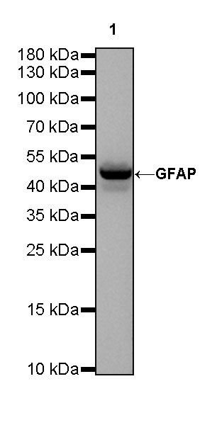
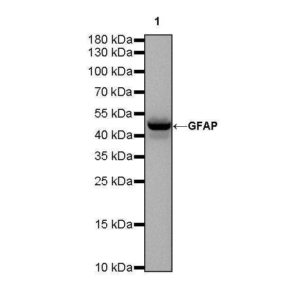
WB result of GFAP Rabbit mAb
Primary antibody: GFAP Rabbit mAb at 1/1000 dilution
Lane 1: rat brain lysate 20 µg
Secondary antibody: Goat Anti-Rabbit IgG, (H+L), HRP conjugated at 1/10000 dilution
Predicted MW: 50kDa
Observed MW: 50kDa
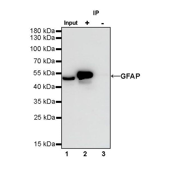
IP
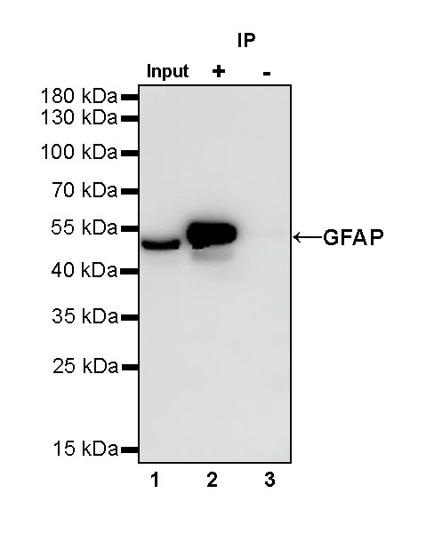
GFAP Rabbit mAb at 1/25 dilution (1 µg) immunoprecipitating GFAP in 0.4 mg rat brain lysate.
Western blot was performed on the immunoprecipitate using GFAP Rabbit mAb at 1/1000 dilution.
Secondary antibody (HRP) for IP was used at 1/400 dilution.
Lane 1: rat brain lysate 10 µg (Input)
Lane 2: GFAP Rabbit mAb IP in rat brain lysate
Lane 3: Rabbit monoclonal IgG IP in rat brain lysate
Predicted MW: 50 kDa
Observed MW: 50 kDa
(This blot was developed with high sensitivity substrate)
Immunohistochemistry
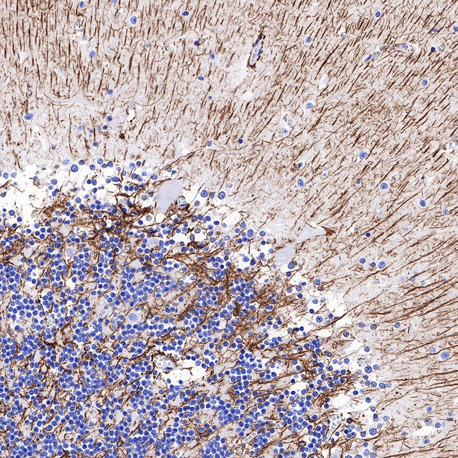
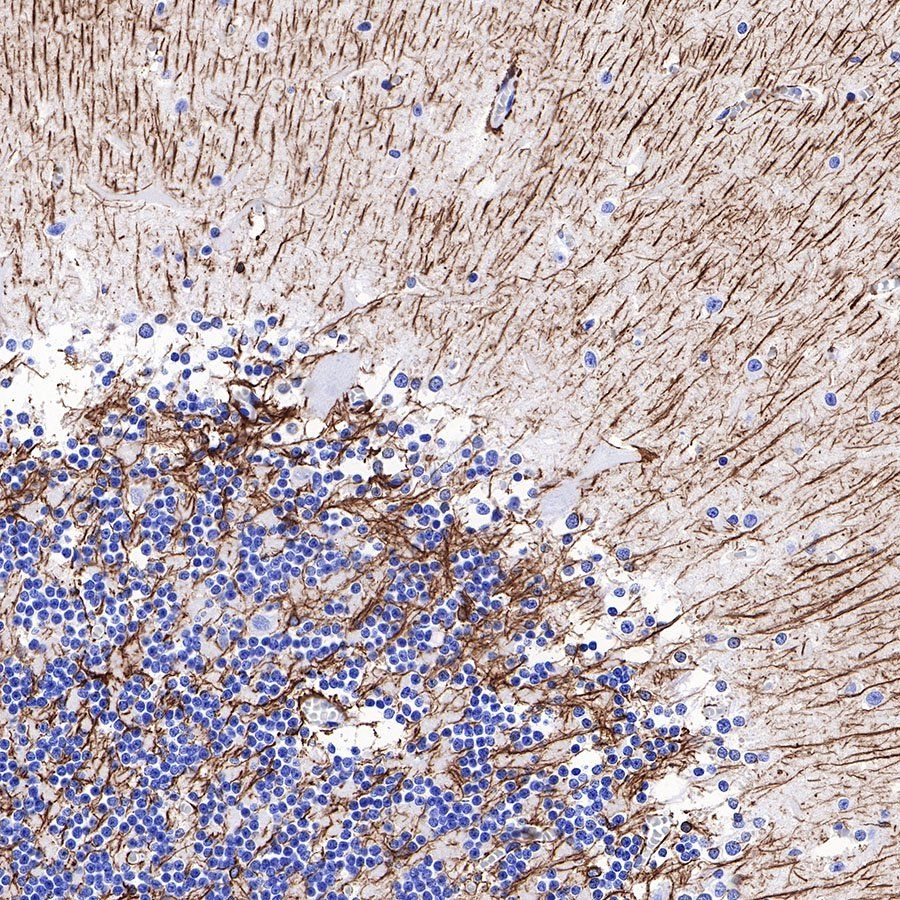
IHC shows positive staining in paraffin-embedded human cerebellum. Anti-GFAP antibody was used at 1/1000 dilution, followed by a HRP Polymer for Mouse & Rabbit IgG (ready to use). Counterstained with hematoxylin. Heat mediated antigen retrieval with Tris/EDTA buffer pH9.0 was performed before commencing with IHC staining protocol.
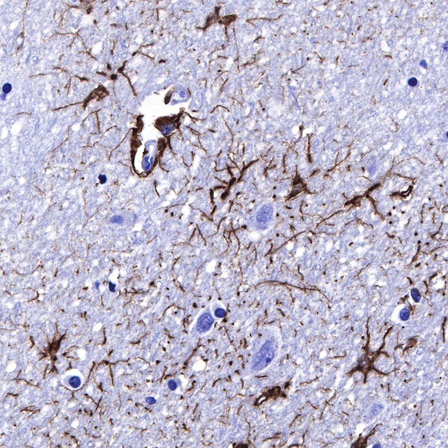
IHC shows positive staining in paraffin-embedded human cerebral cortex. Anti-GFAP antibody was used at 1/1000 dilution, followed by a HRP Polymer for Mouse & Rabbit IgG (ready to use). Counterstained with hematoxylin. Heat mediated antigen retrieval with Tris/EDTA buffer pH9.0 was performed before commencing with IHC staining protocol.
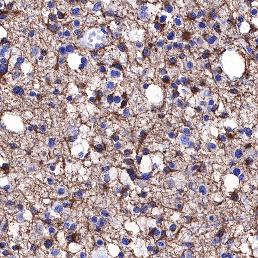
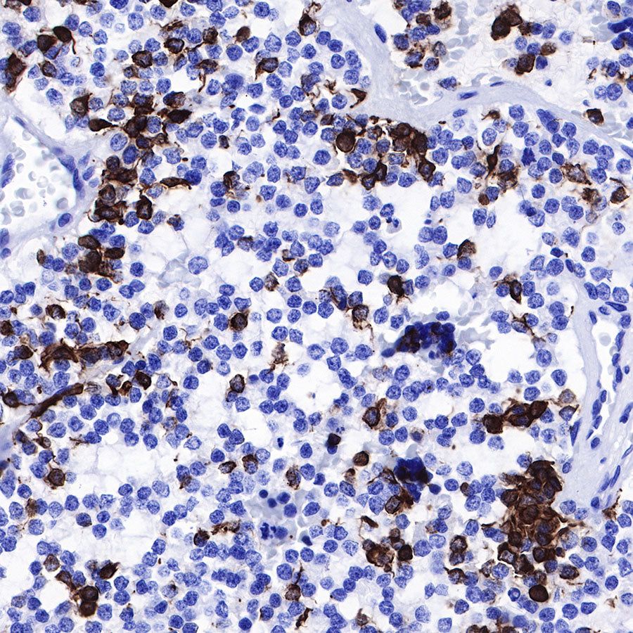
IHC shows positive staining in paraffin-embedded human glioma. Anti-GFAP antibody was used at 1/1000 dilution, followed by a HRP Polymer for Mouse & Rabbit IgG (ready to use). Counterstained with hematoxylin. Heat mediated antigen retrieval with Tris/EDTA buffer pH9.0 was performed before commencing with IHC staining protocol.

IHC shows positive staining in paraffin-embedded human astrocytoma. Anti-GFAP antibody was used at 1/1000 dilution, followed by a HRP Polymer for Mouse & Rabbit IgG (ready to use). Counterstained with hematoxylin. Heat mediated antigen retrieval with Tris/EDTA buffer pH9.0 was performed before commencing with IHC staining protocol.
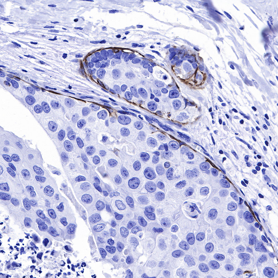
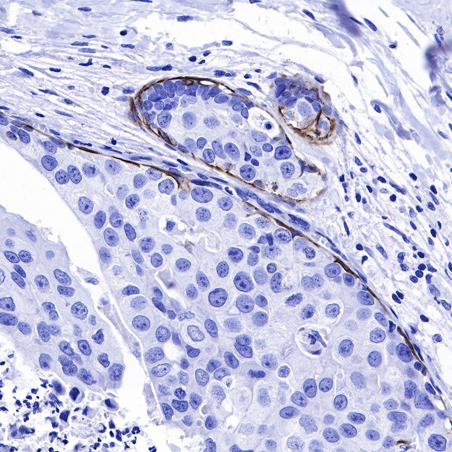
IHC shows positive staining in paraffin-embedded human breast cancer. Anti-GFAP antibody was used at 1/1000 dilution, followed by a HRP Polymer for Mouse & Rabbit IgG (ready to use). Counterstained with hematoxylin. Heat mediated antigen retrieval with Tris/EDTA buffer pH9.0 was performed before commencing with IHC staining protocol.
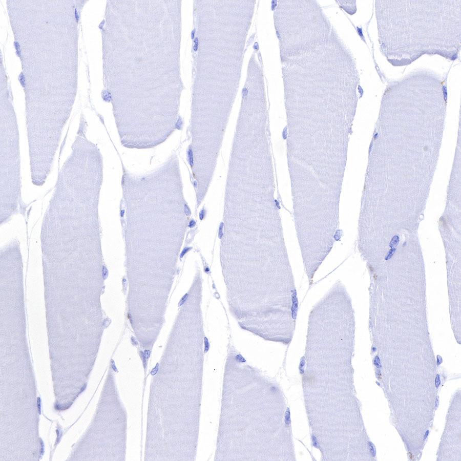
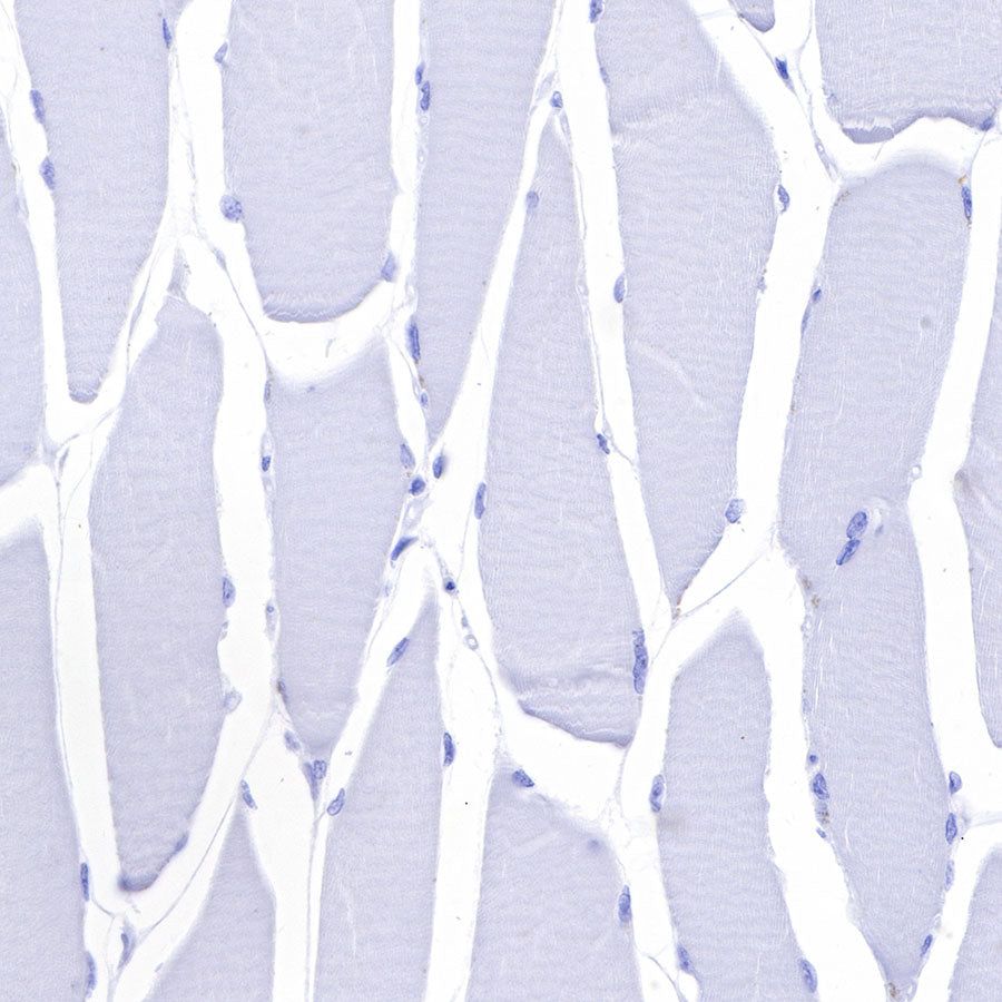
Negative control: IHC shows negative staining in paraffin-embedded human skeletal muscle. Anti-GFAP antibody was used at 1/1000 dilution, followed by a HRP Polymer for Mouse & Rabbit IgG (ready to use). Counterstained with hematoxylin. Heat mediated antigen retrieval with Tris/EDTA buffer pH9.0 was performed before commencing with IHC staining protocol.
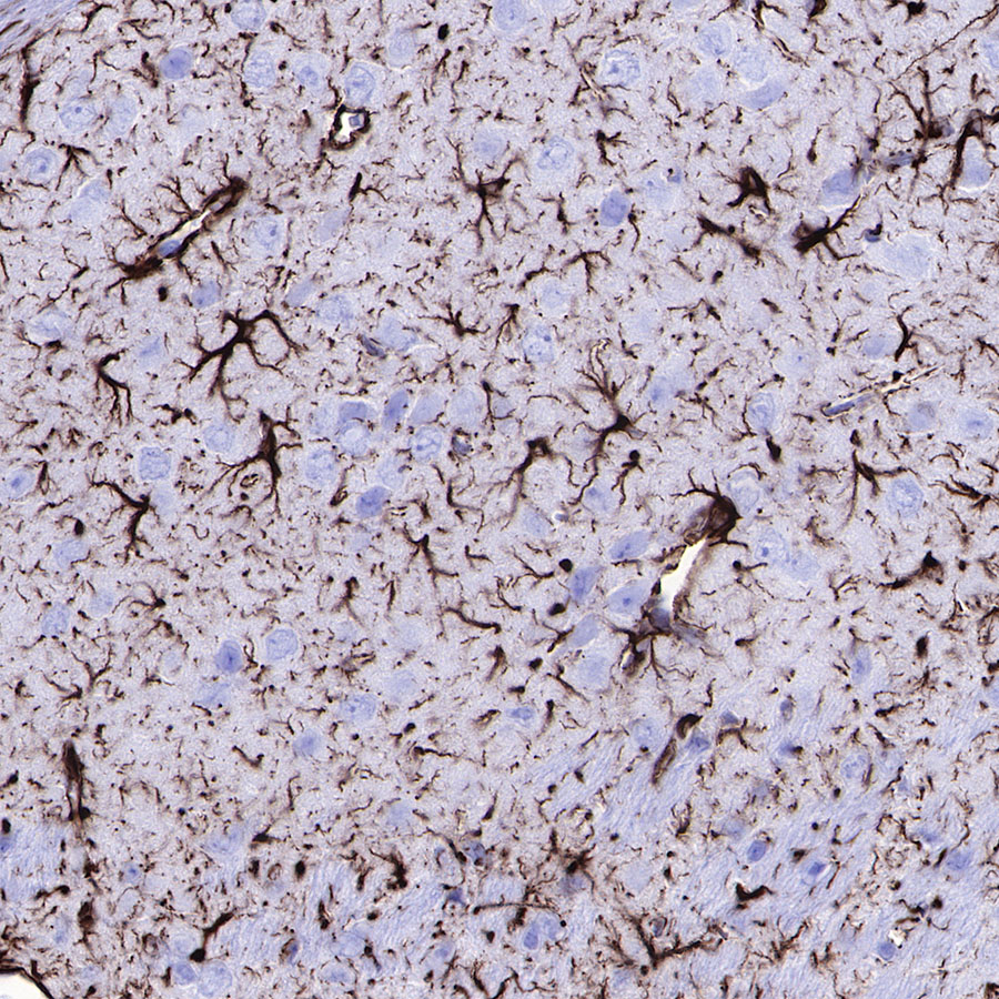
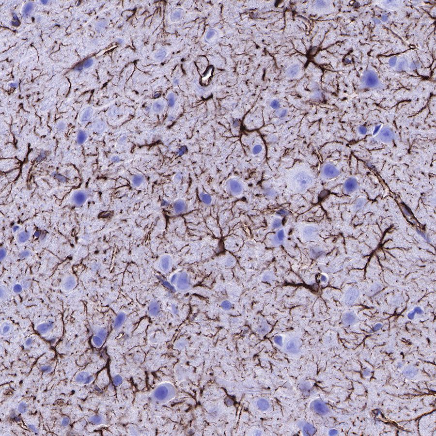
IHC shows positive staining in paraffin-embedded mouse cerebral cortex. Anti-GFAP antibody was used at 1/1000 dilution, followed by a HRP Polymer for Mouse & Rabbit IgG (ready to use). Counterstained with hematoxylin. Heat mediated antigen retrieval with Tris/EDTA buffer pH9.0 was performed before commencing with IHC staining protocol.
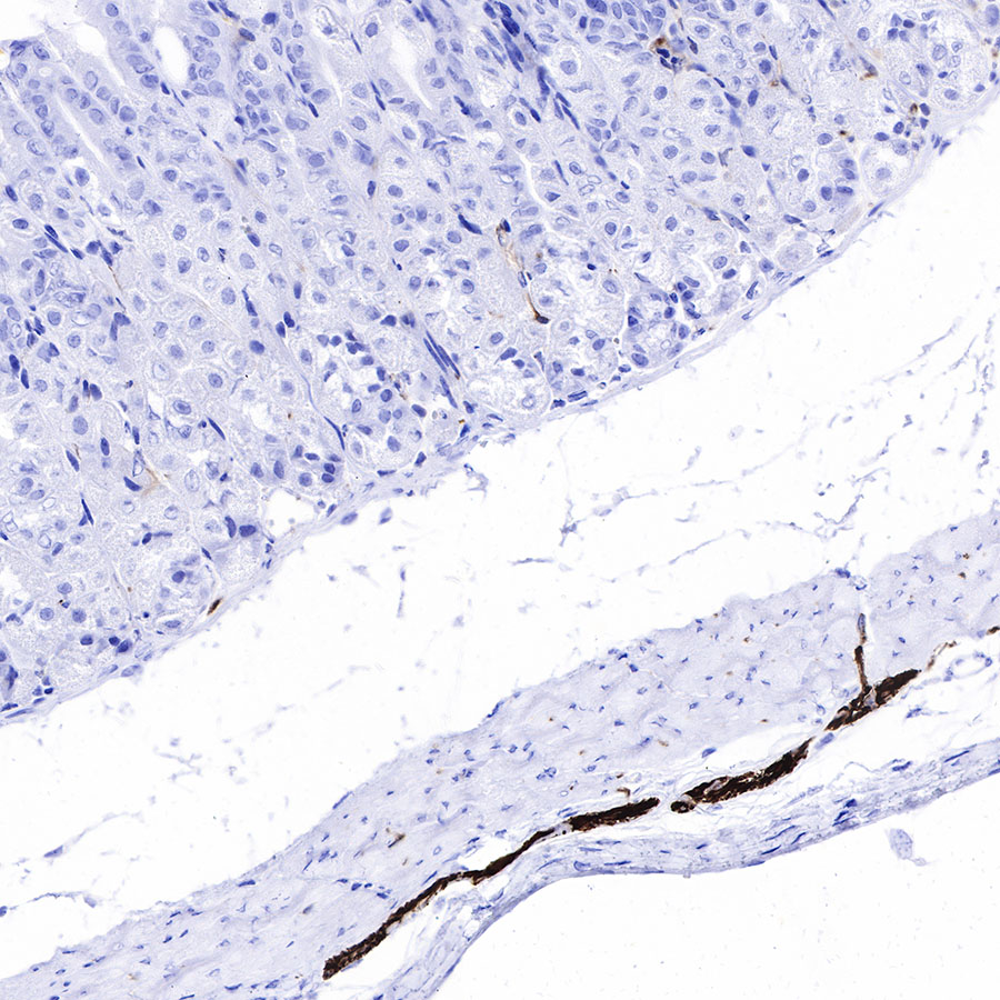
IHC shows positive staining in paraffin-embedded mouse stomach. Anti-GFAP antibody was used at 1/1000 dilution, followed by a HRP Polymer for Mouse & Rabbit IgG (ready to use). Counterstained with hematoxylin. Heat mediated antigen retrieval with Tris/EDTA buffer pH9.0 was performed before commencing with IHC staining protocol.
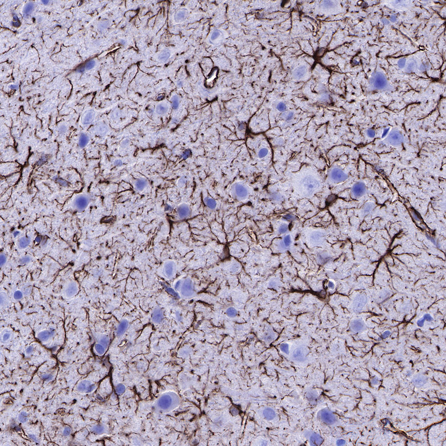
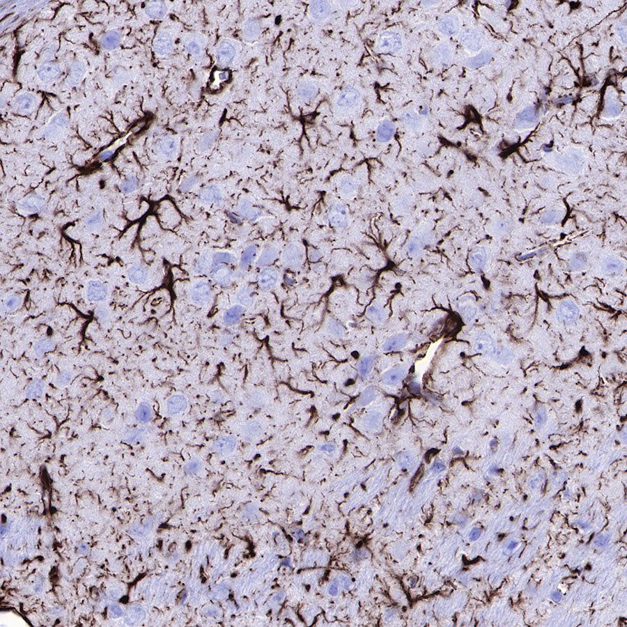
IHC shows positive staining in paraffin-embedded rat cerebral cortex. Anti-GFAP antibody was used at 1/1000 dilution, followed by a HRP Polymer for Mouse & Rabbit IgG (ready to use). Counterstained with hematoxylin. Heat mediated antigen retrieval with Tris/EDTA buffer pH9.0 was performed before commencing with IHC staining protocol.

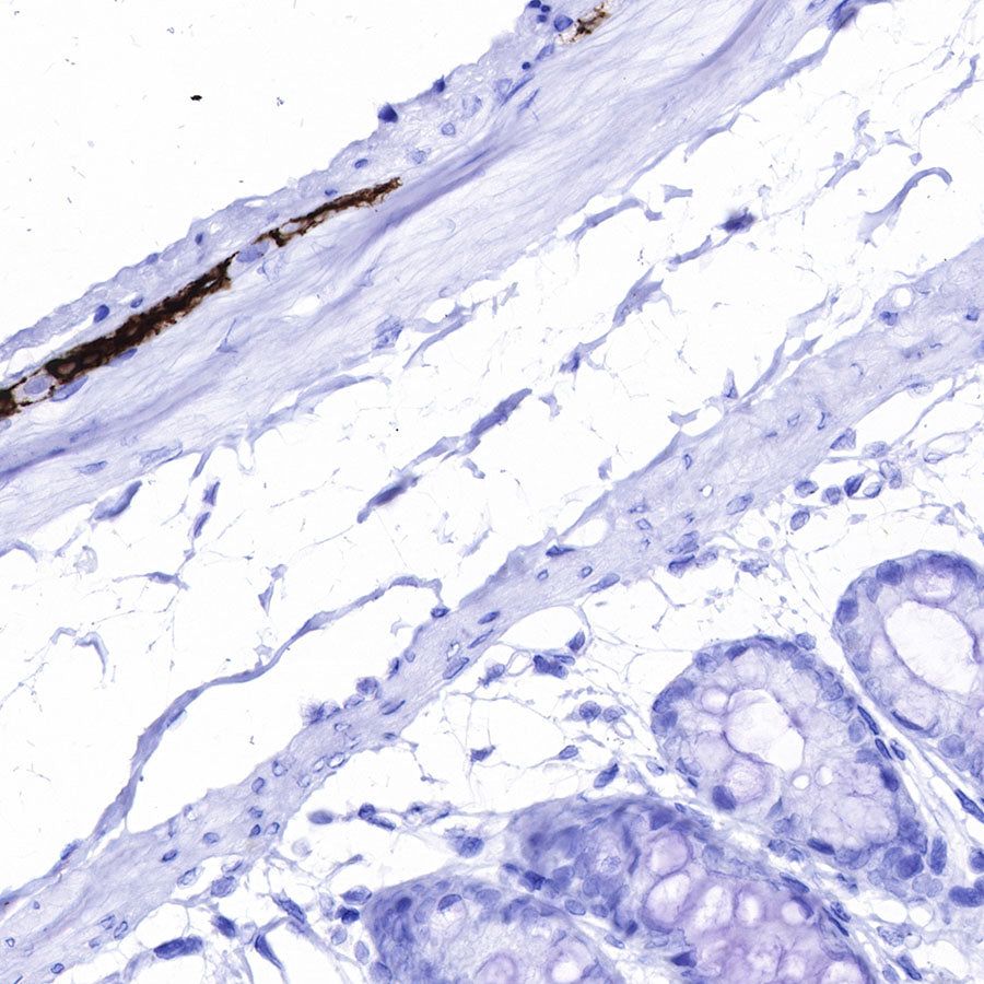
IHC shows positive staining in paraffin-embedded rat colon. Anti-GFAP antibody was used at 1/1000 dilution, followed by a HRP Polymer for Mouse & Rabbit IgG (ready to use). Counterstained with hematoxylin. Heat mediated antigen retrieval with Tris/EDTA buffer pH9.0 was performed before commencing with IHC staining protocol.
Immunofluorescence
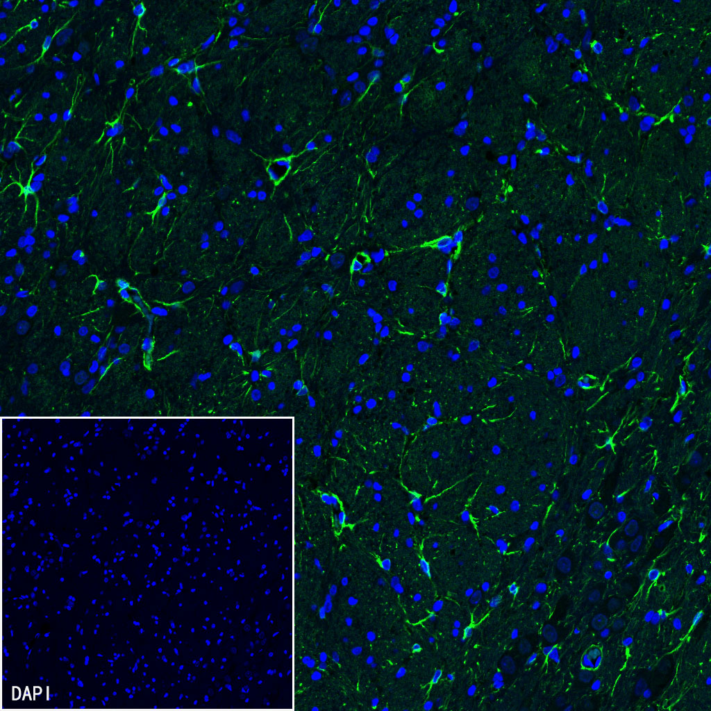
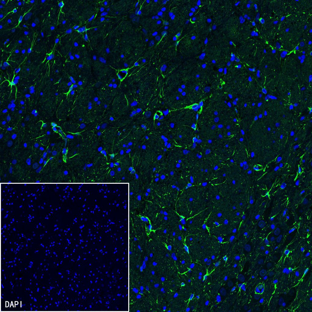
IF shows positive staining in paraffin-embedded rat cerebellum. Anti-GFAP antibody was used at 1/2000 dilution (Green) and incubated overnight at 4°C. Goat polyclonal Antibody to Rabbit IgG - H&L (Alexa Fluor® 488) was used as secondary antibody at 1/1000 dilution. Counterstained with DAPI (Blue). Heat mediated antigen retrieval with EDTA buffer pH9.0 was performed before commencing with IF staining protocol.

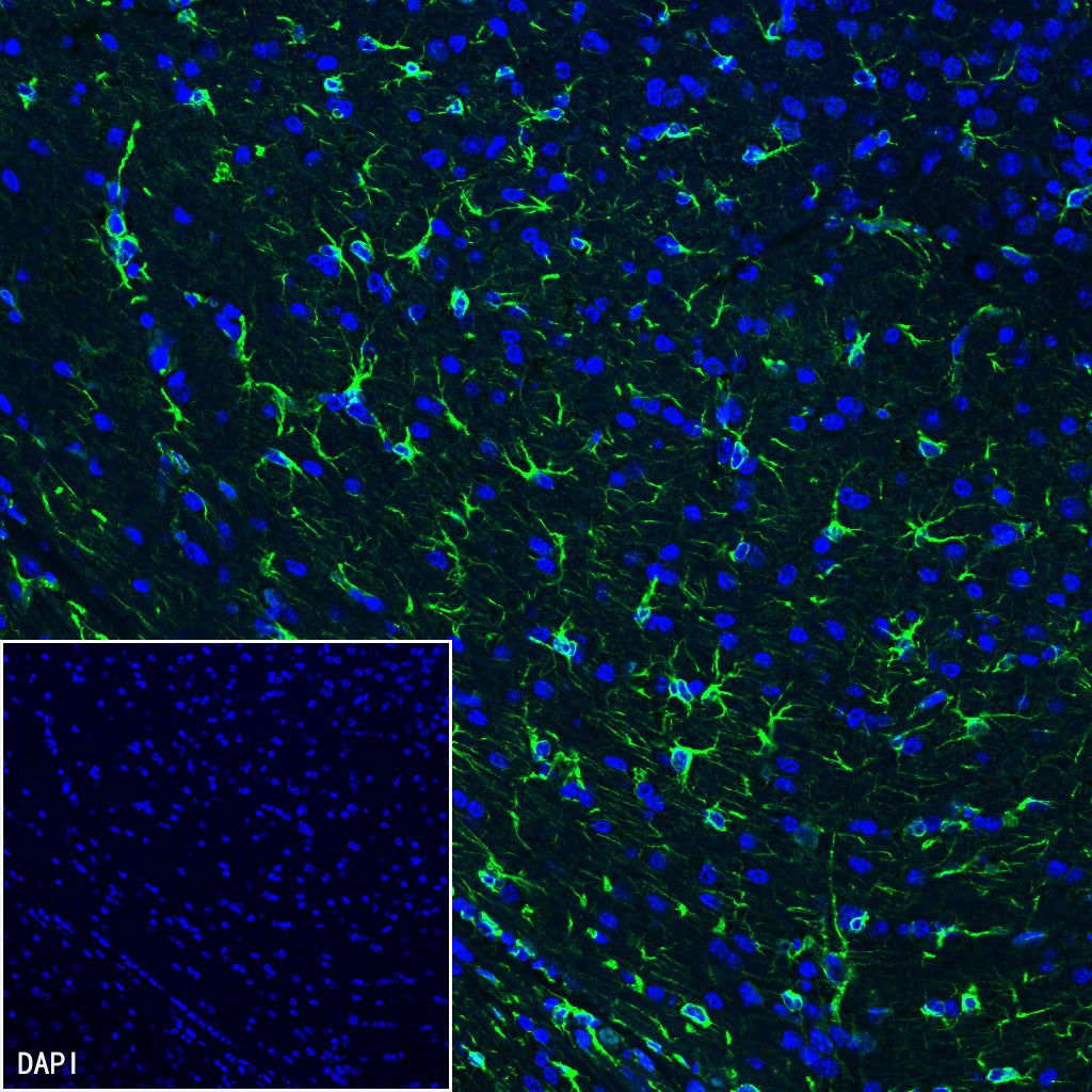
IF shows positive staining in paraffin-embedded mouse cerebellum. Anti-GFAP antibody was used at 1/2000 dilution (Green) and incubated overnight at 4°C. Goat polyclonal Antibody to Rabbit IgG - H&L (Alexa Fluor® 488) was used as secondary antibody at 1/1000 dilution. Counterstained with DAPI (Blue). Heat mediated antigen retrieval with EDTA buffer pH9.0 was performed before commencing with IF staining protocol.
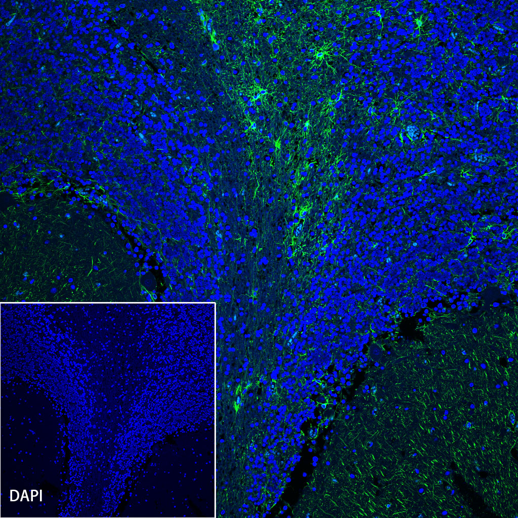
IF shows positive staining in paraffin-embedded human cerebellum. Anti-GFAP antibody was used at 1/2000 dilution (Green) and incubated overnight at 4°C. Goat polyclonal Antibody to Rabbit IgG - H&L (Alexa Fluor® 488) was used as secondary antibody at 1/1000 dilution. Counterstained with DAPI (Blue). Heat mediated antigen retrieval with EDTA buffer pH9.0 was performed before commencing with IF staining protocol.