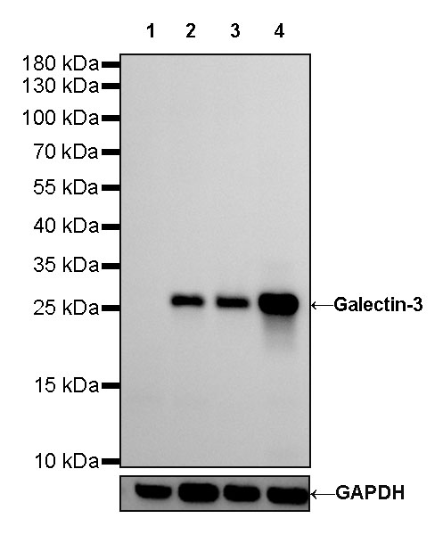
WB result of Galectin-3 Rabbit mAb
Primary antibody: Galectin-3 Rabbit mAb at 1/500 dilution
Lane 1: LNCaP whole cell lysate 20 µg
Lane 2: HeLa whole cell lysate 20 µg
Lane 3: SW480 whole cell lysate 20 µg
Lane 4: MCF7 whole cell lysate 20 µg
Negative control: LNCaP whole cell lysate
Secondary antibody: Goat Anti-Rabbit IgG, (H+L), HRP conjugated at 1/10000 dilution
Predicted MW: 26 kDa
Observed MW: 28 kDa
Galectin-3 Recombinant Rabbit mAb (SDT-370-111)
Galectin-3 Recombinant Rabbit mAb (SDT-370-111)
Price:
Regular price
$100 USD
Regular price
Sale price
$100 USD
Unit price
per
For shipping services or bulk orders, you may request a quotation.
Secure checkout with

View full details
Product Details
Product Details
Product Specification
| Host | Rabbit |
| Antigen | Galectin-3 |
| Synonyms | Gal-3, 35 kDa lectin, CBP 35, Galactose-specific lectin 3, GALBP, IgE-binding protein, L-31, Laminin-binding protein, Lectin L-29, Mac-2 antigen |
| Immunogen | Recombinant Protein |
| Location | Cytoplasm, Nucleus, Secreted |
| Accession | P17931 |
| Clone Number | SDT-370-111 |
| Antibody Type | Rabbit mAb |
| Application | WB, IHC-P, ICC, IP |
| Reactivity | Hu |
| Purification | Protein A |
| Concentration | 0.25 mg/ml |
| Conjugation | Unconjugated |
| Physical Appearance | Liquid |
| Storage Buffer | PBS, 40% Glycerol, 0.05% BSA, 0.03% Proclin 300 |
| Stability & Storage | 12 months from date of receipt / reconstitution, -20 °C as supplied |
Dilution
| application | dilution | species |
| WB | 1:500 | null |
| WB | 1:500 | null |
| IHC-P | 1:2500-1:5000 | null |
| IP | 1:25 | null |
| ICC | 1:250 | null |
Background
Galectin-3 (Gal-3; formally named MAC-2) is a β-galactoside-binding lectin. Various cell types produce Gal-3 under either normal conditions and/or pathological conditions. Gal-3 can be present in cells' nuclei and cytoplasm, secreted from producing cells, and associated with cells' plasma membranes [PMID: 36274989].
Picture
Picture
Western Blot
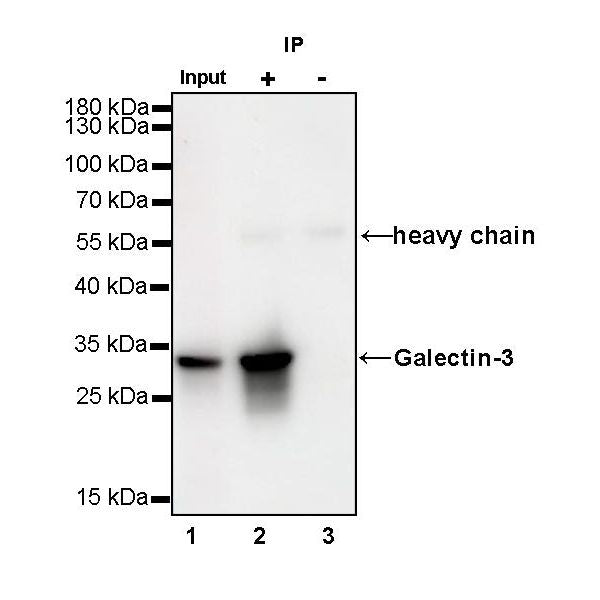
IP
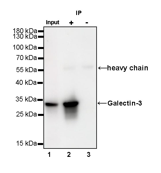
Galectin-3 Rabbit mAb at 1/25 dilution (1 µg) immunoprecipitating Galectin-3 in 0.4 mg SW480 whole cell lysate.
Western blot was performed on the immunoprecipitate using Galectin-3 Rabbit mAb at 1/1000 dilution.
Secondary antibody (HRP) for IP was used at 1/400 dilution.
Lane 1: SW480 whole cell lysate 20 µg (Input)
Lane 2: Galectin-3 Rabbit mAb IP in SW480 whole cell lysate
Lane 3: Rabbit monoclonal IgG IP in SW480 whole cell lysate
Predicted MW: 26 kDa
Observed MW: 30 kDa
Immunohistochemistry
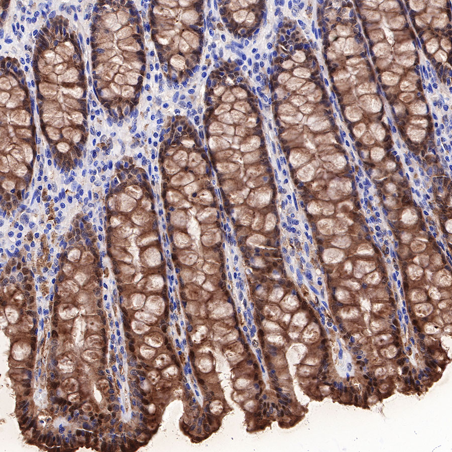
IHC shows positive staining in paraffin-embedded human colon. Anti-Galectin-3 antibody was used at 1/2500 dilution, followed by a HRP Polymer for Mouse & Rabbit IgG (ready to use). Counterstained with hematoxylin. Heat mediated antigen retrieval with Tris/EDTA buffer pH9.0 was performed before commencing with IHC staining protocol.
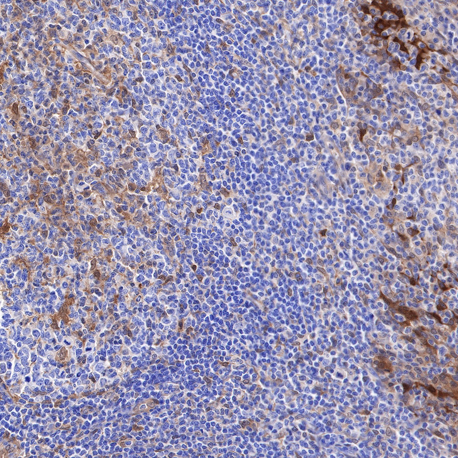
IHC shows positive staining in paraffin-embedded human tonsil. Anti-Galectin-3 antibody was used at 1/2500 dilution, followed by a HRP Polymer for Mouse & Rabbit IgG (ready to use). Counterstained with hematoxylin. Heat mediated antigen retrieval with Tris/EDTA buffer pH9.0 was performed before commencing with IHC staining protocol.
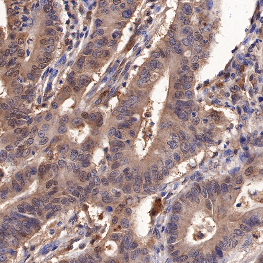
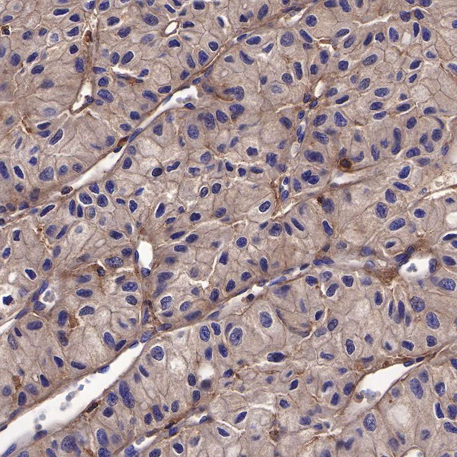
IHC shows positive staining in paraffin-embedded human colon cancer. Anti-Galectin-3 antibody was used at 1/2500 dilution, followed by a HRP Polymer for Mouse & Rabbit IgG (ready to use). Counterstained with hematoxylin. Heat mediated antigen retrieval with Tris/EDTA buffer pH9.0 was performed before commencing with IHC staining protocol.
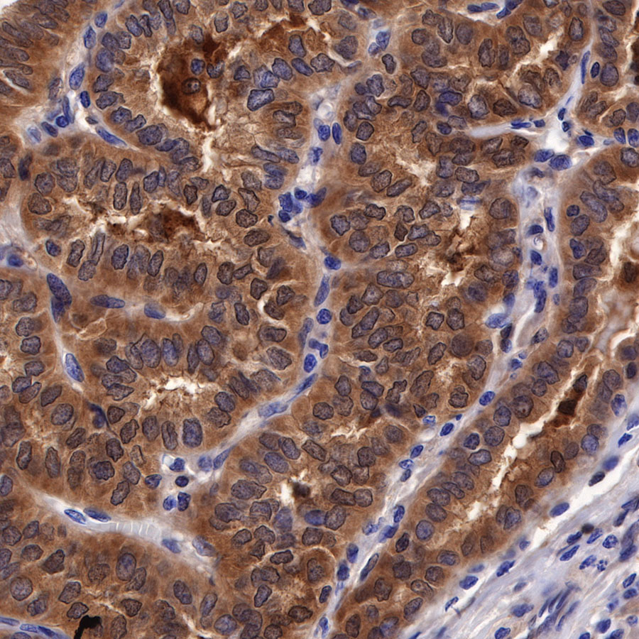
IHC shows positive staining in paraffin-embedded human papillary thyroid carcinoma. Anti-Galectin-3 antibody was used at 1/2500 dilution, followed by a HRP Polymer for Mouse & Rabbit IgG (ready to use). Counterstained with hematoxylin. Heat mediated antigen retrieval with Tris/EDTA buffer pH9.0 was performed before commencing with IHC staining protocol.
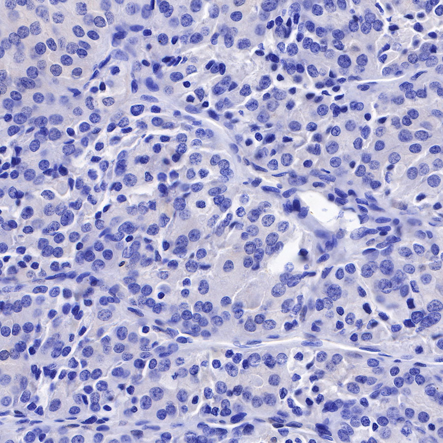
Negative control: IHC shows negative staining in paraffin-embedded human follicular thyroid carcinoma. Anti-Galectin-3 antibody was used at 1/2500 dilution, followed by a HRP Polymer for Mouse & Rabbit IgG (ready to use). Counterstained with hematoxylin. Heat mediated antigen retrieval with Tris/EDTA buffer pH9.0 was performed before commencing with IHC staining protocol.
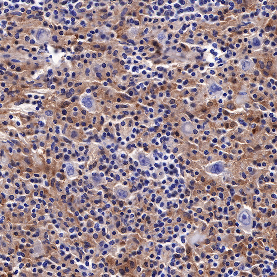
IHC shows positive staining in paraffin-embedded human Hodgkin's lymphoma. Anti-Galectin-3 antibody was used at 1/5000 dilution, followed by a HRP Polymer for Mouse & Rabbit IgG (ready to use). Counterstained with hematoxylin. Heat mediated antigen retrieval with Tris/EDTA buffer pH9.0 was performed before commencing with IHC staining protocol.
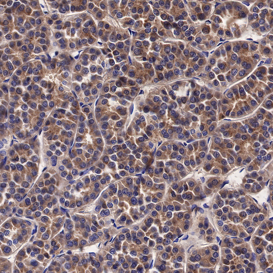
IHC shows positive staining in paraffin-embedded human oncocytic adenoma. Anti-Galectin-3 antibody was used at 1/2500 dilution, followed by a HRP Polymer for Mouse & Rabbit IgG (ready to use). Counterstained with hematoxylin. Heat mediated antigen retrieval with Tris/EDTA buffer pH9.0 was performed before commencing with IHC staining protocol.
IHC shows positive staining in paraffin-embedded human chromophobe renal carcinoma. Anti-Galectin-3 antibody was used at 1/2500 dilution, followed by a HRP Polymer for Mouse & Rabbit IgG (ready to use). Counterstained with hematoxylin. Heat mediated antigen retrieval with Tris/EDTA buffer pH9.0 was performed before commencing with IHC staining protocol.
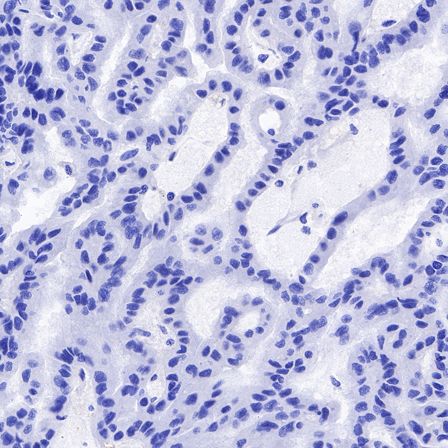
Negative control: IHC shows negative staining in paraffin-embedded human papillary renal cell carcinoma. Anti-Galectin-3 antibody was used at 1/2500 dilution, followed by a HRP Polymer for Mouse & Rabbit IgG (ready to use). Counterstained with hematoxylin. Heat mediated antigen retrieval with Tris/EDTA buffer pH9.0 was performed before commencing with IHC staining protocol.
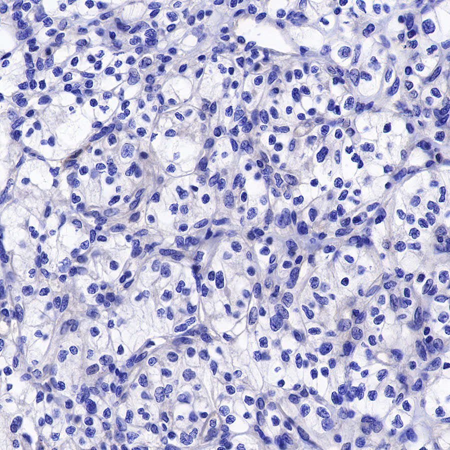
Negative control: IHC shows negative staining in paraffin-embedded human renal clear cell carcinoma. Anti-Galectin-3 antibody was used at 1/2500 dilution, followed by a HRP Polymer for Mouse & Rabbit IgG (ready to use). Counterstained with hematoxylin. Heat mediated antigen retrieval with Tris/EDTA buffer pH9.0 was performed before commencing with IHC staining protocol.
Immunocytochemistry
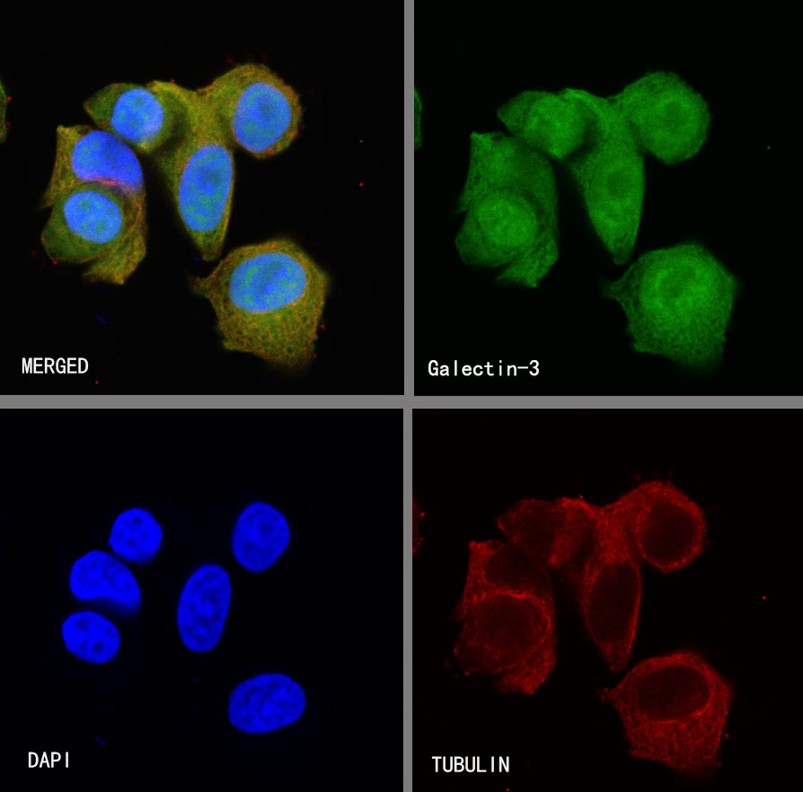
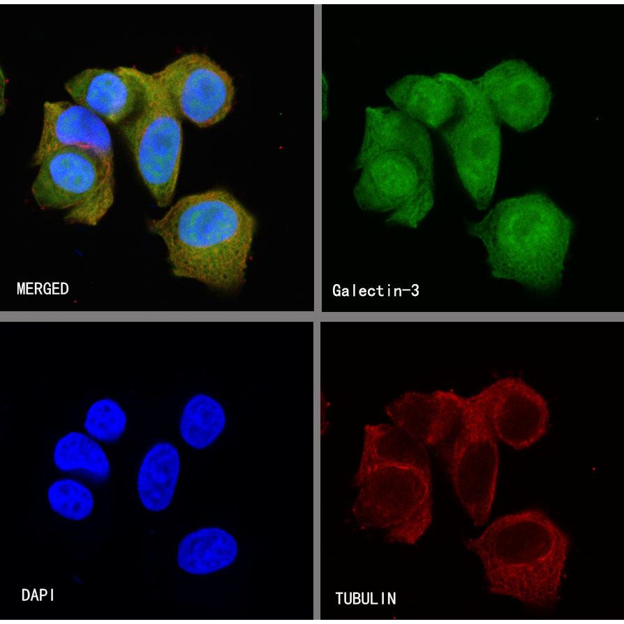
ICC shows positive staining in MCF7 cells. Anti-Galectin-3 antibody was used at 1/250 dilution (Green) and incubated overnight at 4°C. Goat polyclonal Antibody to Rabbit IgG - H&L (Alexa Fluor® 488) was used as secondary antibody at 1/1000 dilution. The cells were fixed with 4% PFA and permeabilized with 0.1% PBS-Triton X-100. Nuclei were counterstained with DAPI (Blue). Counterstain with tubulin (red).
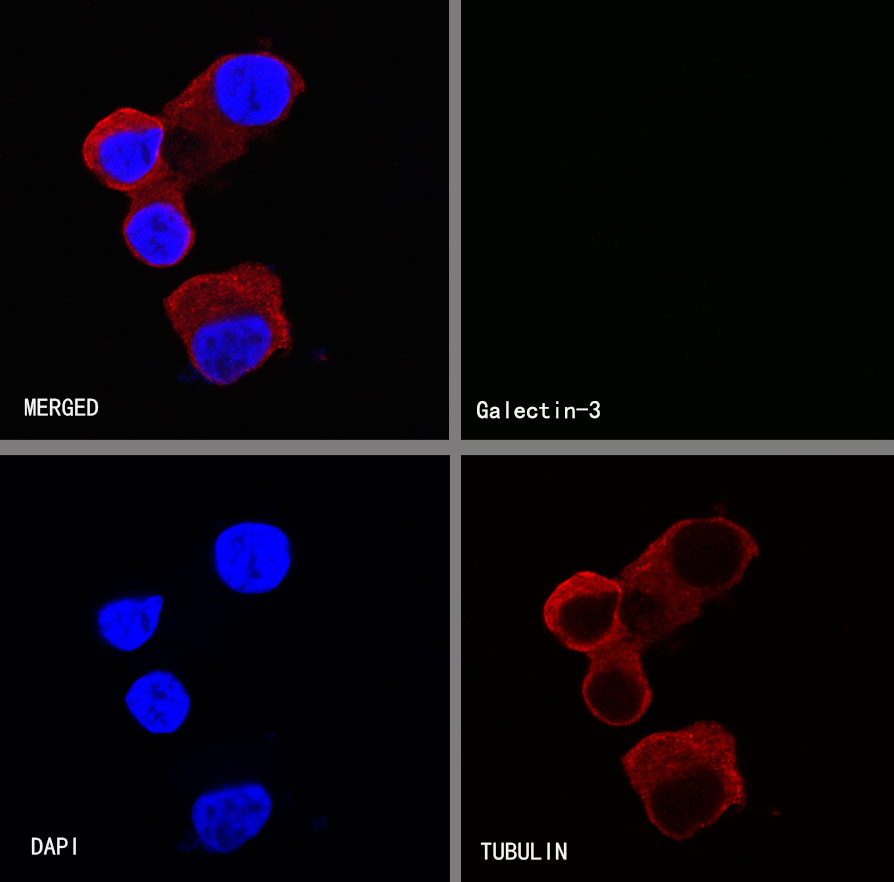
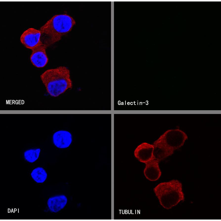
Negative control:ICC shows negative staining in LnCaP cells. Anti-Galectin-3 antibody was used at 1/250 dilution and incubated overnight at 4°C. Goat polyclonal Antibody to Rabbit IgG - H&L (Alexa Fluor® 488) was used as secondary antibody at 1/1000 dilution. The cells were fixed with 4% PFA and permeabilized with 0.1% PBS-Triton X-100. Nuclei were counterstained with DAPI (Blue). Counterstain with tubulin (red).