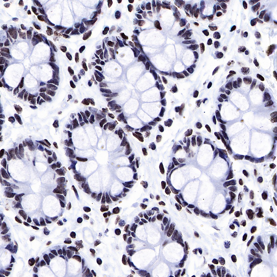
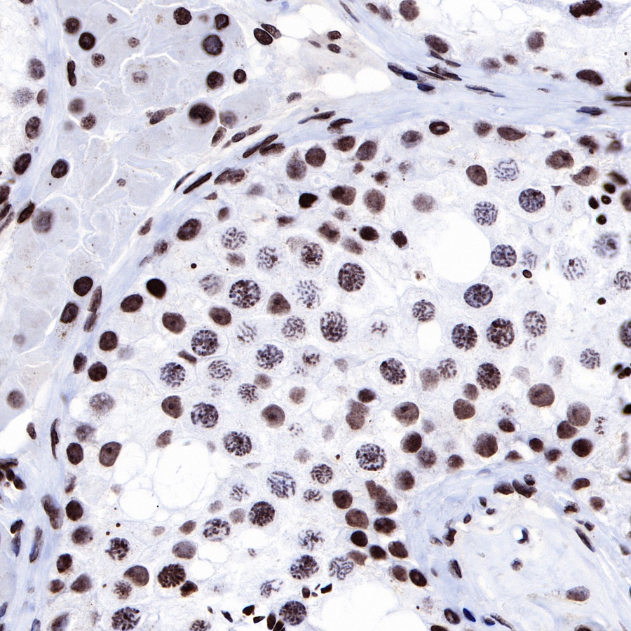
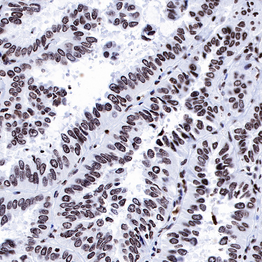
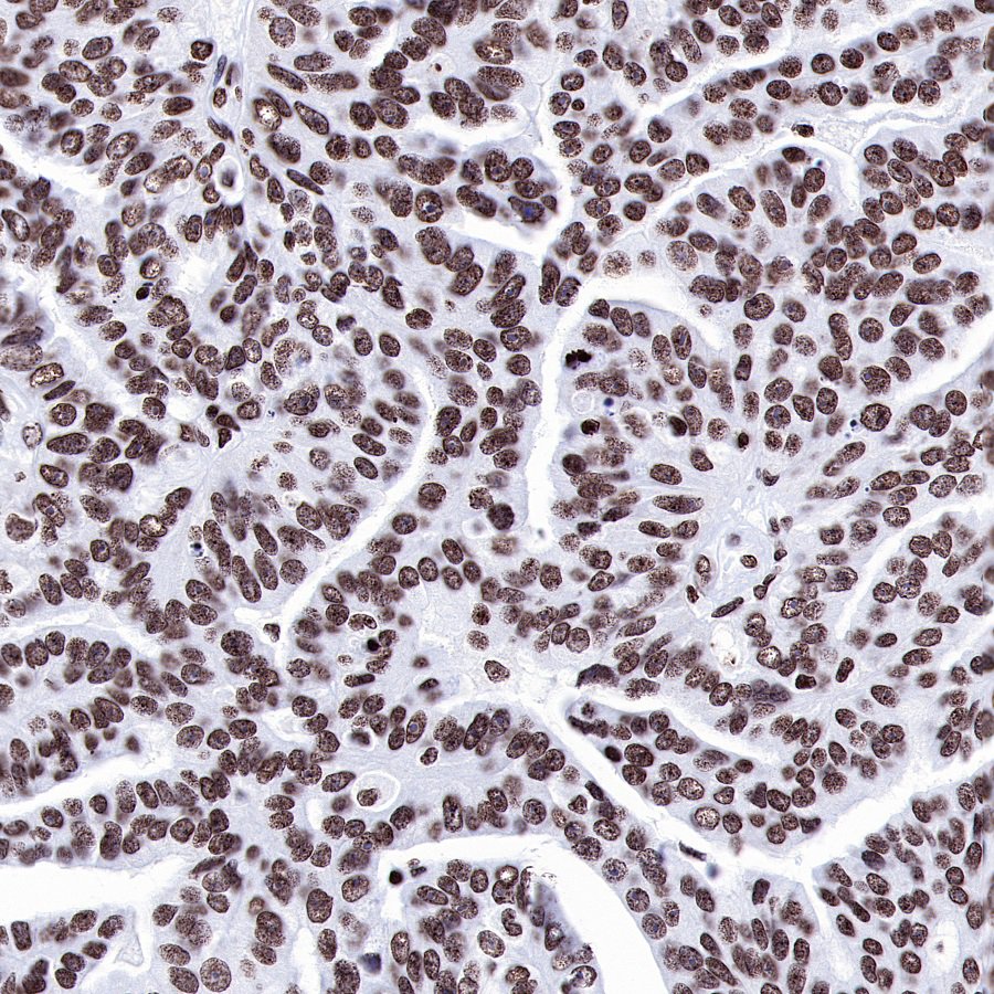
Product Specification
| Host |
Rabbit |
| Antigen |
ERCC1 |
| Synonyms |
DNA excision repair protein ERCC-1 |
| Immunogen |
N/A |
| Location |
Nucleus |
| Accession |
P07992 |
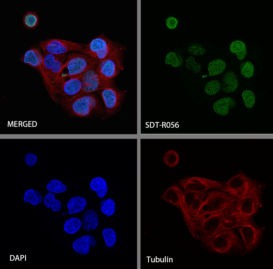
| Clone Number |
SDT-R056 |
| Antibody Type |
Rabbit mAb |
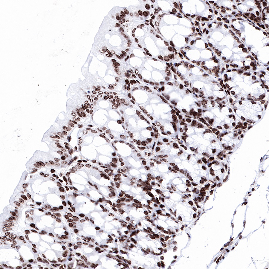
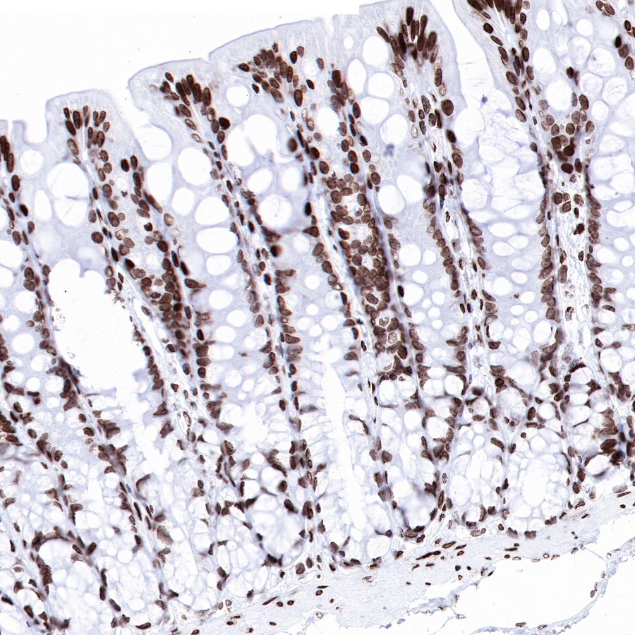
| Application |
IHC-P, ICC |
| Reactivity |
Hu, Ms, Rt |
| Purification |
Protein A |
| Concentration |
0.5 mg/ml |
| Physical Appearance |
Liquid |
| Storage Buffer |
PBS, 40% Glycerol, 0.05%BSA, 0.03% Proclin 300 |
| Stability & Storage |
12 months from date of receipt / reconstitution, -20 °C as supplied |
Dilution
| application |
dilution |
species |
| IHC-P |
1:1000 |
null |
| ICC |
1:50 |
null |
Background
ERCC1 is a non-catalytic component of a structure-specific DNA repair endonuclease responsible for the 5'-incision during DNA repair. It is responsible, in conjunction with SLX4, for the first step in the repair of interstrand cross-links (ICL). It participates in the processing of anaphase bridge-generating DNA structures, which consist in incompletely processed DNA lesions arising during S or G2 phase, and can result in cytokinesis failure. ERCC1 is also required for homology-directed repair (HDR) of DNA double-strand breaks, in conjunction with SLX4.