Product Specification
| Host |
Rabbit |
| Antigen |
Claudin-1 |
| Synonyms |
CLDN1,CLD1, SEMP1 |
| Immunogen |
N/A |
| Location |
Cell membrane |
| Accession |
O95832 |
| Clone Number |
SDT-R024 |
| Antibody Type |
Rabbit mAb |
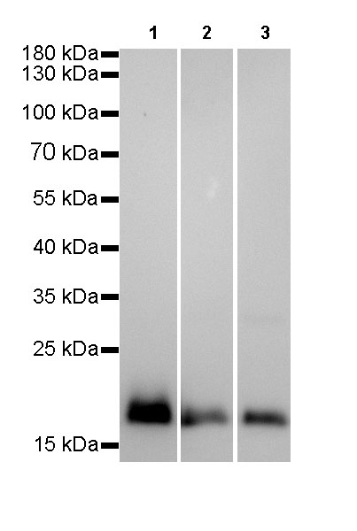
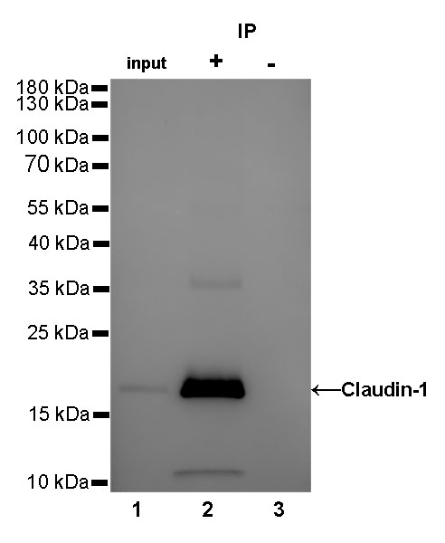
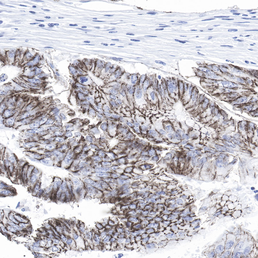
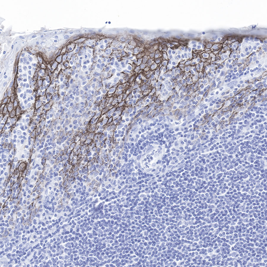
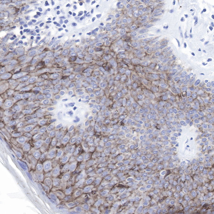
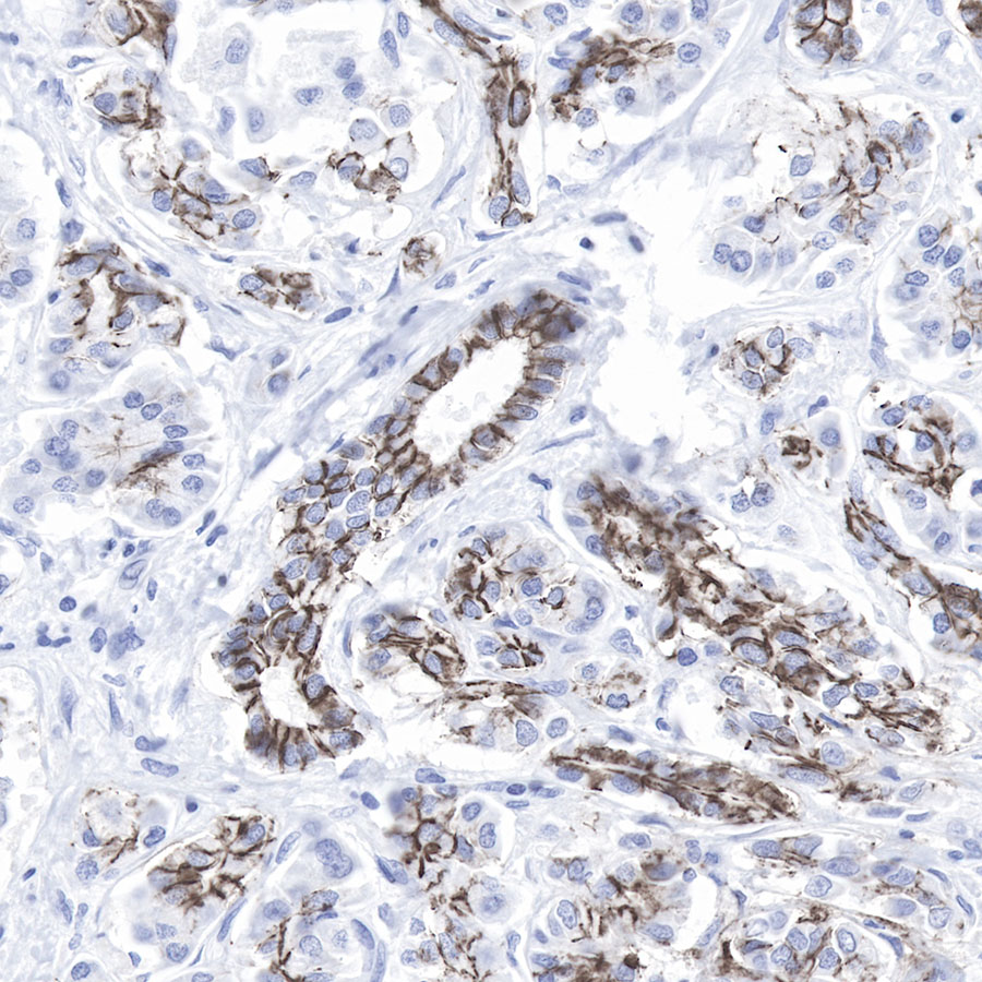
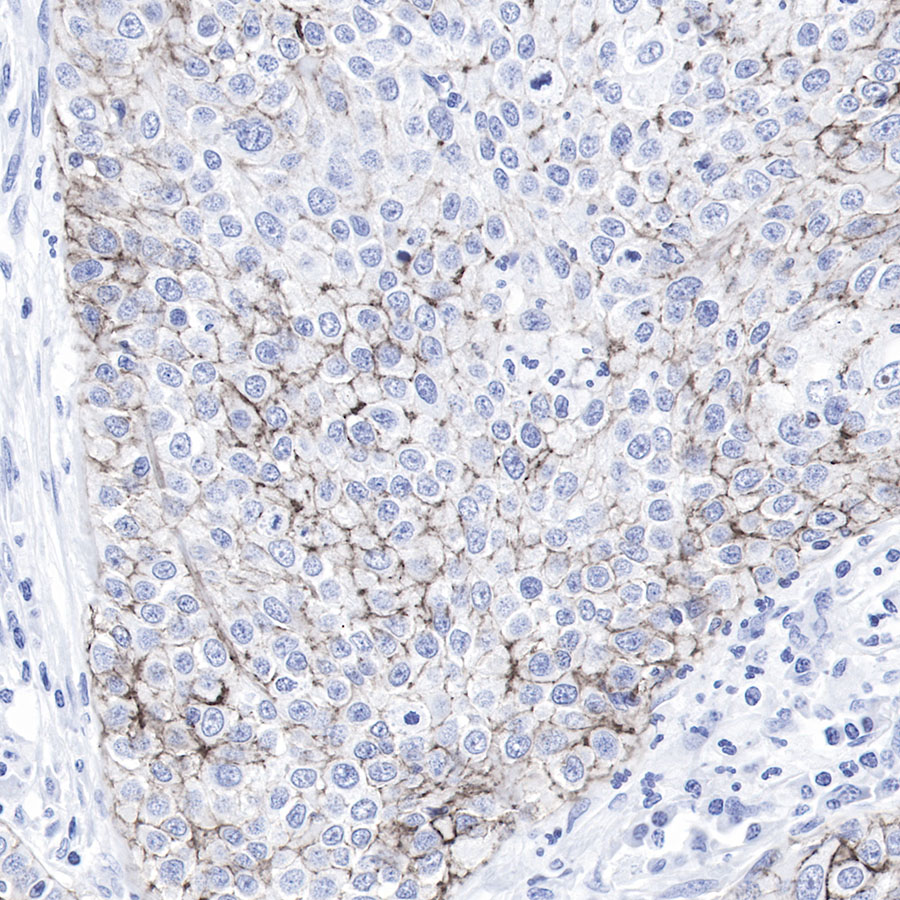
| Application |
WB, IHC-P, IP |
| Reactivity |
Hu |
| Purification |
Protein A |
| Concentration |
0.5mg/ml |
| Physical Appearance |
Liquid |
| Storage Buffer |
PBS, 40% Glycerol, 0.05%BSA, 0.03% Proclin 300 |
| Stability & Storage |
12 months from date of receipt / reconstitution, -20 °C as supplied |
Dilution
| application |
dilution |
species |
| IP |
1:25 |
|
| WB |
1:1000 |
|
| IHC-P |
1:500 |
|
Background
The close connection consists of seal protein and closed protein, and can be connected to the cell skeleton. The Claudin family includes 23 integrated membrane proteins and their expression. Its expression is different from each tissue type, and can determine the intensity and characteristics of epithelial cell barrier. Claudin-1 is expressed in epithelial cells and nerve cysts.The change of Claudin protein expression mode is related to multiple cancer types. It mainly expresses the peripheral neuroma (partial type of negative), derivative skin fibrous sarcoma, low -level malignant fibrous mucus -like sarcoma, hardened fibroblastoma, fibroma, deity fibroma, and neurobetoma.