Product Specification
| Host |
Rabbit |
| Antigen |
CDX2 |
| Synonyms |
CDX-3, Caudal-type homeobox protein 2 |
| Immunogen |
Synthetic Peptide |
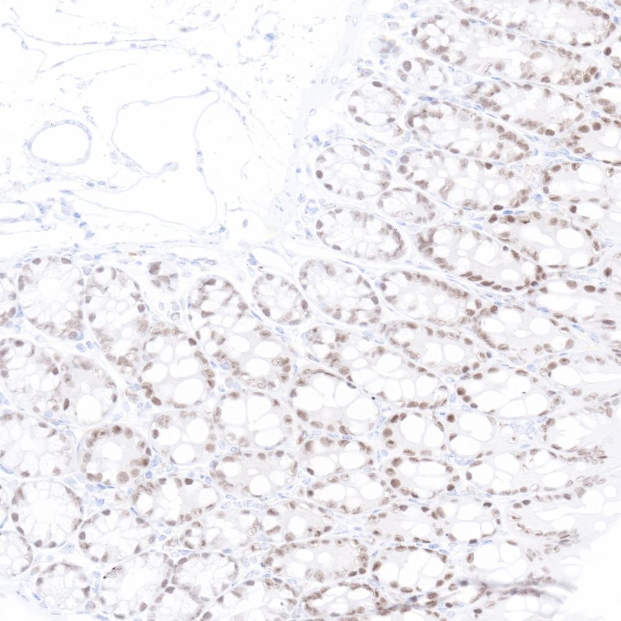
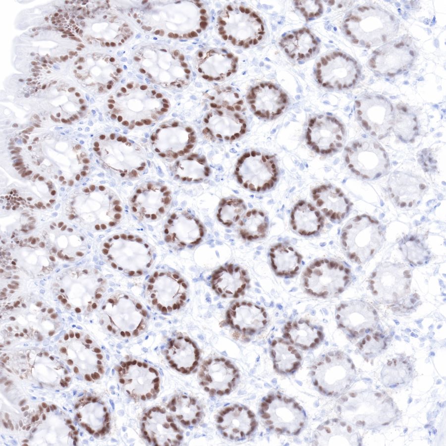
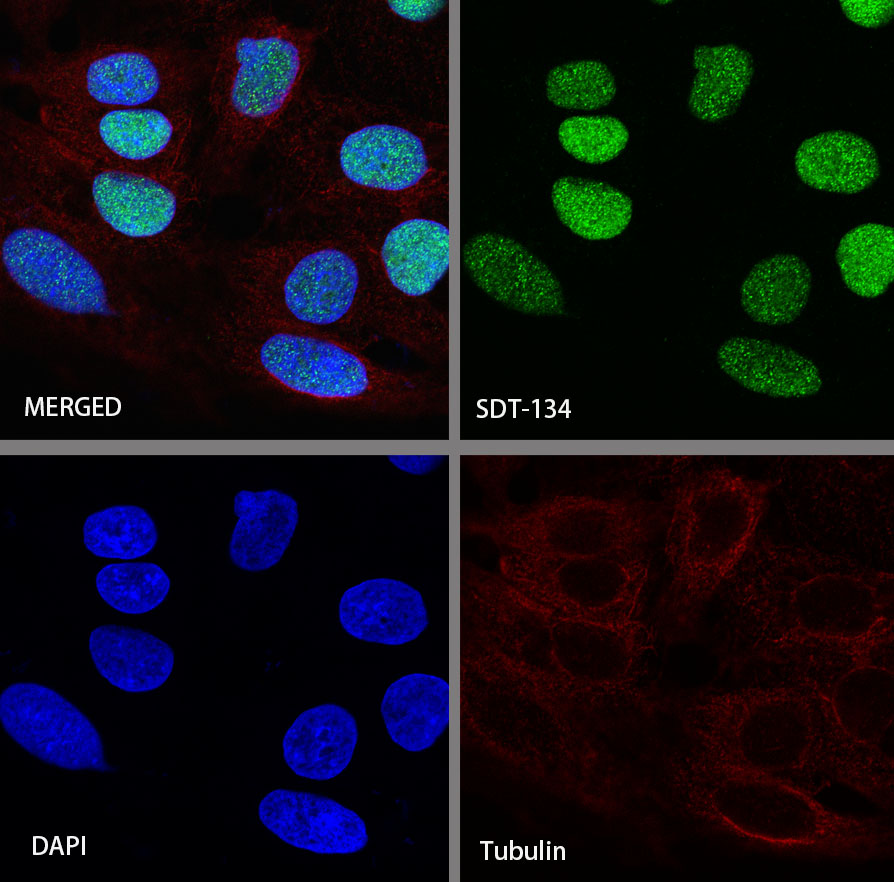
| Location |
Nucleus |
| Accession |
Q99626 |
| Clone Number |
SDT-134-76 |
| Antibody Type |
Rabbit mAb |
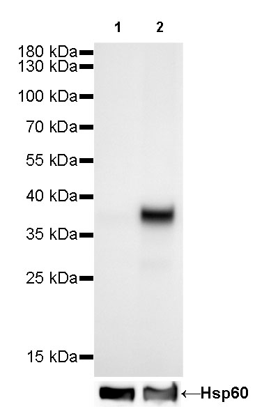
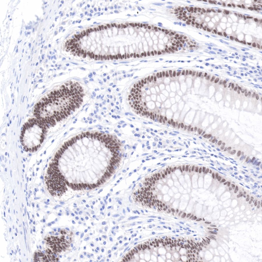
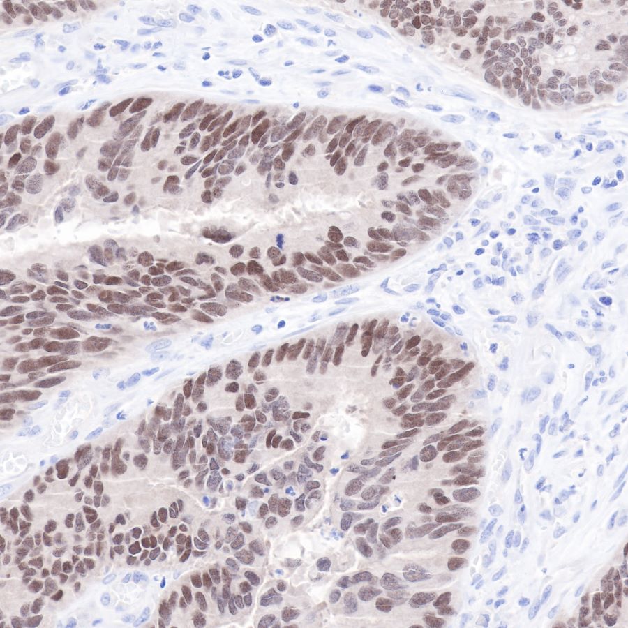
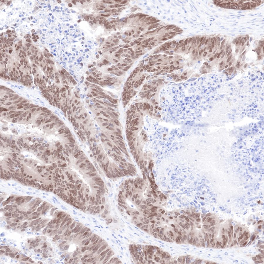
| Application |
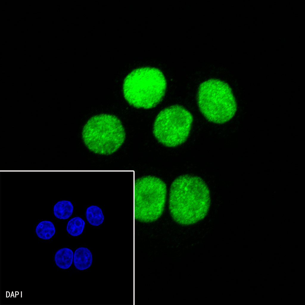
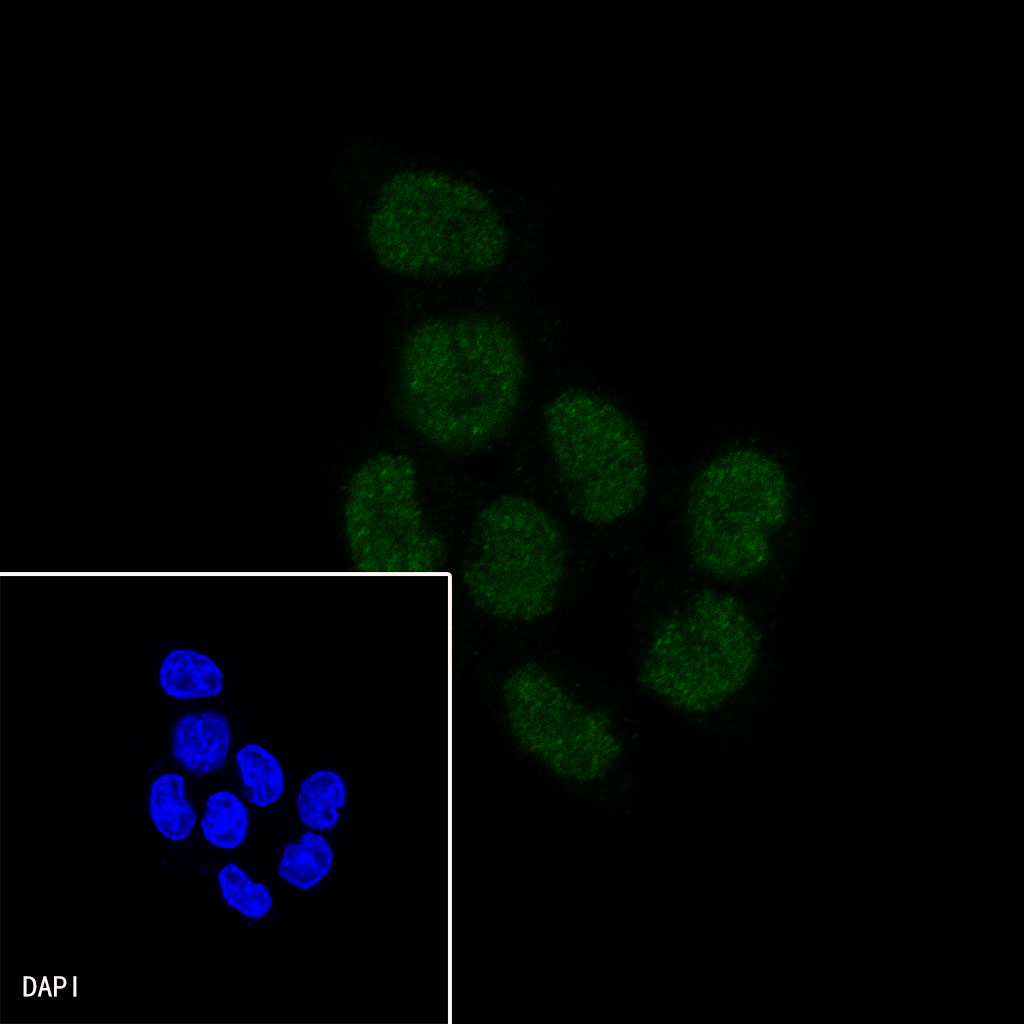
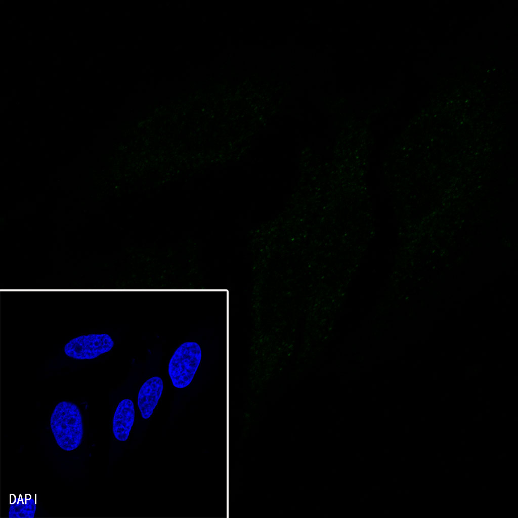
WB, IHC-P, ICC |
| Reactivity |
Hu, Ms, Rt |
| Predicted Reactivity |
Hm |
| Purification |
Protein A |
| Concentration |
0.5 mg/ml |
| Physical Appearance |
Liquid |
| Storage Buffer |
PBS, 40% Glycerol, 0.05%BSA, 0.03% Proclin 300 |
| Stability & Storage |
12 months from date of receipt / reconstitution, -20 °C as supplied |
Dilution
| application |
dilution |
species |
| WB |
1:1000 |
|
| IHC-P |
1:500 |
|
| ICC |
1:200-1:500 |
|
Background
Homeobox protein CDX-2 is a protein that in humans is encoded by the CDX2 gene. The CDX2 protein is a homeobox transcription factor expressed in the nuclei of intestinal epithelial cells, playing an essential role in the development and function of the digestive system. CDX2 is also used in diagnostic surgical pathology as a marker for gastrointestinal differentiation, especially colorectal.