Product Specification
| Host |
Rabbit |
| Antigen |
CD9 |
| Immunogen |
Synthetic Peptide |
| Location |
Cell membrane |
| Accession |
P21926 |
| Clone Number |
SDT-143-53 |
| Antibody Type |
Rabbit mAb |
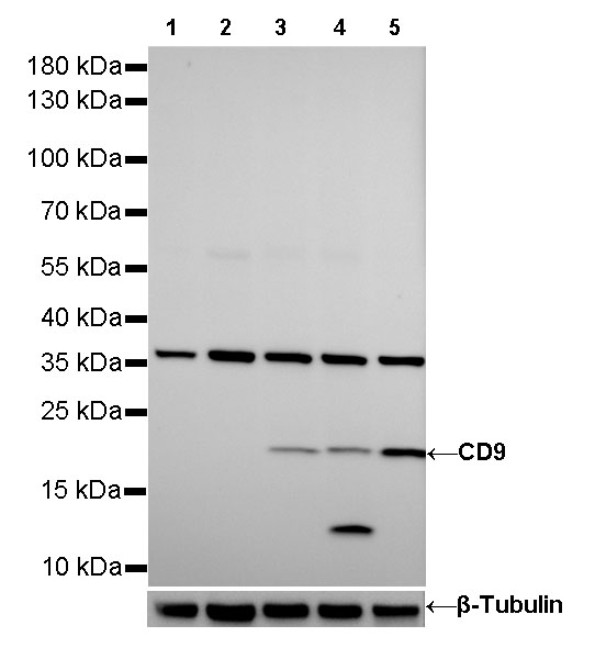
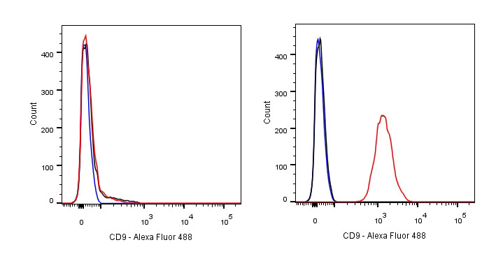
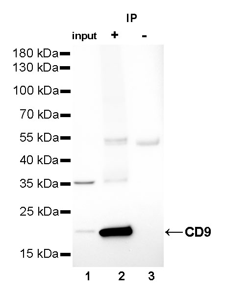
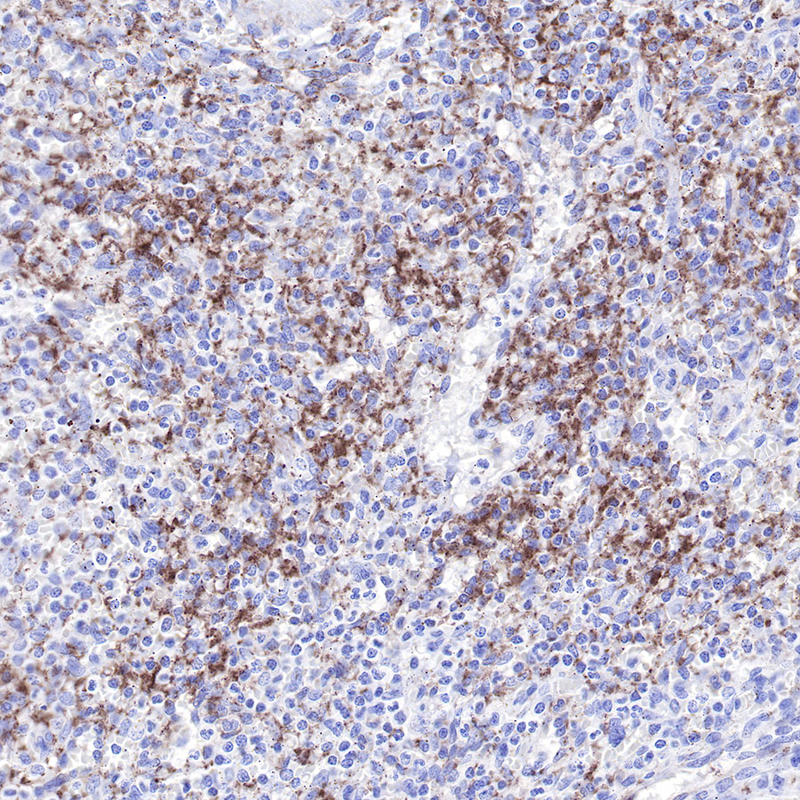
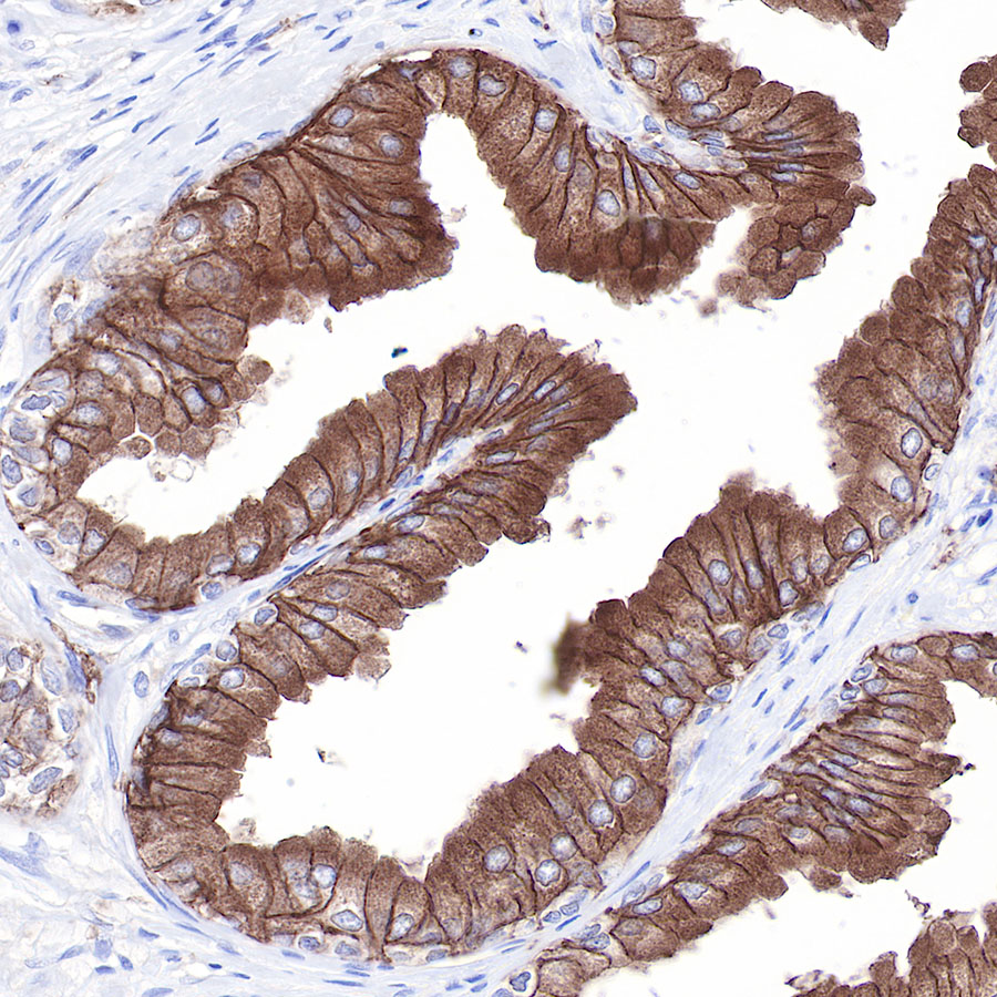
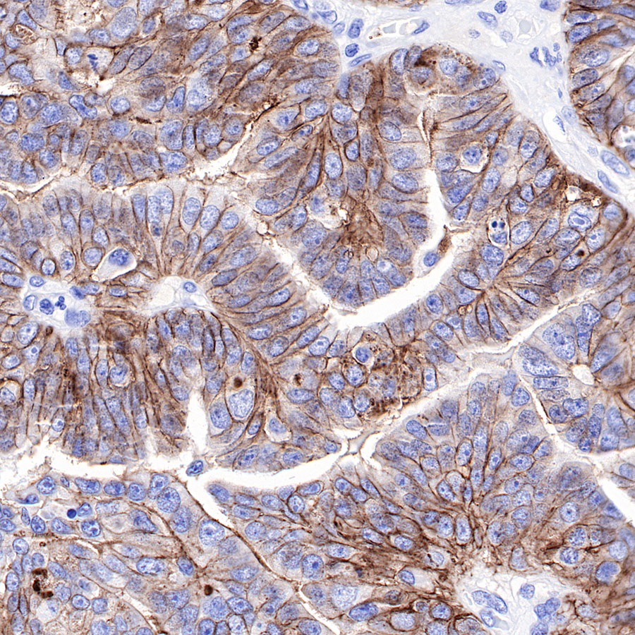
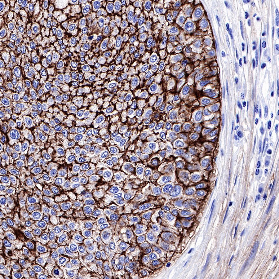
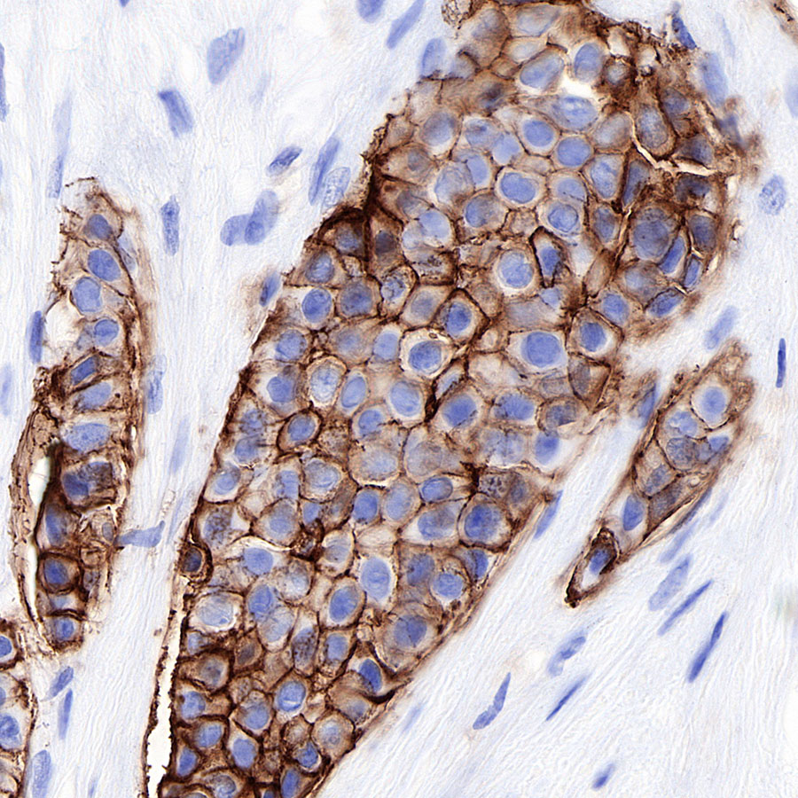
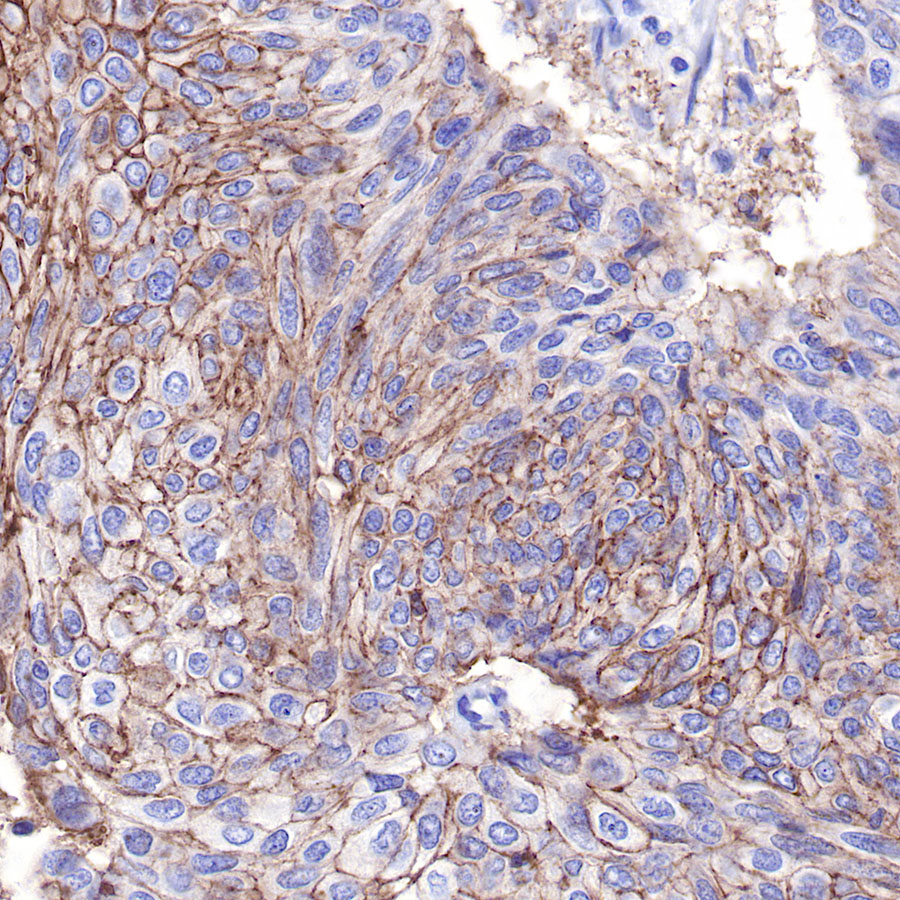
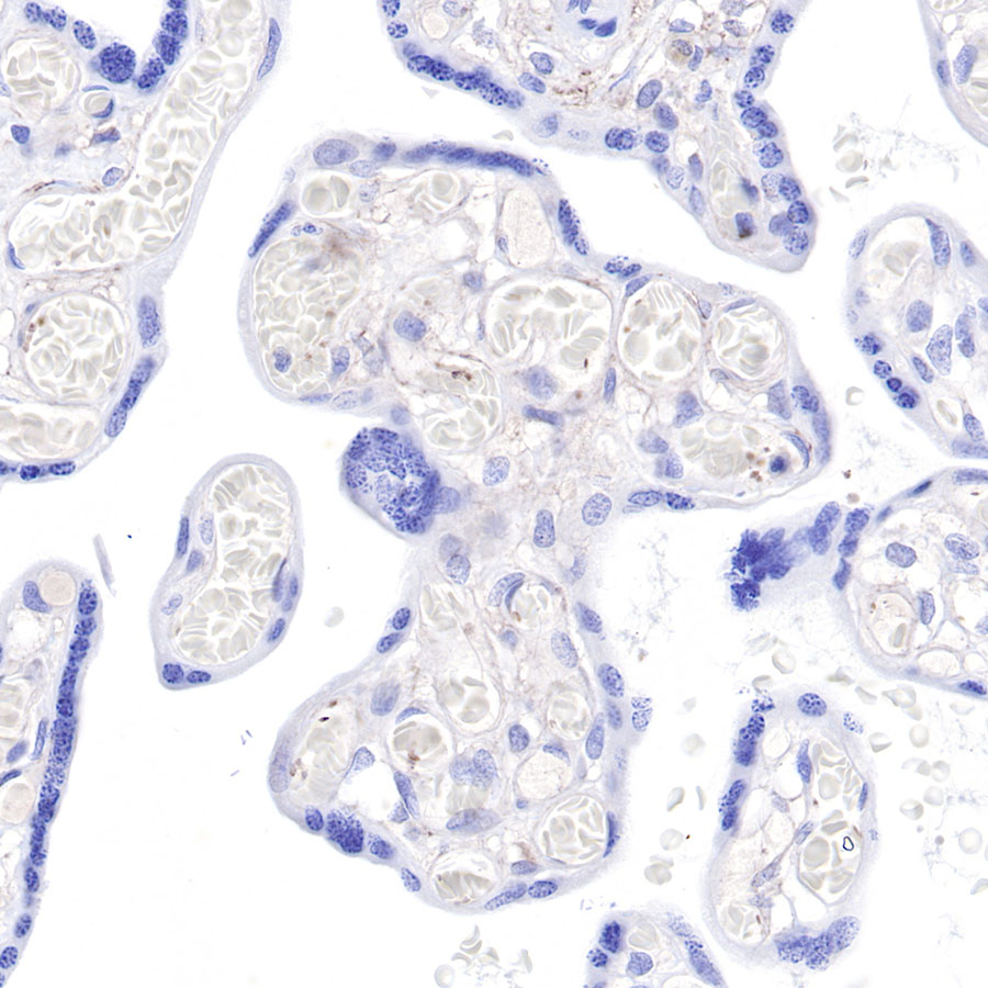
| Application |
WB, IHC-P, FCM, IP |
| Reactivity |
Hu |
| Purification |
Protein A |
| Concentration |
0.5 mg/ml |
| Physical Appearance |
Liquid |
| Storage Buffer |
PBS, 40% Glycerol, 0.05%BSA, 0.03% Proclin 300 |
| Stability & Storage |
12 months from date of receipt / reconstitution, -20 °C as supplied |
Dilution
| application |
dilution |
species |
| WB |
1:1000 |
null |
| IHC-P |
1:500-2000 |
null |
| FCM |
1:500 |
null |
| IP |
1:25 |
null |
Background
CD9 belongs to the cell surface glycoprotein cross -membrane four -protein family, with four cross -membrane domains, a shorter outer domain (ECL1), and a longer extracellular domain (ECL2). It is expressed in a variety of hematopoietic cells and epithelial cells. For example, Pre B cells, B cell subset, activated T cells, basophils, eosinophils, macrophages, megakaryocytes, plasma cells, plasma cell precursors in germinal centers, and platelets. Broadcasting a series of cell processes such as cell adhesion, movement, membrane tissue and signal transfusion. The lowering of CD9 expression is related to the adverse prognosis and progress of various cancers. It is a favorable marker of gallbladder cancer, gastic GIST, malignant cortex and oral squamous cell carcinoma.