Product Specification
| Host |
Rabbit |
| Antigen |
CD3G |
| Synonyms |
T-cell surface glycoprotein CD3 gamma chain, T-cell receptor T3 gamma chain, T3G |
| Immunogen |
Synthetic Peptide |
| Accession |
P09693 |
| Clone Number |
SDT-42-18 |
| Antibody Type |
Rabbit mAb |
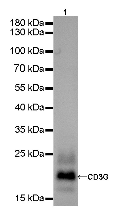
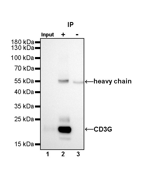
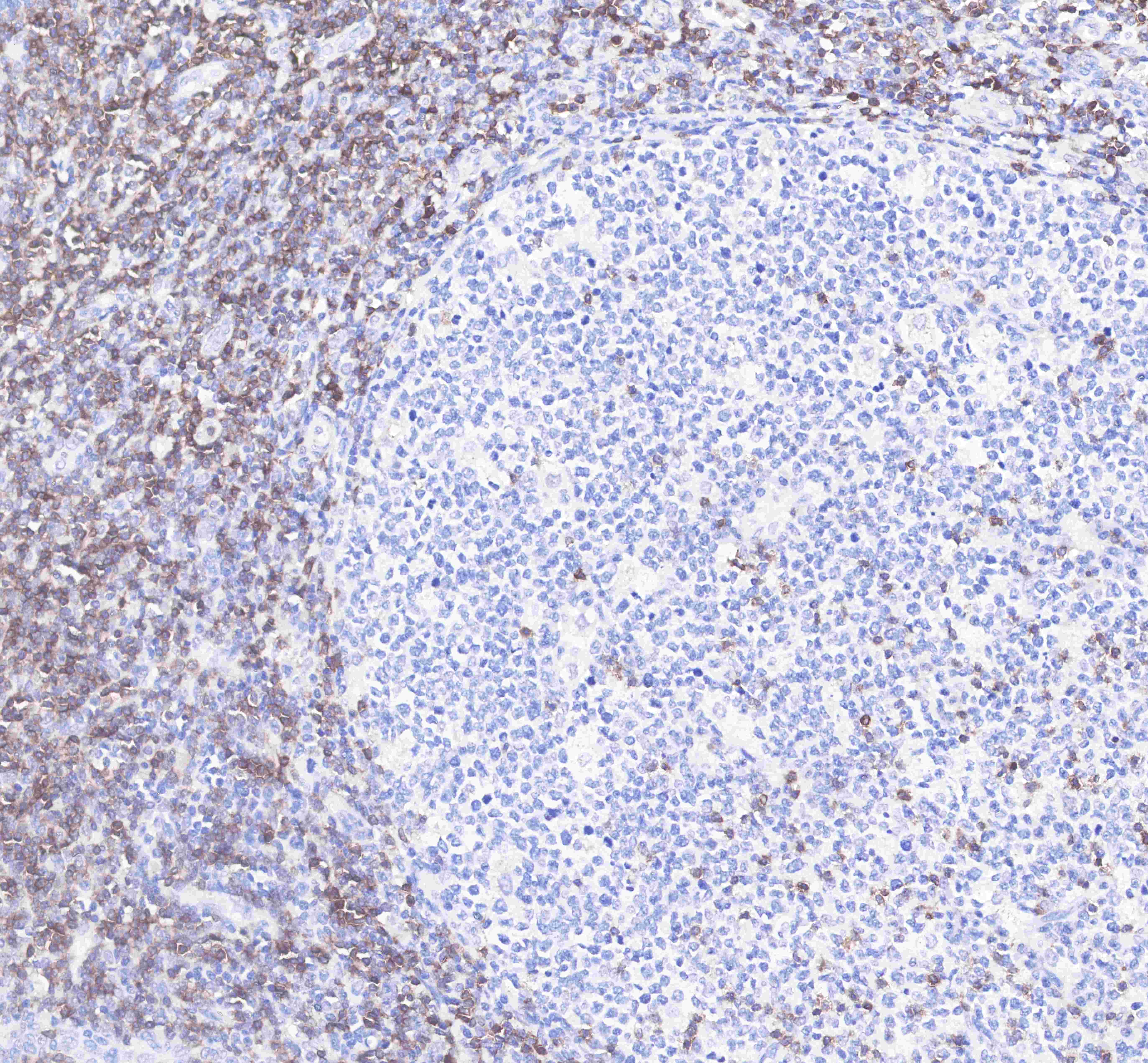
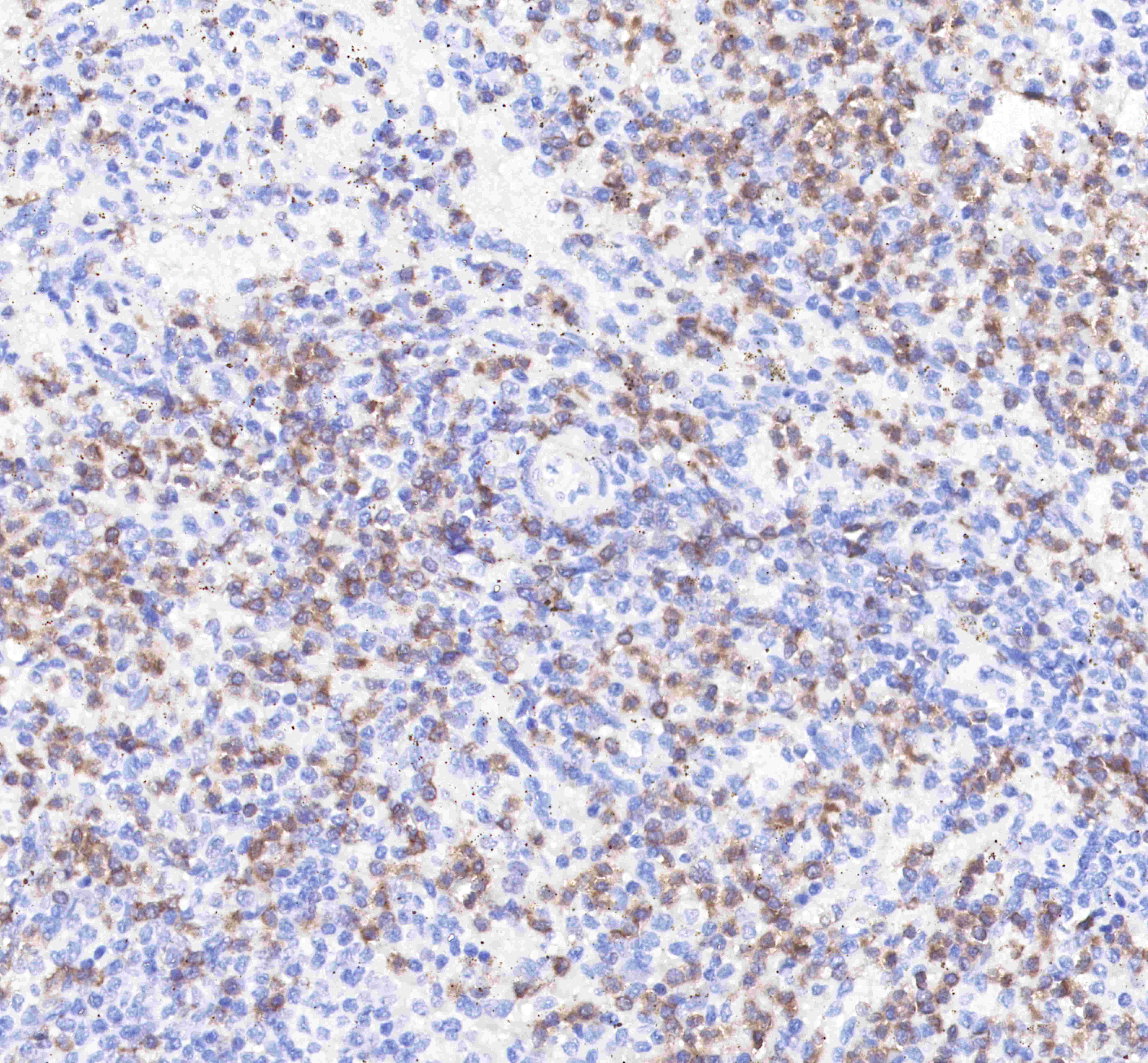
| Application |
WB, IHC-P, ICC, IP |
| Reactivity |
Hu |
| Purification |
Protein A |
| Concentration |
0.5mg/ml |
| Conjugation |
Unconjugated |
| Physical Appearance |
Liquid |
| Storage Buffer |
PBS, 40% Glycerol, 0.05%BSA, 0.03% Proclin 300 |
| Stability & Storage |
12 months from date of receipt / reconstitution, -20 °C as supplied |
Dilution
| application |
dilution |
species |
| IHC-P |
1:500 |
|
| ICC |
1:100 |
|
| WB |
1:1000 |
|
| IP |
1:50 |
|
Background
T-cell surface glycoprotein CD3 gamma chain is a protein that in humans is encoded by the CD3G gene. T cell antigen receptor (TCR) is associated on the T cell surface with a complex of protein called CD3. CD3G (gamma chain) is one of the four peptides (gamma, delta, epsilon and zeta) that form CD3. Defects in CD3G are associated with T cell immunodeficiency.